Comparative Study
. 2025 Jun 2;8(6):e2515672.
doi: 10.1001/jamanetworkopen.2025.15672.

AI-Assisted vs Unassisted Identification of Prostate Cancer in Magnetic Resonance Images

Collaborators, Affiliations
Comparative Study

AI-Assisted vs Unassisted Identification of Prostate Cancer in Magnetic Resonance Images

Jasper J Twilt et al. JAMA Netw Open. .

Abstract

Importance: Artificial intelligence (AI) assistance in magnetic resonance imaging (MRI) assessment for prostate cancer shows promise for improving diagnostic accuracy but lacks large-scale observational evidence.

Objective: To evaluate whether use of AI-assisted assessment for diagnosing clinically significant prostate cancer (csPCa) on MRI is superior to unassisted readings.

Design, setting, and participants: This diagnostic study was conducted between March and July 2024 to compare unassisted and AI-assisted diagnostic performance using the AI system developed within the international Prostate Imaging-Cancer AI (PI-CAI) Consortium. The study involved 61 readers (34 experts and 27 nonexperts) from 53 centers across 17 countries. Readers assessed prostate magnetic resonance images both with and without AI assistance, providing Prostate Imaging Reporting and Data System (PI-RADS) annotations from 3 to 5 (higher PI-RADS indicated a higher likelihood of csPCa) and patient-level suspicion scores ranging from 0 to 100 (higher scores indicated a greater likelihood of harboring csPCa). Biparametric prostate MRI examinations were included for 780 men from the PI-CAI study who were included in the newly-conducted observer study. All men within the PI-CAI study had suspicion of harboring prostate cancer, sufficient diagnostic image quality, and no prior clinically significant cancer findings. Disease presence was defined by histopathology, and absence was determined by 3 or more years of follow-up. The AI system was recalibrated using 420 Dutch examinations to generate lesion-detection maps, with AI scores ranging from 1 to 10, in which 10 indicates the highest likelihood of csPCa. The remaining 360 examinations, originating from 3 Dutch centers and 1 Norwegian center, were included in the observer study.

Main outcomes and measures: The primary outcome was diagnosis of csPCa, evaluated using the area under the receiver operating characteristic curve and sensitivity and specificity at a PI-RADS threshold of 3 or more. The secondary outcomes included analysis at alternate operating points and reader expertise.

Results: Among the 360 examinations of 360 men (median age, 65 years [IQR, 62-70 years]) who were included for testing, 122 (34%) harbored csPCa. AI assistance was associated with significantly improved performance, achieving a 3.3% increase in the area under the receiver operating characteristic curve (95% CI, 1.8%-4.9%; P < .001), from 0.882 (95% CI, 0.854-0.910) in unassisted assessments to 0.916 (95% CI, 0.893-0.938) with AI assistance. Sensitivity improved by 2.5% (95% CI, 1.1%-3.9%; P < .001), from 94.3% (95% CI, 91.9%-96.7%) to 96.8% (95% CI, 95.2%-98.5%), and specificity increased by 3.4% (95% CI, 0.8%-6.0%; P = .01), from 46.7% (95% CI, 39.4%-54.0%) to 50.1% (95% CI, 42.5%-57.7%), at a PI-RADS score of 3 or more. Secondary analyses demonstrated similar performance improvements across alternate operating points and a greater benefit of AI assistance for nonexpert readers.

Conclusions and relevance: The findings of this diagnostic study of patients suspected of harboring prostate cancer suggest that AI assistance was associated with improved radiologic diagnosis of clinically significant disease. Further research is required to investigate the generalization of outcomes and effects on workflow improvement within prospective settings.


Conflict of interest statement

Conflict of Interest Disclosures: Mr Saha reported receiving personal fees from Guerbet and Health~Holland outside the submitted work. Prof Padhani reported receiving research funding from Siemens Healthineers and Bayer and having stock options in Lucida Medical. Dr Bonekamp reported receiving personal fees from Bayer outside the submitted work. Dr Giannarini reported receiving personal fees from Astellas, Curium, Ferring, Hauora Med, Ipsen, Janssen, Johnson and Johnson, Pierre Fabre, and Recordati outside the submitted work. Dr van den Bergh reported serving as an advisory board member for Janssen; receiving speakers honoraria from Amgen, Astellas, Ipsen, Janssen, and MSD and research support from Astellas and Janssen; and participating in trials run by Janssen. Dr Kasivisvanathan reported receiving speakers honoraria from the European Association of Urology and the Singapore Urological Association and research funding from Prostate Cancer UK and The John Black Charitable Foundation. Dr Obuchowski reported providing statistical consultation to Siemens Healthineers, Takeda, and Qure and serving as a committee member of the Eastern Cooperative Oncology Group, the American College of Radiology Imaging Network, the Tomosynthesis Mammographic Imaging Screening Trial, and the National Cancer Institute’s Clinical Imaging Steering Committee. Dr Yakar reported receiving research grants from Siemens Healthineers, Health~Holland, The Dutch Research Council (NWO), and Hanarth; consulting fees from Astellas; speakers fees from Bayer; and a travel grant from the Multidisciplinary Digital Publishing Institute. Dr Elschot reported receiving grants from the Norwegian Cancer Society during the conduct of the study. Dr Huisman reported receiving research funding from Siemens Healthineers and Canon Medical Systems. Dr de Rooij reported receiving personal fees from Siemens Healthineers outside the submitted work. No other disclosures were reported.

Figures

Figure 1.
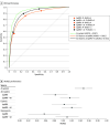
Figure 1.. The Area Under the Receiver Operating Characteristic Curve (AUROC) Diagnostic Performance at Biparametric Magnetic Resonance Imaging (bpMRI) and at bpMRI With Artificial Intelligence Assistance (bpMRI + AI)
A, ROCs of the performances of the AI system and the pool of 61 readers at bpMRI and bpMRI + AI. The diagonal dashed line indicates a random classifier. B, AUROC performance for the stand-alone AI system, all readers (N = 61), and subgroups considering expert (n = 34) and nonexpert (n = 27) readers for assessment made at bpMRI and bpMRI + AI at a Prostate Imaging Reporting and Data System (PI-RADS) score of 3 or more. Expert readers are readers with more than 1000 cases read in total and more than 200 cases per year, following 2020 consensus statements from the European Society of Urogenital Radiology and the European Association of Urology. Markers indicate mean AUROC; error bars, 95% CIs.
Figure 2.
Figure 2.. Sensitivity and Specificity at Biparametric Magnetic Resonance Imaging (bpMRI) Assessments and at bpMRI Assessments With Artificial Intelligence Assistance (bpMRI + AI)
Sensitivities (A) and specificities (B) for all 61 readers and subgroups considering experts (n = 34) and nonexperts (n = 27) at a Prostate Imaging Reporting and Data System operating point of 3 or more. Expert readers are readers with more than 1000 cases read in total and more than 200 cases per year, following 2020 consensus statements from the European Society of Urogenital Radiology and the European Association of Urology. Markers indicate mean percentages; error bars, 95% CIs.
Figure 3.
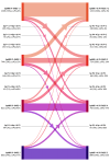
Figure 3.. Diagram of Patient-Level Prostate Imaging Reporting and Data System (PI-RADS) Scores From Unassisted Biparametric Magnetic Resonance Imaging (bpMRI; Left) and Artificial Intelligence (AI)-Assisted bpMRI (bpMRI + AI; Right) Assessments in the Observer Study
The diagram highlights interrater consistencies and changes (upgrades and downgrades) between the 2 configurations. Each scoring category and pair is presented with occurrence numbers, percentages, and clinically significant prostate cancer (csPCa) prevalence. Of all readings, 2465 of the 3660 total assessments (67%) remained unchanged between assessments, 278 (8%) involved reclassification from negative (PI-RADS [P] <3) to positive (PI-RADS [P] ≥3) MRI, and 330 (9%) involved reclassification from positive to negative MRI. The remaining 587 (16%) involved reclassification within the positive MRI group. The overall PI-RADS score distribution was similar across both reading configurations, while csPCa prevalence changed due to scoring updates.
Figure 4.
Figure 4.. Proportion of Prostate Imaging Reporting and Data System (PI-RADS) Scores Observed for Unassisted and Artificial Intelligence (AI)-Assisted Assessments by AI Scores of Examinations
Among the 3660 total assessments, AI assistance (right) compared with unassisted assessment (left) was associated with increases in the PI-RADS score of 1 to 2 in lower AI score categories and with decreases in higher AI score categories. Similarly, the proportion of PI-RADS scores of 4 to 5 increased in high AI score categories with AI assistance.

Comment in

References

    1. Bray F, Laversanne M, Sung H, et al. Global cancer statistics 2022: GLOBOCAN estimates of incidence and mortality worldwide for 36 cancers in 185 countries. CA Cancer J Clin. 2024;74(3):229-263. doi: 10.3322/caac.21834 - DOI - PubMed
    1. Drost FH, Osses DF, Nieboer D, et al. Prostate MRI, with or without MRI-targeted biopsy, and systematic biopsy for detecting prostate cancer. Cochrane Database Syst Rev. 2019;4(4):CD012663. doi: 10.1002/14651858.CD012663.pub2 - DOI - PMC - PubMed
    1. Kasivisvanathan V, Rannikko AS, Borghi M, et al. ; PRECISION Study Group Collaborators . MRI-targeted or standard biopsy for prostate-cancer diagnosis. N Engl J Med. 2018;378(19):1767-1777. doi: 10.1056/NEJMoa1801993 - DOI - PMC - PubMed
    1. Ahmed HU, El-Shater Bosaily A, Brown LC, et al. ; PROMIS study group . Diagnostic accuracy of multi-parametric MRI and TRUS biopsy in prostate cancer (PROMIS): a paired validating confirmatory study. Lancet. 2017;389(10071):815-822. doi: 10.1016/S0140-6736(16)32401-1 - DOI - PubMed
    1. van der Leest M, Cornel E, Israël B, et al. Head-to-head comparison of transrectal ultrasound-guided prostate biopsy versus multiparametric prostate resonance imaging with subsequent magnetic resonance-guided biopsy in biopsy-naïve men with elevated prostate-specific antigen: a large prospective multicenter clinical study. Eur Urol. 2019;75(4):570-578. doi: 10.1016/j.eururo.2018.11.023 - DOI - PubMed

Publication types