

Enzymatic carbon-fluorine bond cleavage by human gut microbes
- PMID: 40512801
- PMCID: PMC12184663
- DOI: 10.1073/pnas.2504122122
Enzymatic carbon-fluorine bond cleavage by human gut microbes
Abstract
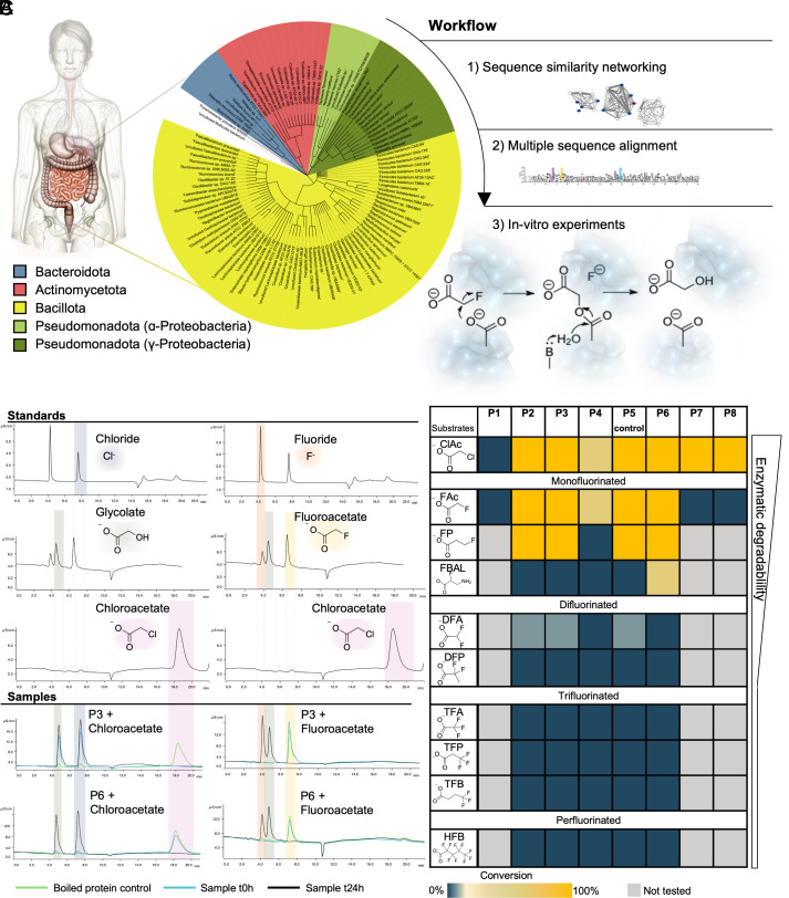
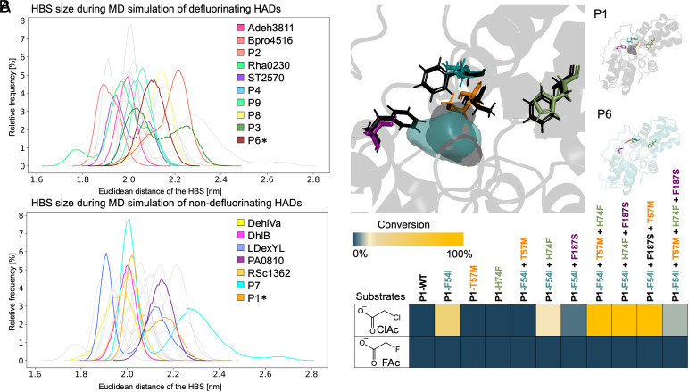
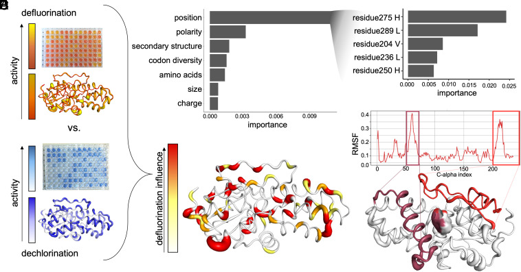
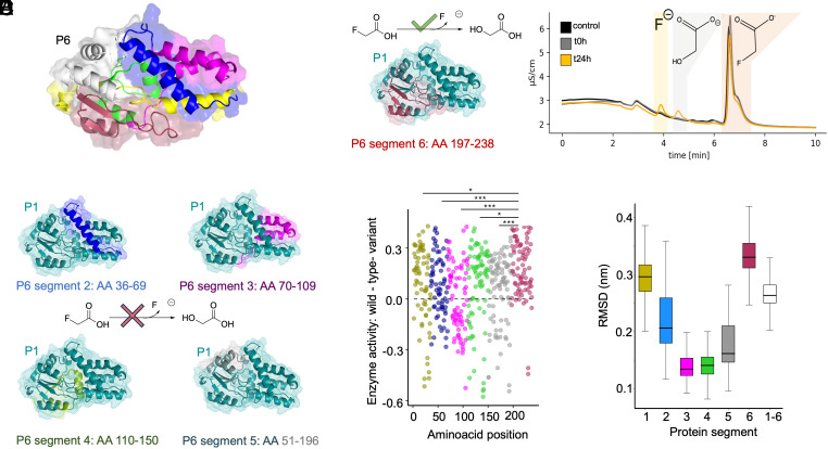
Fluorinated compounds are used for agrochemical, pharmaceutical, and numerous industrial applications, resulting in global contamination. In many molecules, fluorine is incorporated to enhance the half-life and improve bioavailability. Fluorinated compounds enter the human body through food, water, and xenobiotics including pharmaceuticals, exposing gut microbes to these substances. The human gut microbiota is known for its xenobiotic biotransformation capabilities, but it was not previously known whether gut microbial enzymes could break carbon-fluorine bonds, potentially altering the toxicity of these compounds. Here, through the development of a rapid, miniaturized fluoride detection assay for whole-cell screening, we identified active gut microbial defluorinases. We biochemically characterized enzymes from diverse human gut microbial classes including Clostridia, Bacilli, and Coriobacteriia, with the capacity to hydrolyze (di)fluorinated organic acids and a fluorinated amino acid. Whole-protein alanine scanning, molecular dynamics simulations, and chimeric protein design enabled the identification of a disordered C-terminal protein segment involved in defluorination activity. Domain swapping exclusively of the C-terminus conferred defluorination activity to a nondefluorinating dehalogenase. To advance our understanding of the structural and sequence differences between defluorinating and nondefluorinating dehalogenases, we trained machine learning models which identified protein termini as important features. Models trained on 41-amino acid segments from protein C termini alone predicted defluorination activity with 83% accuracy (compared to 95% accuracy based on full-length protein features). This work is relevant for therapeutic interventions and environmental and human health by uncovering specificity-determining signatures of fluorine biochemistry from the gut microbiome.
Keywords: defluorination; haloacid dehalogenases; human gut microbiome; molecular dynamics; protein engineering.
Conflict of interest statement
Competing interests statement:The authors declare no competing interest.
Figures
Similar articles
-
Computational Studies of Enzymes for C-F Bond Degradation and Functionalization.Chemphyschem. 2025 May 5;26(9):e202401130. doi: 10.1002/cphc.202401130. Epub 2025 Mar 6. Chemphyschem. 2025. PMID: 39962931 Free PMC article. Review.
-
Immunogenicity and seroefficacy of pneumococcal conjugate vaccines: a systematic review and network meta-analysis.Health Technol Assess. 2024 Jul;28(34):1-109. doi: 10.3310/YWHA3079. Health Technol Assess. 2024. PMID: 39046101 Free PMC article.
-
Integrating Gut Microbiome and Metabolomics with Magnetic Resonance Enterography to Advance Bowel Damage Prediction in Crohn's Disease.J Inflamm Res. 2025 Jun 11;18:7631-7649. doi: 10.2147/JIR.S524671. eCollection 2025. J Inflamm Res. 2025. PMID: 40535353 Free PMC article.
-
Signs and symptoms to determine if a patient presenting in primary care or hospital outpatient settings has COVID-19.Cochrane Database Syst Rev. 2022 May 20;5(5):CD013665. doi: 10.1002/14651858.CD013665.pub3. Cochrane Database Syst Rev. 2022. PMID: 35593186 Free PMC article.
-
Nivolumab for adults with Hodgkin's lymphoma (a rapid review using the software RobotReviewer).Cochrane Database Syst Rev. 2018 Jul 12;7(7):CD012556. doi: 10.1002/14651858.CD012556.pub2. Cochrane Database Syst Rev. 2018. PMID: 30001476 Free PMC article.
References
-
- Yamashita K., Yada H., Ariyoshi T., Neurotoxic effects of alpha-fluoro-beta-alanine (FBAL) and fluoroacetic acid (FA) on dogs. J. Toxicol. Sci. 29, 155–166 (2004). - PubMed
-
- Okeda R., et al. , Experimental neurotoxicity of 5-fluorouracil and its derivatives is due to poisoning by the monofluorinated organic metabolites, monofluoroacetic acid and alpha-fluoro-beta-alanine. Acta Neuropathol. 81, 66–73 (1990). - PubMed
MeSH terms
Substances
Grants and funding
- 2022-YIG-090/Helmut Horten Stiftung (Helmut Horten Foundation)
- PZPGP2_209124/Schweizerischer Nationalfonds zur Förderung der Wissenschaftlichen Forschung (SNF)
- PZPGP2_209124/Schweizerischer Nationalfonds zur Förderung der Wissenschaftlichen Forschung (SNF)
- N/A/Peter und Traudl Engelhorn Stiftung (Peter and Traudl Engelhorn Foundation)
- 230-2024/Uniscientia Foundation
