. 2025 Jun 13;11(24):eadu5791.
doi: 10.1126/sciadv.adu5791. Epub 2025 Jun 13.

Switchable client specificity in a dual functional chaperone coordinates light-harvesting complex biogenesis

Affiliations

Switchable client specificity in a dual functional chaperone coordinates light-harvesting complex biogenesis

Alex R Siegel et al. Sci Adv. .

Abstract

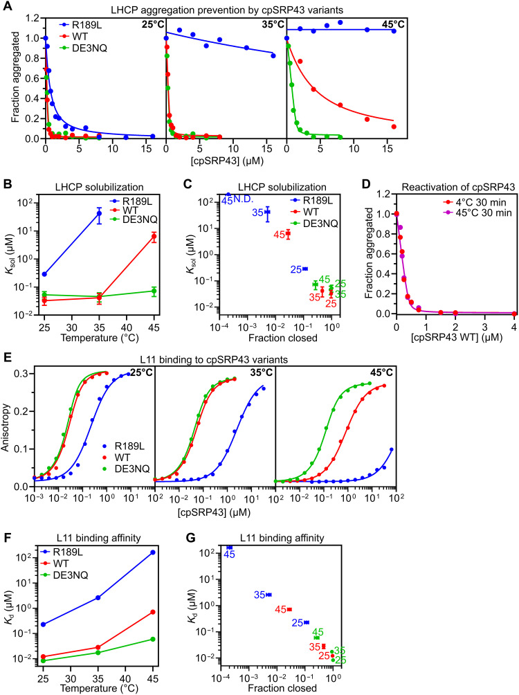
The proper assembly of light-harvesting complexes (LHCs) is critical for photosynthesis and requires the biogenesis of light-harvesting chlorophyll a,b-binding proteins (LHCPs) to be coordinated with chlorophyll (Chl) biosynthesis. The mechanism underlying this coordination is not well understood. Here, we show that a conserved molecular chaperone, chloroplast signal recognition particle 43-kDa protein (cpSRP43), provides a molecular thermostat that helps maintain this coordination. cpSRP43 undergoes a conformational rearrangement between a well-folded closed state and a partially disordered open state. Closed cpSRP43 is dedicated to the biogenesis of LHCPs, whereas open cpSRP43 protects multiple Chl biosynthesis enzymes from heat-induced destabilization. Rising temperature shifts cpSRP43 to the open state, enabling it to protect heat-destabilized Chl biosynthesis enzymes. Our results reveal the molecular basis of a posttranslational mechanism for the thermoadaptation of LHC biogenesis. They also demonstrate how an adenosine triphosphate-independent chaperone uses conformational dynamics to switch its activity and client selectivity, thereby adapting to different proteostatic demands under shifting environmental conditions.


Figures

Fig. 1.
Fig. 1.. Identification of mutations that stabilize the open or closed state of cpSRP43.
(A) 19F-NMR spectra of BTFA-labeled WT cpSRP43 at the indicated temperatures. The open (red “O”) and closed (green “C”) state peaks were assigned in (27). Spectra were collected in CD buffer with 50 mM NaCl. ppm, parts per million. (B) The fraction of cpSRP43 variants in the open state at different temperatures determined by 19F-NMR. Data were from the spectra shown in (A) and fig. S1. “*” denotes the data collected after the completion of one heating cycle, where 10 to 20% of cpSRP43 did not return to the closed state. (C) CD spectra of WT cpSRP43 at 25°C (solid) and 55°C (dashed). Data were collected in CD buffer with 50 mM NaCl and repeated for three heat cycles (dark to light red). Values represent mean residue ellipticity ± SE from n = 3 technical replicates. (D) Temperature dependence of helical content for R189L, WT, DE3NQ, and DE6NQ cpSRP43, measured as per-residue molar ellipticity (θ) at 222 nm by CD in CD buffer with 50 mM NaCl. Representative data from three technical replicates are shown. Values represent mean residue ellipticity ± SD. The lines are fits of the data to Eq. 2, and the obtained Tm values from n = 3 technical replicates are summarized in Table 1. (E and F) Closed state structure of the SBD of A. thaliana cpSRP43 [Protein Data Bank (PDB): 3DEP (9)] showing the hydrogen bonding interactions of R189 (E) or the seven closely spaced acidic residues (F). Sequence conservation of these acidic residues across 1000 land plants is shown in WebLogo [(F), top right] (51). The charge-neutralizing mutations are indicated [(F), bottom right].
Fig. 2.
Fig. 2.. Closed cpSRP43 mediates chaperone activity toward the LHCPs.
(A) Turbidity assays to measure the protection of LHCP by increasing concentrations of cpSRP43 variants at 25°C (left), 35°C (middle), and 45°C (right) in CD buffer with 50 mM NaCl. Optical density at 360 nm (A360) was normalized to that without cpSRP43. Representative data from three technical replicates are shown. Lines are fits of the data to Eq. 4A. (B and C) Obtained Ksol values for LHCP plotted as a function of temperature (B) or fraction of chaperone in the closed state (C). Values represent mean fitted values ± SE of n = 3 technical replicates. (D) Turbidity assays of reactivated WT cpSRP43 heated at 45°C for 30 min and then returned to ice (violet) or unheated (red) carried out toward LHCP at 25°C as in (A). A360 was normalized to that without cpSRP43. Representative data from three technical replicates are shown. Lines are fits of the data to Eq. 4A. (E) Equilibrium titrations to measure the binding of the indicated cpSRP43 variants to HiLyte Fluor 488–labeled L11 peptide at 25°C (left), 35°C (middle), and 45°C (right) in CD buffer with 50 mM NaCl. Representative data from three technical replicates are shown. Lines are fits of the data to Eq. 6A. (F and G) Obtained dissociation constant (Kd) values for L11 peptide binding were plotted as a function of temperature (F) or fraction of chaperone in the closed state (G). Values represent mean fitted values ± SE of n = 3 technical replicates. The numbers next to the data points in (C) and (G) indicate the temperatures at which the measurement was made. The fraction of chaperone in the closed state was from the measurements in Fig. 1D.
Fig. 3.
Fig. 3.. Open cpSRP43 mediates the thermoprotection of TBS enzymes.
(A) Turbidity assays to measure the heat-induced aggregation of 10 μΜ GUN4 and its protection by the indicated cpSRP43 variants in the low-salt (LS) buffer with 50 (left), 100 (middle), and 200 (right) mM NaCl. A360 was normalized to that without cpSRP43. Representative data from three technical replicates are shown. The lines are fits of the data to Eq. 4B. (B) Same as (A), except with 2.7 μM GluTR as the client protein. (C and D) The fitted Ksol values from the data in (A) (see Materials and Methods) are plotted as a function of NaCl concentration (C) and fraction of chaperone in the closed state (D). The numbers next to the data indicate the NaCl concentration at which the measurements were made. Values represent mean fitted values ± SE of n = 3 technical replicates.
Fig. 4.
Fig. 4.. cpSRP54 and C-terminal chromodomains modulate TBS thermoprotection by open cpSRP43.
(A) Equilibrium titrations to measure the binding affinity of the 54C peptide for cpSRP43 variants at rising temperatures. Reactions were carried out in CD buffer with 50 mM NaCl. Representative data from three technical replicates are shown. The lines are fits of the data to Eqs. 6A and 6B. (B) The obtained mean Kd values ± SE from n = 3 technical replicates are plotted as a function of temperature. (C) Temperature dependence of the helical content for R189L, WT, and DE3NQ cpSRP43 (SBD), measured as per-residue molar ellipticity (θ) at 222 nm by CD in CD buffer with 200 mM NaCl and fit as in Fig. 1D. Representative data from three technical replicates are shown. Values represent mean residue ellipticity ± SD of the signal over 10 s. (D and E) Comparison of the Ksol values between FL and SBD constructs in the thermoprotection of GUN4 (D) and GluTR (E) for cpSRP43 variants. Ksol values shown are from fits of the data in Fig. 3 (A and B) and fig. S9 (B and C) to Eq. 4B for n = 3 technical replicates.
Fig. 5.
Fig. 5.. Characterization of transgenic cpSRP43 Arabidopsis lines.
(A) Representative images of 18-day-old seedlings of WT (Ler-0), chaos, and transgenic lines expressing the indicated cpSRP43 variants using the endogenous cpSRP43 promoter. Seedlings were grown under normal conditions. Scale bars, 0.5 cm. (B to D) Steady-state levels of Chl (B) and Chl precursors including Mg-porphyrins (C) and protochlorophyllide [PChlide; (D)] in 18-day-old seedlings shown in (A). MgP, Mg-protoporphyrin IX; MgPMME, MgP monomethylester; FW, fresh weight. (E) Steady-state levels of cpSRP43, cpSRP54, and GUN4 in 18-day-old seedlings shown in (A) were analyzed by immunoblotting using the indicated antibodies. The Ponceau S–stained large subunit of ribulose-1,5-bisphosphate carboxylase/oxygenase (RbcL) and actin is shown as loading control.
Fig. 6.
Fig. 6.. Open cpSRP43 stabilizes the levels of TBS enzymes in planta during short-term heat stress.
(A) Steady-state levels of the indicated proteins in 10-day-old transgenic seedlings before and after 0.5 and 1 hour of heat treatment at 42°C in the presence of CHX were analyzed by immunoblotting using the indicated antibodies. The Ponceau S–stained RbcL and actin are shown as loading controls. (B and C) Semiquantitative analysis with the ImageJ software of the immunoblots in (A). The relative amounts of GUN4 (B) and CHLH (C) were normalized to their levels before exposure to elevated temperature (0 hours at 42°C), with actin serving as the loading control for quantification. An independent biological replicate is shown in fig. S11.
Fig. 7.
Fig. 7.. Model for cpSRP43’s dual chaperone activities.
Under normal conditions, cpSRP43 primarily populates the closed conformation in which its ARMs in the SBD are tightly folded and further stabilized by interactions with cpSRP54 (left). Closed cpSRP43 confers high-affinity recognition and protection of newly imported LHCP but has no activity toward TBS enzymes. At elevated temperatures, a fraction of cpSRP43 transitions to the open conformation in which the C-terminal ARMs become partially disordered (right). Open cpSRP43 does not recognize LHCP but effectively protects mature TBS enzymes from misfolding, aggregation, and resulting proteolytic degradation. cpSRP54, which inhibits the TBS protection activity of cpSRP43, also dissociates from the chaperone at elevated temperatures. These changes unleash the thermoprotection activity of cpSRP43 under conditions where this activity is needed.

Similar articles

References

    1. Jarvis P., López-Juez E., Biogenesis and homeostasis of chloroplasts and other plastids. Nat. Rev. Mol. Cell Biol. 14, 787–802 (2013). - PubMed
    1. Leutwiler L. S., Meyerowitz E. M., Tobin E. M., Structure and expression of three light-harvesting chlorophyll a/b-binding protein genes in Arabidopsis thaliana. Nucleic Acids Res. 14, 4051–4064 (1986). - PMC - PubMed
    1. Plumley G. F., Schmidt G. W., Light-harvesting chlorophyll a/b complexes: Interdependent pigment synthesis and protein assembly. Plant Cell 7, 689–704 (1995). - PMC - PubMed
    1. Dall’Osto L., Bressan M., Bassi R., Biogenesis of light harvesting proteins. Biochim. Biophys. Acta Bioenerg. 1847, 861–871 (2015). - PubMed
    1. Paulsen H., Finkenzeller B., Kühlein N., Pigments induce folding of light-harvesting chlorophyll a/b-binding protein. Eur. J. Biochem. 215, 809–816 (1993). - PubMed

MeSH terms

LinkOut - more resources