. 2025 Oct:271:112975.
doi: 10.1016/j.jinorgbio.2025.112975. Epub 2025 Jun 10.

Mutation of an active site-adjacent residue in VIM indirectly dictates interactions with and blunts inhibition by D-captopril

Affiliations

Mutation of an active site-adjacent residue in VIM indirectly dictates interactions with and blunts inhibition by D-captopril

Surendra Bikram Silwal et al. J Inorg Biochem. 2025 Oct.

Abstract

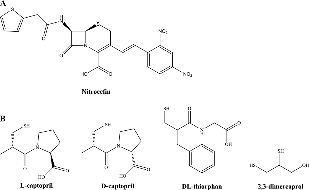
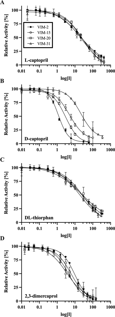
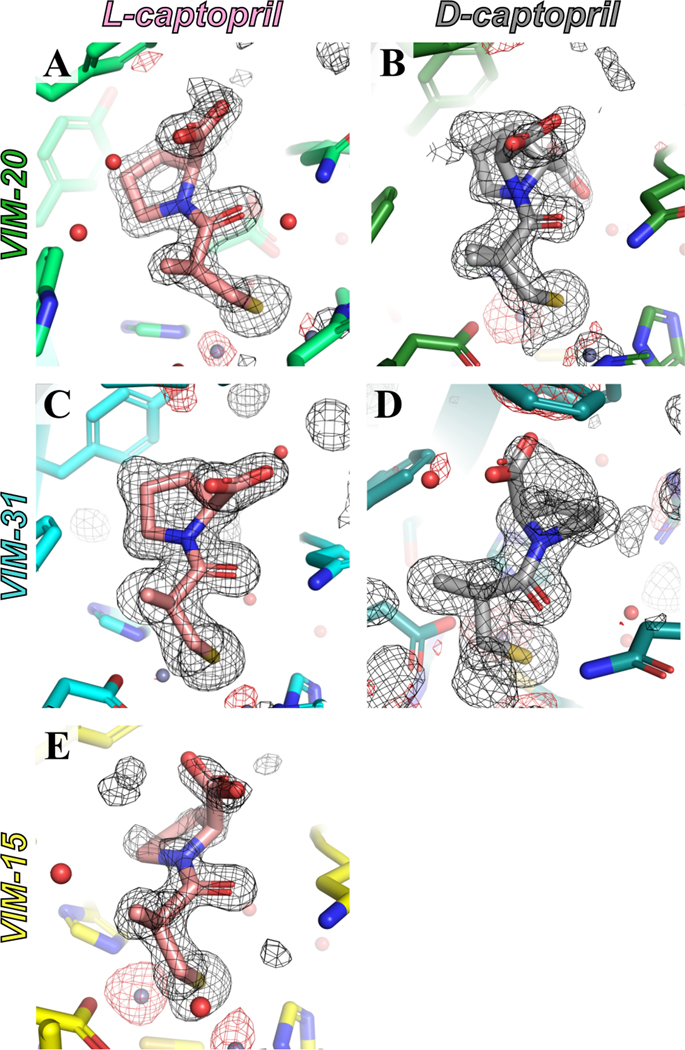
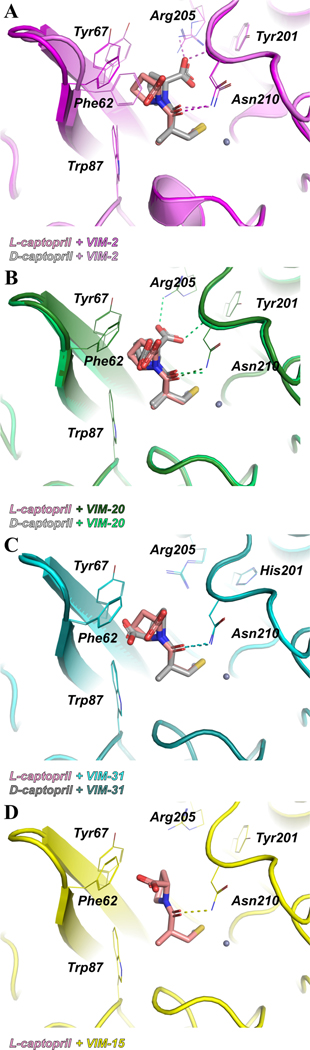
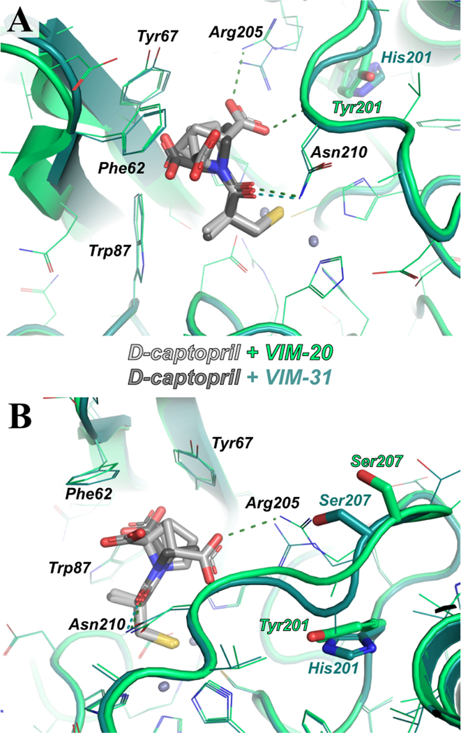
Activity assays and X-ray crystallographic studies were undertaken to elucidate the inhibitory mechanism of captopril stereoisomers on Verona integron-encoded metallo-β-lactamases, specifically VIM-20, VIM-31, and VIM-15. All three VIM-2-like variants (VIM-20, VIM-31, and VIM-15) and VIM-2 expressed in Escherichia coli exhibited catalytic activity with comparable steady-state kinetic parameters. Among the tested thiol drugs (L- and D-captopril, D,L-thiorphan, and 2,3-dimercaprol), IC50 analyses indicated that D-captopril and 2,3-dimercaprol were more potent inhibitors against the VIM enzymes examined in this study. Notably, the IC50 value of D-captopril against VIM-31 was an exception, closely resembling that of L-captopril. To elucidate this exceptional inhibitory potency of D-captopril and its binding mode in the active site of VIM-31, high-resolution crystal structures of VIM-20, VIM-31, and VIM-15 in complex with both L- and D-captopril are reported. These findings will help evaluate whether the identified potent inhibitor D-captopril could be further developed as a pan inhibitor targeting the VIM-family enzymes.

Keywords: Antibiotic resistance; Enzyme inhibition; Metallo-β-lactamase; Protein crystallography.


Conflict of interest statement

Declaration of competing interest Richard Page reports financial support was provided by National Institute of General Medical Sciences. If there are other authors, they declare that they have no known competing financial interests or personal relationships that could have appeared to influence the work reported in this paper.

Figures

Figure 1:
Figure 1:
Chemical structures of (A) the chromogenic cephalosporin substrate nitrocefin and (B) the thiol-containing compounds tested as MBL inhibitors in this study.
Figure 2:
Figure 2:
IC50 plots for (A) L-captopril, (B) D-captopril, (C) DL-thiorphan, and (D) 2,3-dimercaprol, respectively, taking nitrocefin as a substrate and enzyme concentration at 5.4 nM. All data points represent the average slope of three experiments, and error bars indicate the standard deviation.
Figure 3:
Figure 3:
Captopril isomers as inhibitors against VIM enzymes, with X-ray crystallographic electron density omit maps for (A) L-captopril (salmon) with VIM-20 (lime green), (B) D-captopril (grey) with VIM-20 (forest green), (C) L-captopril (salmon) with VIM-31 (cyan), (D) D-captopril (grey) with VIM-31 (dark teal), and (E) L-captopril (salmon) with VIM-15 (in yellow). The Fo-Fc electron density omit maps were calculated using POLDER and are drawn at 3.0 σ throughout, with positive density in black and negative density in red.
Figure 4:
Figure 4:
X-ray crystal structures of VIM enzymes in complex with D-captopril or L-captopril. (A) Overlay of VIM-2 (pink) in complex with D-captopril (grey) and VIM-2 (purple) in complex with L-captopril (salmon). (B) Overlay of VIM-20 (lime green) in complex with D-captopril (grey) and VIM-20 (forest green) in complex with L-captopril (salmon). (C) Overlay of VIM-31 (dark teal) in complex with D-captopril (grey) and VIM-31 (cyan) in complex with L-captopril (salmon). (D)VIM-15 (yellow) in complex with L-captopril (salmon).
Figure 5:
Figure 5:
An overlay of X-ray crystallographic structures featuring VIM-20 and VIM-31 with L-captopril and D-captopril. (A) VIM-20 with D-captopril and VIM-3 with D-captopril; and (B) rotated view of A highlighting the Tyr-201-His mutation and altered position of Ser-207. In each panel, VIM-31 is colored dark teal, D-captopril bound to VIM-31 is colored dark grey, VIM-20 is colored lime green, and D-captopril bound to VIM-20 is colored light grey.

Similar articles

References

    1. Uluseker C, Kaster KM, Thorsen K, et al. (2021) A Review on Occurrence and Spread of Antibiotic Resistance in Wastewaters and in Wastewater Treatment Plants: Mechanisms and Perspectives. Front Microbiol 12:717809. 10.3389/fmicb.2021.717809 - DOI - PMC - PubMed
    1. Carfi A, Pares S, Duée E, et al. (1995) The 3-D structure of a zinc metallo-beta-lactamase from Bacillus cereus reveals a new type of protein fold. EMBO J 14:4914–4921. 10.1002/j.1460-2075.1995.tb00174.x - DOI - PMC - PubMed
    1. Yan Y-H, Li G, Li G-B (2020) Principles and current strategies targeting metallo-β-lactamase mediated antibacterial resistance. Med Res Rev 40:1558–1592. 10.1002/med.21665 - DOI - PubMed
    1. Pandey N, Cascella M (2023) Beta-Lactam Antibiotics. In: StatPearls. StatPearls Publishing, Treasure Island (FL) - PubMed
    1. Kohanski MA, Dwyer DJ, Collins JJ (2010) How antibiotics kill bacteria: from targets to networks. Nat Rev Microbiol 8:423–435. 10.1038/nrmicro2333 - DOI - PMC - PubMed

MeSH terms