. 2025 Jun 13;13(6):e011776.
doi: 10.1136/jitc-2025-011776.

Inhibiting CMTM4 reverses the immunosuppressive function of myeloid-derived suppressor cells and augments immunotherapy response in cervical cancer

Affiliations

Inhibiting CMTM4 reverses the immunosuppressive function of myeloid-derived suppressor cells and augments immunotherapy response in cervical cancer

Jianyi Ding et al. J Immunother Cancer. .

Abstract

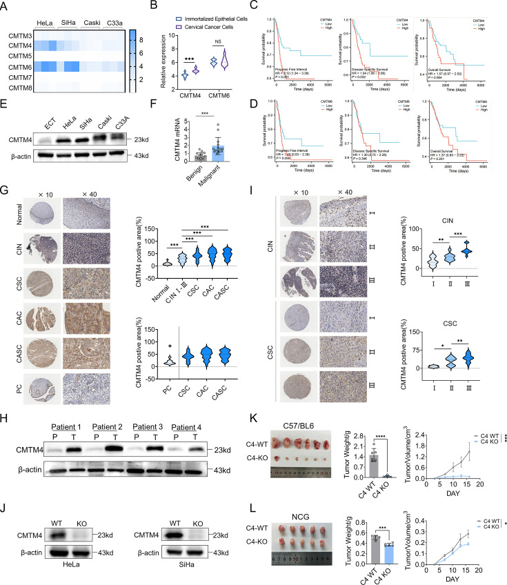
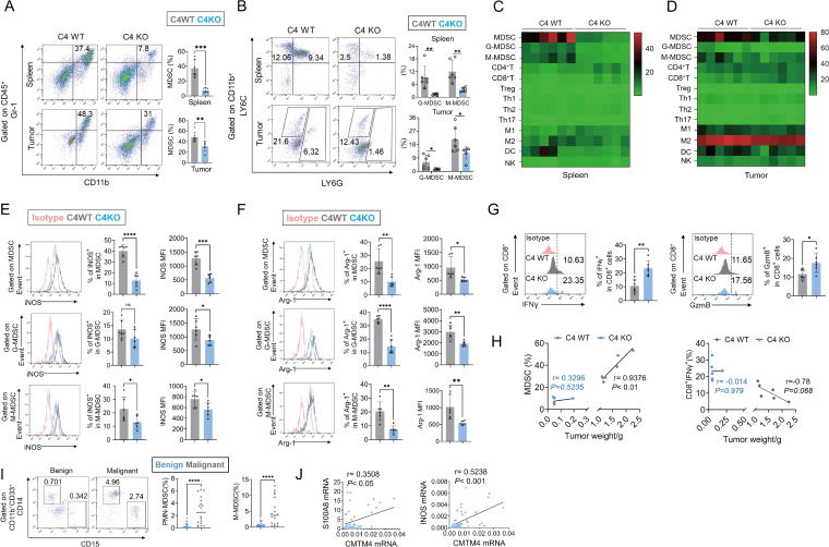
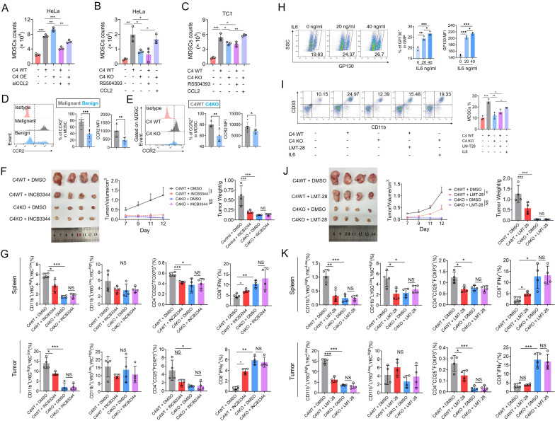
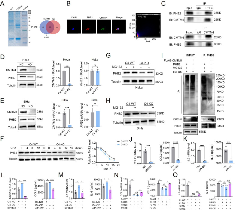
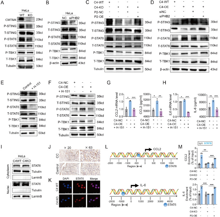
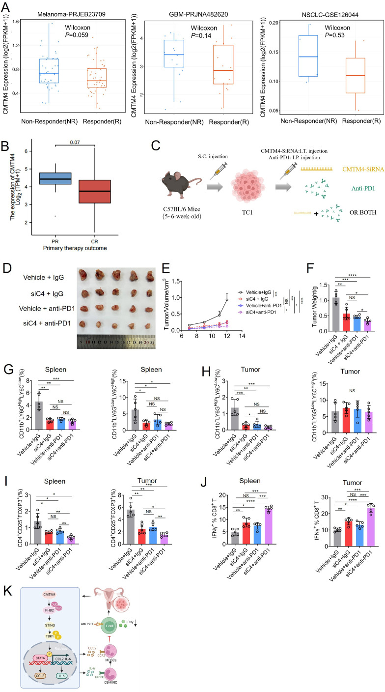
CKLF (chemokine-like factor)-like MARVEL transmembrane domain-containing family member 4 (CMTM4), belonging to the CMTM family of transmembrane domain proteins, plays a significant role in the initiation, progression, and metastasis of cancer. Nevertheless, its involvement in tumor immunity remains elusive. In the present investigation, we observed an upregulation of CMTM4 expression in patients with cervical cancer (CC), which also serves as a prognostic indicator for patients with CC. In vitro experiments and therapeutic models have demonstrated that CMTM4 upregulates the expansion of myeloid-derived suppressor cells (MDSCs) in the tumor microenvironment via the CCL2 (C-C motif chemokine ligand 2)/CCR2 (C-C motif chemokine ligand 2) and IL-6 (interleukin-6)/GP130 (glycoprotein 130) axes. This process exerts immunosuppressive effects and promotes the occurrence and progression of CC. Mechanistically, CMTM4 interacts and stabilizes PHB2 (prohibitin 2) through post-translational modification, which further induces activation of the STING (stimulator of interferon genes)/TBK1 (TANK-binding kinase 1)/STAT6 (signal transducer and activator of transcription 6) pathway, facilitating the nuclear translocation of STAT6 which binds to the CCL2/IL-6 promoter, leading to the upregulation of CCL2/IL-6 transcription expression. Importantly, targeting CMTM4 with CMTM4-small interfering RNA enhanced the effectiveness of anti-programmed cell death protein 1 (anti-PD-1) therapy. Our study identifies CMTM4 as a crucial determinant guiding the homing of MDSCs to CC, thereby contributing to MDSCs-mediated immune suppression and tumor progression. The combination of CMTM4 inhibition and anti-PD-1 treatment shows promising antitumor efficacy against CC. These findings offer novel insights into the tumor microenvironment and have the potential to inform the development of innovative immunotherapy approaches for CC.

Keywords: Cervical Cancer; Immunosuppression; Immunotherapy; Myeloid-derived suppressor cell - MDSC; Tumor microenvironment - TME.


Conflict of interest statement

Competing interests: None declared.

Figures

Figure 1
Figure 1. CMTM4 is highly expressed in CC and mediates its malignant biological behaviors. (A) Heatmap of CMTM3–8 of mRNA expression in cervical cancer cells. (B) Cancer Cell Line Encyclopedia database analysis of CMTM4 and CMTM6 expression in normal cervical epithelium and cervical cancer cell lines. (C–D) TCGA survival analysis of high CMTM4 (C) or CMTM6 (D) expression. (E) WB analysis of CMTM4 expression in normal cervical epithelial cells and cervical cancer cells. (F) CMTM4 expression in benign controls (n=14) and cervical cancer (n=14). (G) Immunohistochemical staining of human cervical tissue arrays using specific antibodies for CMTM4. Scale bar for×10 images: 200 µm; scale bar for×40 images: 50 µm. Statistical diagram (above) of positive proportion of CMTM4 in normal cervical epithelial cells (n=16), CSC (n=32), CAC (n=41) and CASC (n=40). Statistical diagram (below) of the positive proportion of CMTM4 in paracancerous tissues and tumor tissues. (H) WB analysis of CMTM4 expression in cervical cancer tissues and adjacent normal tissues. p, paracancerous tissues; T, tumor tissues. (I), immunohistochemical staining of I–III stages of CIN (left) or CSC (right) using specific antibodies for CMTM4. Scale bar for×10 images: 200 μm; scale bar for×40 images: 50 µm. Statistical diagram of the positive proportion of CMTM4 in I (n=13), II (n=10), III (n=11) stages of CIN. Statistical diagram of the positive proportion of CMTM4 in I (n=3), II (n=18), III (n=25) stages of CSC. (J) Knockout of CMTM4 in HeLa, SiHa was examined by WB analysis. (K) Intradermal tumor volume (n=6) of C57/BL6 mice injected with TC1 CMTM4 KO or control cells. Images of TC1 allografts, tumor weight and tumor growth are shown. (L) Intradermal tumor volume (n=5) of NCG mice injected with TC1 CMTM4 KO or control cells. Images of TC1 allografts, tumor weight, and tumor growth are shown. *p<0.05, **p<0.01, ***p<0.001, ****p<0.0001. CAC, cervical adenocarcinoma; CASC, cervical adenocarcinoma squamous cell carcinoma; CC, cervical cancer; CIN,cervical intraepithelial neoplasia; CKLF, chemokine-like factor; CMTM, CKLF-like MARVEL transmembrane domain-containing family; CSC, cervical squamous cell carcinoma; KO, knockout; mRNA, messenger RNA; PC, paracancerous; TCGA, The Cancer Genome Atlas; WB, western blot; WT, wild type.
Figure 2
Figure 2. CMTM4 increases MDSCs infiltration and inhibits CD8+ T cell accumulation in CC. (A–B) Flow cytometric anaysis of MDSCs (A), G-MDSC or M-MDSC (B) in TC1-C4KO and TC1-C4WT tumor-bearing mice. (C–D) Composition of immune cells in the TC1 spleen (C) and tumor (D) was shown in the heatmap. (E–F) Flow cytometric analysis of the ability of MDSC, G-MDSC, and M-MDSC to secrete INOS (E) or Arg-1 (F). INOS and Arg-1 expressions were described by percentage and mean fluorescence intensity (MFI). (G) Flow cytometric analysis of the infiltration of IFN-γ and GzmB CD8+ T cells. (H) Correlation between tumor weight with tumorous MDSCs abundance, tumor-infiltrating CD8+ T cells IFN-γ level in TC1-C4KO and TC1-C4WT tumor-bearing mice model (All n=6). (I) Flow cytometric analysis of PMN-MDSC and M-MDSC in PBMCs isolated from patients with benign tumors (n=21) and cervical cancer (n=18). (J) Correlation analysis of S100A8 or INOS expression and CMTM4 expression in clinical samples. *p<0.05, **p<0.01, ***p<0.001, ****p<0.0001. Arg-1, arginase-1; CC, cervical cancer; CKLF, chemokine-like factor; CMTM, CKLF-like MARVEL transmembrane domain-containing family; DC, dendritic cell; G-MDSC, granulocyte-like MDSC; GzmB, granzyme B; IFN, interferon; iNOS, inducible nitric oxide synthase; KO, knockout; MDSC, myeloid-derived suppressor cell; M-MDSC, monocyte-like MDSC; mRNA, messenger RNA; NK, natural killer; PMN-MDSC, polymorphonuclear myeloid-derived suppressor cell; Th1, T helper type 1 cell; Treg, regulatory T cell; WT, wild type.
Figure 3
Figure 3. Knockdown of CMTM4 results in the dysregulation of immune-associated cytokines and chemokines. (A–B) CM collected from CMTM4 KO-HeLa and CMTM4 KO-SiHa or from control were placed in the lower chambers. Freshly isolated PBMCs MDSCs were seeded in the upper chambers and allowed to transwell for 24 hours. Total numbers of transwelled MDSCs found in the lower chambers were counted. Data are presented as the mean±SEM (n=3). (C–D) CM collected from CMTM4 KO-HeLa and CMTM4 KO-SiHa or control was cocultured with mononuclear cells from cord blood for 24 hours. The proportion of MDSCs was detected by flow cytometry (n=3). (E) After treating umbilical cord blood mononuclear cells with CM from CMTM4 KO HeLa and CMTM4 KO SiHa or the control group for 24 hours, the coculture supernatant was collected and the secretion of Arg-1 or INOS was detected by ELISA. (F) Heatmap of 43 different human cytokine expression levels with a concentration greater than 100 pg/mL in CMTM4 KO-HeLa and CMTM4 NC-HeLa using Luminex-MultiDTX-43-Human. (G–H) RT-qPCR and ELISA assays analyze expression of IL-6 and CCL2 in CMTM4 KO-HeLa and CMTM4 KO-SiHa (n=3). (I–J) ELISA analysis of CCL2 and IL-6 expression in the serum of patients with benign tumors (CCL2 n=17; IL-6 n=10) and cervical cancer (CCL2 n=11; IL-6 n=18). (K–L) Correlation analysis of PMN-MDSC or M-MDSC expression and CCL2 or IL-6 expression in clinical samples. (M) GEPIA overall survival curves analysis of patients with CC with high or low expression of CCL2 or IL-6. N. Tumor mRNA level and serum protein concentration of CCL2 and IL-6 in TC1 tumor-bearing mice models (n=6). (O) Correlation analysis of CCL2 or IL-6 mRNA expression and CMTM4 mRNA expression in clinical cervical tissue. *p<0.05, **p<0.01, ***p<0.001, ****p<0.0001. Arg-1, arginase-1; CCL, C-C motif chemokine ligand; CM, conditioned medium; CKLF, chemokine-like factor; CMTM, CKLF-like MARVEL transmembrane domain-containing family; CXCL, C-X-C motif chemokine ligand; GEPIA, Gene Expression Profiling Interactive Analysis; IL-6, interleukin-6; iNOS, inducible nitric oxide synthase; KO, knockout; MDSC, myeloid-derived suppressor cell; mRNA, messenger RNA; PB, peripheral blood; PBMCs, peripheral blood mononuclear cells; PDGF-AA, platelet-derived growth factor-AA; PD-L1, programmed death-ligand 1; RT-qPCR, real-time quantitative PCR; Th1, T helper type 1 cell; TPM, transcripts per million; WT, wild type.
Figure 4
Figure 4. CMTM4 facilitates CCL2/CCR2-mediated MDSCs chemotaxis and IL-6/GP130-induced MDSCs differentiation. (A) MDSCs were seeded in the top chamber of the transwell containing 100 µL RPMI medium and the CM derived from different groups placed in the bottom. (B–C) MDSCs were seeded in the top chamber of the transwell containing 100 µL RPMI medium with or without CCR2 inhibitor (RS504393, 10 mM) in CMTM4-NC HeLa or CMTM4-NC TC1 group. On the other hand, the bottom chamber contained 600 µL of CMTM4-KO HeLa or CMTM4-KO TC1 CM with or without recombinant CCL2 protein (1 ng/mL). After 24-hour incubation, cells that have completely migrated to the bottom chamber were counted (n=3). (D) Flow cytometric analysis of CCR2 expression on PBMCs derived MDSCs in patients with benign tumors (n=6) and cervical cancer (n=6). CCR2 expressions were described by percentage and MFI. (E) Flow cytometric analysis of CCR2 expression on tumor-bearing mice-derived MDSCs in spleen. CCR2 expressions were described by percentage and MFI (n=6). (F) Tumor growth in mice intradermally injected with TC1 CMTM4 KO cells or control cells and treated with INCB3344 (10 mg/kg body weight) or DMSO daily following tumor inoculation. Images of TC1 allografts, tumor growth tumor, and weight are shown (n=4). (G) Flow cytometric analysis of G-MDSC, M-MDSC, Treg, and CD8+IFN-γ+ T cells in the treated tumor-bearing mice models. Data are presented as the mean±SEM (n=4). (H) Flow cytometric analysis of GP130 expression on cord blood mononuclear cells treated with IL-6 recombinant factor at different concentrations. GP130 expression was described by percentage and MFI (n=3). I. CM collected from CMTM4 NC-HeLa with or without GP130 inhibitor (LMT-28, 10 mM) and CMTM4 KO-HeLa with or without recombinant IL-6 protein (40 ng/mL) were cocultured with mononuclear cells from cord blood for 24 hours. Each group is treated by GM-CSF (40 ng/mL). The proportion of MDSCs was detected by flow cytometry. Data are presented as the mean±SEM (n=3). (J) Tumor growth in mice intradermally injected with TC1 CMTM4 KO cells or control cells and treated with LMT-28 (10 mg/kg body weight) or DMSO daily following tumor inoculation. Images of TC1 allografts, tumor growth tumor, and weight are shown (n=4). (K) Flow cytometric analysis of G-MDSC, M-MDSC, Treg, and CD8+IFN-γ+ T cells in the treated tumor-bearing mice models. Data are presented as the mean±SEM (n=4). *p<0.05, **p<0.01, ***p<0.001. NS, not significant. CCL2, C-C motif chemokine ligand 2; CCR2, C-C motif chemokine receptor 2; CM, conditioned medium; CKLF, chemokine-like factor; CMTM, CKLF-like MARVEL transmembrane domain-containing family; DMSO, dimethyl sulfoxide; G-MDSC, granulocyte-like MDSC; GM-CSF, granulocyte-macrophage colony-stimulating factor; GMP, granulocyte–monocyte progenitor; GP130, glycoprotein 130; IFN, interferon; IL-6, interleukin-6; KO, knockout; LMT-28, small-molecule inhibitor of the IL-6/GP130 signaling pathway; MDSC, myeloid-derived suppressor cell; MFI, mean fluorescence intensity; M-MDSC, monocyte-like MDSC; OE, overexpression; PBMCs, peripheral blood mononuclear cells; RPMI, Roswell Park Memorial Institute medium; siCCL2, small interfering RNA targeting CCL2; Treg, regulatory T cell; WT, wild type.
Figure 5
Figure 5. CMTM4 regulates the stability of PHB2. (A) Co-IP of HeLa protein lysates with anti-CMTM4 or anti-IgG, followed by Coomassie staining and LC-MS/MS identification of 699 potential interactors. (B) Immunofluorescence analysis of colocalization of CMTM4 and PHB2. HeLa is transfected with Flag-tagged CMTM4 (green) and HA-tagged PHB2 (red). ImageJ analysis of localization correlation between CMTM4 and PHB2. (C) Endogenous Co-IP showing CMTM4-PHB2 interaction. (D–E) PHB2 protein or mRNA levels in CMTM4-KO HeLa and SiHa. (F) CHX chase assay in HeLa-C4KO cells showing PHB2 half-life. (G–H) MG132 (10 µM, 6 hours) rescues PHB2 degradation in CMTM4-KO HeLa and SiHa. (I) Reduced PHB2 ubiquitination in 293T cells co-expressing CMTM4. (J–K) The expression of CCL2 or IL-6 in HeLa with or without PHB2 knockdown. (L–M) The expression of CCL2 or IL-6 in CMTM4-OE HeLa with or without PHB2 knockdown. (N–O) The expression of CCL2 or IL-6 in CMTM4-KO HeLa with or without PHB2 overexpression. *p<0.05, **p<0.01, ***p<0.001, ****p<0.0001, NS, not significant. CCL2, C-C motif chemokine ligand 2; CHX, cycloheximide; CKLF, chemokine-like factor; CMTM, CKLF-like MARVEL transmembrane domain-containing family; Co-IP, co-immunoprecipitation; DAPI, 4′,6-diamidino-2-phenylindole; FLAG, FLAG epitope tag; HA-Ub, hemagglutinin-tagged ubiquitin; IB, immunoblotting; IL-6, interleukin-6; IP, immunoprecipitation; KO, knockout; LC-MS/MS, liquid chromatography-tandem mass spectrometry; mRNA, messenger RNA; NC, negative control; OE, overexpression; PHB2, prohibitin 2; siPHB2, small interfering RNA targeting prohibitin 2; WT, wild type.
Figure 6
Figure 6. CMTM4 regulates CCL2/IL-6 expression through PHB2 and activation of the STING/TBK1/STAT6 pathway. (A) WB analysis of STING/TBK1/STAT6 pathway in HeLa cells with or without CMTM4 knockout. Tubulin was used as a control. (B) WB analysis of STING/TBK1/STAT6 pathway in HeLa cells with or without PHB2 knockdown. β-actin was used as a control. (C) WB analysis of STING/TBK1/STAT6 pathway in CMTM4-KO HeLa with or without PHB2 overexpression. β-actin was used as a control. (D) WB analysis of STING/TBK1/STAT6 pathway in CMTM4-OE HeLa with or without PHB2 knockdown. β-actin was used as a control. (E) WB analysis of STING/TBK1/STAT6 pathway in HeLa cells with or without treatment by H-151. β-actin was used as a control. (F) WB analysis of STING/TBK1/STAT6 pathway in CMTM4-OE HeLa with or without treatment by H-151. Tubulin was used as a control. (G–H) The expression of CCL2 or IL-6 in CMTM4-OE HeLa with or without PHB2 knockdown. (I) STAT6 level is tested in the nuclear and cytoplasmic fractions of CMTM4 NC-HeLa and CMTM4 KO-HeLa cells. Protein expression levels in the nuclear and cytosolic fractions were normalized to LaminB and Tubulin, respectively. (J) IHC analysis of benign controls and cervical cancer using specific antibodies for STAT6. Scale bar for×20 images: 50 µm; scale bar for×63 images: 20 µm. (K) IHC analysis of subcellular distribution of endogenous STAT6 in CMTM4 NC-HeLa and CMTM4 KO-HeLa cells. (L) The potential binding site of STAT6 to the CCL2 (up) or IL-6 (below) promoter is predicted by using the hTFtarget website. (M) ChIP assay analysis of STAT6 binding to the CCL2 (up) or IL-6 (below) promoter via the CMTM4/PHB2 pathway. *p<0.05, **p<0.01, ***p<0.001. CC, cervical cancer; CCL2, C-C motif chemokine ligand 2; ChIP, chromatin immunoprecipitation; CKLF, chemokine-like factor; CMTM, CKLF-like MARVEL transmembrane domain-containing family; DAPI, 4′,6-diamidino-2-phenylindole; IHC, immunohistochemical; IL-6, interleukin-6; KO, knockout; mRNA, messenger RNA; NC, negative control; OE, overexpression; PHB2, prohibitin 2; siNC, small interfering negative control; siPHB2, small interfering RNA targeting prohibitin 2; STAT6, signal transducer and activator of transcription 6; STING, stimulator of interferon genes; TBK1, TANK-binding kinase 1; WB, western blot; WT, wild type.
Figure 7
Figure 7. Targeted CMTM4 treatment enhances the efficacy of PD-1 blockade. (A) ICBatlas website analysis of expression level of CMTM4 in different subgroups (NR and R) of anti-PD-1 therapy-treated clinical cohort. (B) Expression level of CMTM4 in different subgroups (CR and PR) of prime therapy-treated TCGA clinical cohort. (C–D) Schematic showing the treatment plan: Tumor growth of TC1 tumor-bearing mice treated with CMTM4 siRNA, anti-PD-1 mAb alone or the anti-PD-1 mAb combined with the CMTM4 siRNA (Up, n=5/group). Images of TC1 allografts are shown (D). (E–F) Analysis of tumor growth and tumor weight is shown. (G–H) Flow cytometric analysis of CD11b+LY6GHighLY6CLow G-MDSCs and CD11b+LY6GLowLY6CHigh M-MDSCs in spleens and tumors isolated from tumor-bearing mice. (I) Flow cytometric analysis of CD4+CD25+FOXP3+ Tregs in spleens and tumors isolated from tumor-bearing mice. (J) Flow cytometric analysis of the infiltration of IFN-γ CD8+ T cells in spleens and tumors. (K) Schematic representations of the role of CMTM4 in driving immune suppression in CC. *p<0.05, **p<0.01. ***p<0.001, ****p<0.0001. NS=No significance. CC, cervical cancer; CCL2, C-C motif chemokine ligand 2; CCR2, C-C motif chemokine receptor 2; CKLF, chemokine-like factor; CMTM, CKLF-like MARVEL transmembrane domain-containing family; CN-MNC, cord blood-derived mononuclear cell; CR, complete responders; GBM, glioblastoma multiforme; GP130, glycoprotein 130; IFN, interferon; IL-6, interleukin-6; I.P., intraperitoneal; I.T., intertumoral; mAb, monoclonal antibody; MDSC, myeloid-derived suppressor cell; NSCLC, non-small cell lung cancer; PD-1, programmed cell death protein 1; PHB2, prohibitin 2; PR, partial responder; S.C., subcutaneous; siRNA, small interfering RNA; STAT6, signal transducer and activator of transcription 6; STING, stimulator of interferon genes; TBK1, TANK-binding kinase 1; TCGA, The Cancer Genome Atlas.

Similar articles

References

    1. Bray F, Ferlay J, Soerjomataram I, et al. Global cancer statistics 2018: GLOBOCAN estimates of incidence and mortality worldwide for 36 cancers in 185 countries. CA Cancer J Clin. 2018;68:394–424. doi: 10.3322/caac.21492. - DOI - PubMed
    1. Chambers D, Simpson L, Hill-Briggs F, et al. Proceedings of the 8th Annual Conference on the Science of Dissemination and Implementation: Washington, DC, USA. 14-15 December 2015. Implement Sci. 2016;11 Suppl 2:100. doi: 10.1186/s13012-016-0452-0. - DOI - PMC - PubMed
    1. Han W, Ding P, Xu M, et al. Identification of eight genes encoding chemokine-like factor superfamily members 1-8 (CKLFSF1-8) by in silico cloning and experimental validation. Genomics. 2003;81:609–17. doi: 10.1016/s0888-7543(03)00095-8. - DOI - PubMed
    1. Plate M, Li T, Wang Y, et al. Identification and characterization of CMTM4, a novel gene with inhibitory effects on HeLa cell growth through Inducing G2/M phase accumulation. Mol Cells. 2010;29:355–61. doi: 10.1007/s10059-010-0038-7. - DOI - PubMed
    1. Mezzadra R, Sun C, Jae LT, et al. Identification of CMTM6 and CMTM4 as PD-L1 protein regulators. Nature New Biol. 2017;549:106–10. doi: 10.1038/nature23669. - DOI - PMC - PubMed

MeSH terms