Case Reports
. 2025 Jun 13;20(1):74.
doi: 10.1186/s13000-025-01672-9.

Bilateral synchronous salivary gland tumors: report of three cases

Affiliations
Case Reports

Bilateral synchronous salivary gland tumors: report of three cases

Jacqueline E van der Wal et al. Diagn Pathol. .

Abstract

Background: Bilateral salivary gland tumors, both benign and malignant and synchronous or metachronous are very rare.

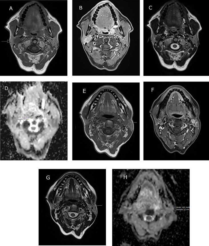
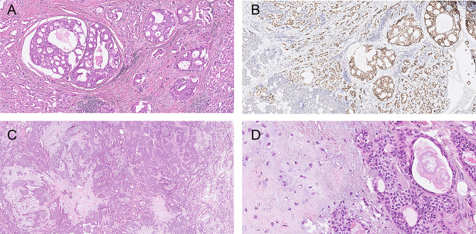
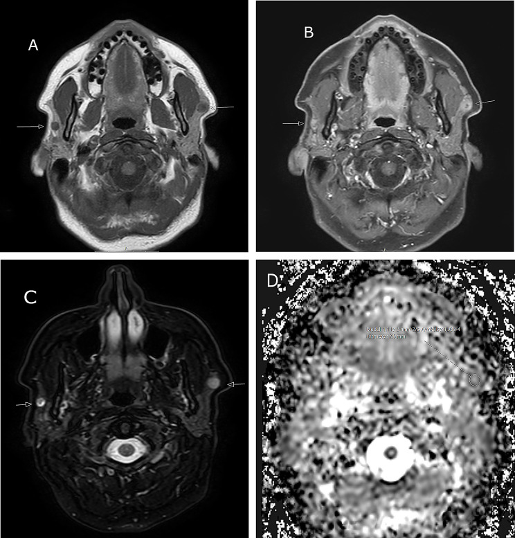
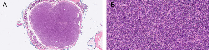
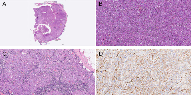
Case presentation: Here three cases of synchronous bilateral salivary gland tumors are described and discussed. Recognizing the entity is important for diagnostics and treatment planning. The first patient was a 56-year-old female with a bilateral parotid tumor, a malignant tumor, salivary duct carcinoma on the right side and a benign tumor, pleomorphic adenoma on the left side. The second patient was a 50-year old female with a bilateral benign parotid tumor, a pleomorphic adenoma. The third patient was a 51-year old female with a bilateral malignant tumor, an acinic cell carcinoma. Details on the diagnostic work-up, histopathology and treatment are described and discussed.

Conclusions: In the case of a unilateral salivary gland tumor, especially of the major glands, the contralateral gland is always included in the clinical and radiological (MRI) head and neck evaluation prior to surgery, to detect or exclude possible bilateral occurrence.

Keywords: Acinic cell carcinoma; Pleomorphic adenoma; Salivary duct carcinoma; Salivary gland tumor.


Conflict of interest statement

Declarations. Ethical approval: According to Swedish regulations studies regarding diagnostics and treatment are not considered research and must thus not be ethically permitted. Competing interests: The authors declare no competing interests.

Figures

Fig. 1
Fig. 1
Patient 1, MR image of bilateral salivary gland tumors in a 56-year old female. A-D show a right sided intraparotid lesion with irregular borders. A) Low signal on T1W (arrow), B) rich contrast enhancement on fat-suppressed TIW, C) low signal on T2W and D) restricted diffusion on the ADC-map, ADC = 1,11 × 10− 3 mm2/s. E-H Left sided intraparotid lesion in the deep lobe. E) Low signal on T1W (arrow), F) rich contrast enhancement on fat-suppressed TIW, G) mixed signal on T2W (arrow) and H) moderate diffusion restriction on the ADC-map, ADC = 1,42 × 10− 3 mm2/s
Fig. 2
Fig. 2
Patient 1, A. salivary duct carcinoma, H&E; B salivary duct carcinoma, androgen receptor; C,D. pleomorphic adenoma, H&E;
Fig. 3
Fig. 3
Patient 2, MR image of bilateral salivary gland tumours in a 50-year old female. A-D Demonstration of bilateral intraparotid lesions in the superficial portions. A) Low signal on T1W (arrows), B) rich, irregular contrast enhancement on fat-suppressed TIW (arrows), C) high signal on fat-suppressed T2W (arrows) and D) restricted diffusion of the left sided lesion on the ADC-map, ADC = 1,14 × 10− 3 mm2/s
Fig. 4
Fig. 4
Patient 2, A and B pleomorphic adenoma on the left side, H&E. Both pleomorphic adenomas had the same morphology
Fig. 5
Fig. 5
Patient 3, MR image of bilateral salivary gland tumors in a 51-year old female. A-D Bilateral intraparotid lesions with irregular borders and similar textures on the left side in the deep lobe and on the right side in the superficial lobe (arrows). A) Low signal on T1W, B) heterogeneous contrast enhancement on fat-suppressed TIW, C) mixed signal on T2W and D) restricted diffusion on DWI; the ADC was bilaterally approximately 1,1 × 10− 3 mm2/s (not shown). E) MR examination 15 months after surgery reveals a small subcutaneous recurrence (arrow) on the right side (fat-suppressed T1W with contrast enhancement)
Fig. 6
Fig. 6
Patient 3, A and B. Overview and detail of acinic cell carcinoma on the left side, H&E;. C. Growth in the parotid lymph node on the right side. D. DOG-1 staining Both acinic cell carcinomas had the same morphology

Similar articles

Cited by

References

    1. Colangeli W, Facchini V, Kapitonov A, Zappala M, Belli E. Multiple synchronous bilateral neoplasms of Parotid glands. Clin Ter. 2022;173:203–6. - PubMed
    1. Yu GY, Ma DQ, Zhang Y, et al. Multiple primary tumours of the Parotid gland. Int J Oral Maxillofac Surg. 2004;33:531–4. - PubMed
    1. Park KS, Jang HB, Lee DH, Lee JK, Lim SC. Multiple synchronous neoplasms in the ipsilateral Parotid gland: case series. Ann Palliat Med. 2022;11:2641–5. - PubMed
    1. Colangeli W, Kapitonov A, Facchini V, Zappala M, Belli E. Synchronous ipsilateral pleomorphic adenomas of Parotid and submandibular glands: an unusual finding. Case Rep Otolaryngol. 2020;2020:88857867. - PMC - PubMed
    1. Seifert G, Donath K. Multiple tumours of the salivary glands– Terminology and nomenclature. Oral Oncol Eur J Cancer. 1996;32B:3–7. - PubMed

Publication types

MeSH terms

LinkOut - more resources