. 2025 Jun 13;13(1):131.
doi: 10.1186/s40478-025-02044-6.

ATRX loss in adult gliomas lacking H3 alterations or IDH mutations, an exceptional situation for exceptional diagnoses: the experience of Sainte-Anne hospital

Affiliations

ATRX loss in adult gliomas lacking H3 alterations or IDH mutations, an exceptional situation for exceptional diagnoses: the experience of Sainte-Anne hospital

Arnault Tauziède-Espariat et al. Acta Neuropathol Commun. .

Abstract

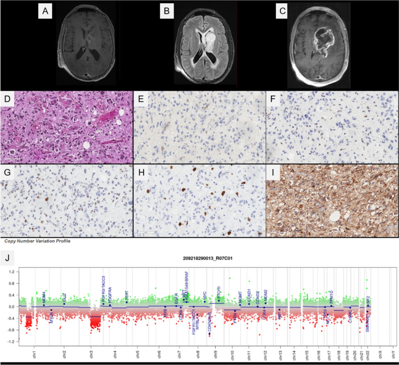
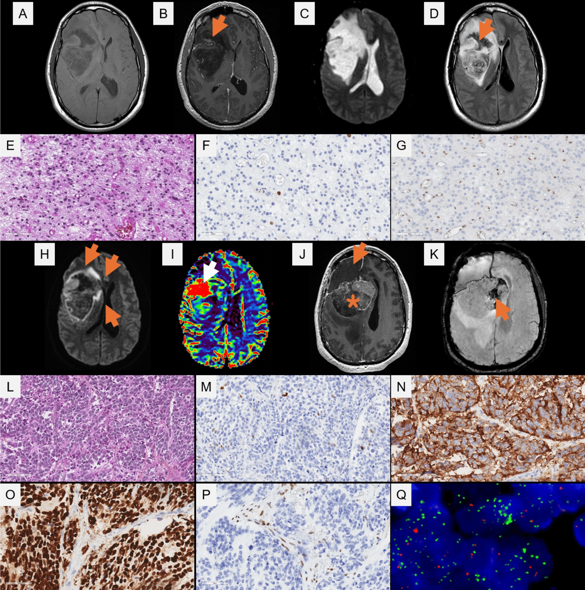
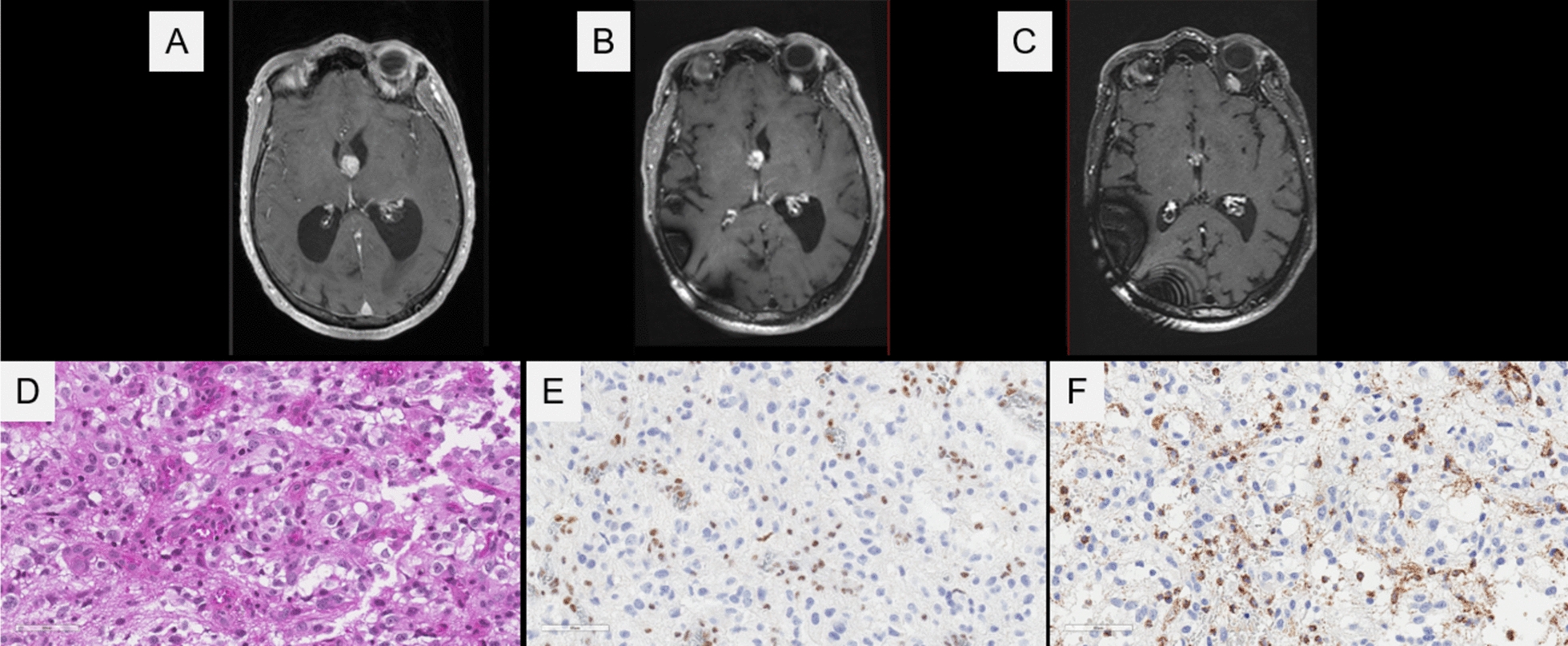
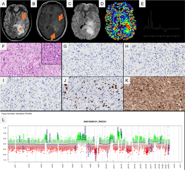
ATRX immunostaining constitutes a routinely used biomarker for the practice of neuropathology. The loss of ATRX expression correlating with ATRX gene alterations is implicated in a wide variety of pediatric and adult gliomas, and has been indexed as a desirable or essential diagnostic criterion for four tumor types featured in the latest world health organization classification of central nervous system Tumors. In adult-type diffuse glioma, the loss of ATRX expression is a hallmark of astrocytoma, IDH-mutant. Recently, novel tumor types and alterations have been referenced in the literature. These include the high-grade astrocytoma with piloid features (HGAP), for which no consistent clinicopathological features have been defined, and the presence of other alterations in the Krebs cycle genes (variants of the Fumarate hydratase -FH- gene) found in gliomas resembling astrocytomas, IDH-mutant. Because of this rapidly evolving classification and histomolecular landscape, we retrospectively analyzed adult gliomas diagnosed over a four consecutive year period to identify supratentorial gliomas, lacking H3 alterations or IDH mutations and harboring a loss of ATRX expression, in order to update their diagnoses in terms of histopathology, genetics and epigenetics. Four specimens (from 620 adult gliomas, 0.7%) were reclassified at the end of the molecular workup, as: 1/ one HGAP, 2/ one malignant transformation with a primitive neuronal component of an astrocytoma, IDH-mutant which lost the IDH2 mutation at recurrence, 3/ a glioma, FH-mutant for which the histopathological and epigenetic features were similar to an astrocytoma, IDH-mutant, and 4/ a glioblastoma, IDH-wildtype. To conclude, these exceptional cases extend the spectrum of ATRX loss in gliomas, beyond the astrocytoma, IDH-mutant and the diffuse hemispheric glioma, H3 G34-mutant.

Keywords: ATRX; Diffuse glioma; FH; HGAP.


Conflict of interest statement

Declarations. Ethics approval and consent to participate: This study was approved by GHU Paris Psychiatry and Neurosciences, Sainte-Anne Hospital’s local ethic committee. Consent for publication: The patient signed informed consent forms before treatment was started. Competing interests: The authors declare that they have no conflicts of interest directly related to the topic of this article.

Figures

Fig. 1
Fig. 1
Imaging, histopathological and molecular features of the case #4. (A) A capsulo-thalamic infiltrating lesion without initial enhancement after gadolinium injection but FLAIR hyperintensity (B). This tumor was non-homogeneously enhanced after gadolinium injection during the following MRI of the follow-up (C). (D) A diffuse glial proliferation composed of astrocytic and multinucleated cells with numerous mitoses (HPS, magnification × 400). (E) No immunoexpression for IDH1R132H (magnification × 400). (F) A loss of ATRX expression (magnification × 400). (G) No overexpression of p53 (magnification × 400). (H) Elevated proliferative index (MIB1, magnification × 400). (I) A preserved expression of FH (magnification × 400). (J) Copy number variation analysis showing a homozygous deletion of the CDKN2A gene. Black scale bars represent 60 µm. HPS: Hematoxylin Phloxin Saffron
Fig. 2
Fig. 2
Imaging, histopathological and molecular features of case #2. Axial T1 sequence before (A) and after contrast injection (B) displayed a large right hemispheric lesion with slight subcortical enhancement (arrow in B), associated with mass effect and brain herniation under the falx cerebri. T2-weighted sequence (C) and T2-FLAIR sequence (D) displayed a hyper-intense liquid central contingent (arrow in D) in T2 and signal suppression in FLAIR. (E) A diffuse astrocytoma (HPS, magnification × 400) with a low proliferative index (F, magnification × 400), and loss of ATRX expression (G, magnification × 400). Two years after the initial surgery, diffusion-weighted imaging (H) displayed a rim with diffusion restriction (arrows), suggesting hypercellularity. Relative Cerebral Volume (rCBV) map (I) computed from Perfusion-Weighted Imaging, displayed a hyperperfused area in the frontal region (rCBV = 5), suggesting the presence of an intermediate or high grade component. Axial T1 sequence after contrast injection (J) displayed a large mass (star) posterior to the postoperative cavity (arrow) with partial enhancement suggesting intratumoral necrosis. Susceptibility imaging (K) displayed hemorrhagic areas (arrow). The recurrent tumor was histopathologically different, composed of solid nodules showing immature cells with numerous mitoses (L). (M) Focal expression of OLIG2 (magnification × 400), and diffuse immunoreactivity for synaptophysin (N, magnification × 400). (O) Overexpression of p53 (magnification × 400). P loss of ATRX expression (magnification × 400). Q High-level amplification of MYCN locus (magnification × 400, MYCN locus: green signals, centromere of chromosome 2: orange signals). Black scale bars represent 60 µm. HPS: Hematoxylin Phloxin Saffron
Fig. 3
Fig. 3
Imaging, histopathological and molecular features of case #3. A lesion in the third ventricle with heterogeneous enhancement after the injection of gadolinium (A-C). (D) A diffuse glioma with an astrocytic proliferation, showing several mitoses and a microvascular proliferation (HPS, magnification × 400). (E) A loss of expression of ATRX (magnification × 400). (F) The FH immunostaining showed a complete loss of expression in tumor cells, and a preserved expression of residual glial cells in the parenchyma (magnification × 400). Black scale bars represent 60 µm. HPS: Hematoxylin Phloxin Saffron
Fig. 4
Fig. 4
Imaging, histopathological and molecular features of case #4. Axial T2-FLAIR sequence (A) displayed a mass centered on the left part of the corpus callosum splenium (star in A), associated with a left hemispheric infiltration with high FLAIR intensity (arrow in A). After contrast injection, axial T1 sequence (B) displayed a diffuse, non-necrotic enhancement on the mass bulk (arrows in B). Diffusion Weighted Imaging (C) displayed high intensity areas inside the mass, suggesting tumoral hypercellularity. Relative Cerebral Volume (rCBV) map (D) computed from Perfusion-Weighted Imaging, displayed a hyper-perfused area in the periventricular region (rCBV = 2.5), suggesting the presence of an intermediate or high grade contingent. Spectroscopic analysis with long Echo Time (TE = 144 ms) (E) displayed a myoinositol peak suggestive of a glial origin, an increase of choline, and a decrease of N-Acetyl-Aspartate, suggestive of hypercellularity and lactate peaks, suggestive of anaerobic metabolism. (F) A diffuse glioma with an astrocytic proliferation, showing atypicalities, multinucleated cells and a microvascular proliferation (HPS, magnification × 400, insert HPS magnification × 400). (G) No immunopositivity for IDH1R132H (magnification × 400). (H) Loss of expression of ATRX (magnification × 400). (I) No overexpression of p53 (magnification × 400). (J) Eleavted proliferative index (MIB1, magnification × 400). (K) Preserved expression of FH (magnification × 400). (L) The copy number variation analysis showed a gain of chromosome 7, without a loss of chromosome 10, and amplifications of the MDM2 and CDK4 genes without EGFR amplification. Black scale bars represent 60 µm. HPS: Hematoxylin Phloxin Saffron

Similar articles

References

    1. Reuss DE, Sahm F, Schrimpf D, Wiestler B, Capper D, Koelsche C et al (2015) ATRX and IDH1-R132H immunohistochemistry with subsequent copy number analysis and IDH sequencing as a basis for an “integrated” diagnostic approach for adult astrocytoma, oligodendroglioma and glioblastoma. Acta Neuropathol (Berl) 129(1):133–146 - PubMed
    1. Ebrahimi A, Skardelly M, Bonzheim I, Ott I, Mühleisen H, Eckert F et al (2016) ATRX immunostaining predicts IDH and H3F3A status in gliomas. Acta Neuropathol Commun 4(1):60 - PMC - PubMed
    1. Louis DN, Perry A, Wesseling P, Brat DJ, Cree IA, Figarella-Branger D et al (2021) The 2021 WHO classification of tumors of the central nervous system: a summary. Neuro-Oncol 23(8):1231–1251 - PMC - PubMed
    1. Reinhardt A, Stichel D, Schrimpf D, Sahm F, Korshunov A, Reuss DE et al (2018) Anaplastic astrocytoma with piloid features, a novel molecular class of IDH wildtype glioma with recurrent MAPK pathway, CDKN2A/B and ATRX alterations. Acta Neuropathol (Berl) 136(2):273–291 - PubMed
    1. Bender K, Perez E, Chirica M, Onken J, Kahn J, Brenner W et al (2021) High-grade astrocytoma with piloid features (HGAP): the Charité experience with a new central nervous system tumor entity. J Neurooncol 153(1):109–120 - PMC - PubMed

MeSH terms

LinkOut - more resources