. 2025 Jun 13;7(1):63.
doi: 10.1186/s42523-025-00431-x.

Mitochondrial sirtuin 4 shapes the intestinal microbiota of Drosophila by controlling lysozyme expression

Affiliations

Mitochondrial sirtuin 4 shapes the intestinal microbiota of Drosophila by controlling lysozyme expression

Mirjam Knop et al. Anim Microbiome. .

Abstract

Background: Sirtuins are deacetylases that are highly conserved throughout the animal kingdom. They act as metabolic sensors that coordinate cellular responses, allowing an adapted response to various stressors. Epithelial cells, especially those of the intestine, are directly exposed to a wide range of stressors. Together with the microbiota, they form a complex ecosystem with mutual influences. The significance of sirtuins in this complex system is still waiting to be clarified.

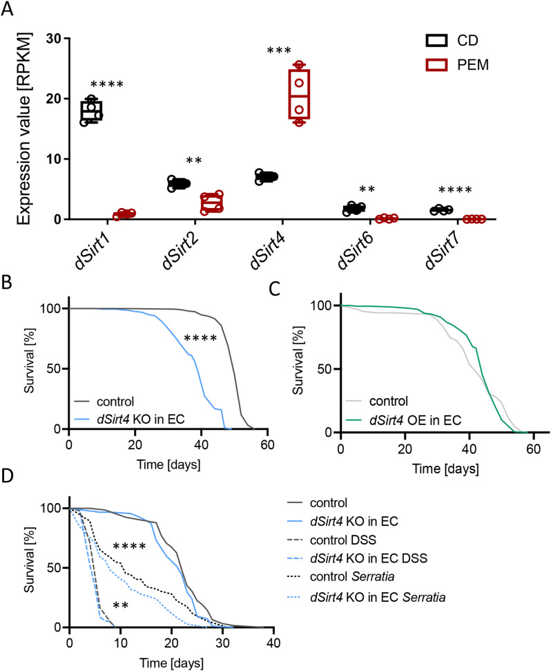
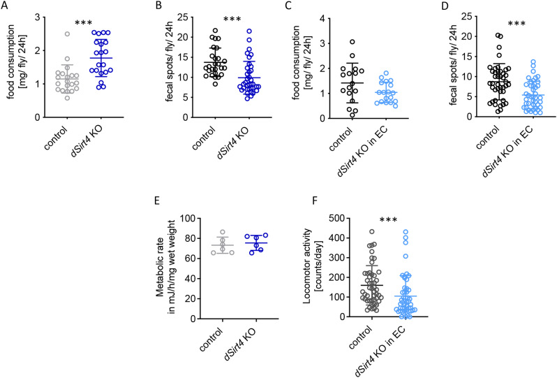
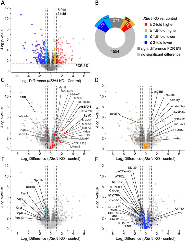
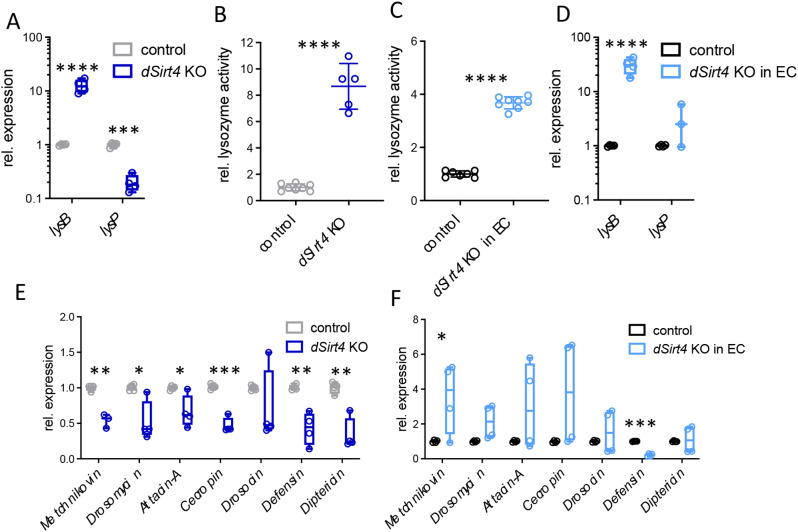
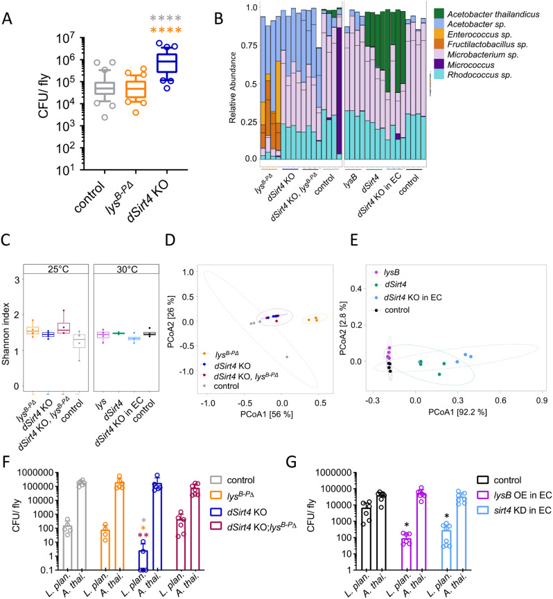
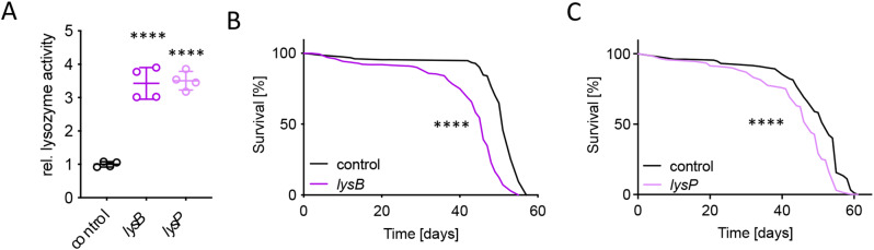
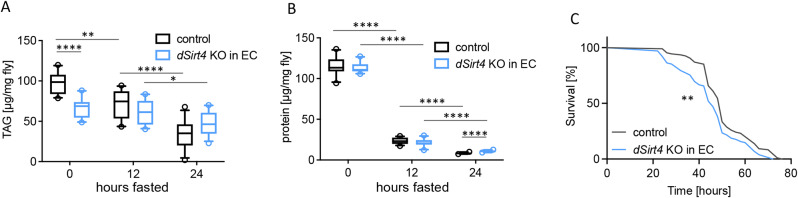
Results: Here, we show that a protein-restricted diet strongly increases the intestinal expression of sirtuin 4 (dSirt4), the only mitochondrial sirtuin in Drosophila. To elucidate the effects of deregulated dSirt4 expression in the intestine, we analyzed dSirt4 knockout flies. These flies showed substantial changes in their intestinal proteome and physiological properties. One of the most striking effects was the strong induction of lysozymes in the intestine, with a corresponding increase in lysozyme activity. This effect was organ-autonomous, as it was also observed in flies with dSirt4 knocked out only in intestinal enterocytes. The significant increase in lysozyme abundance in response to tissue-specific dSirt4 knockdown did not reduce the total number of bacteria in the intestine. However, it did affect the microbiota composition by reducing the number of gram-positive bacteria. This effect on microbiota composition can be attributed to dSirt4-dependent lysozyme expression, which is absent in a lysozyme-deficient background. dSirt4 knockout in the enterocytes shortened the lifespan of the flies, as did ectopic lysozyme overexpression in the enterocytes.

Conclusions: The only mitochondrial sirtuin in Drosophila, dSirt4, is induced by dietary stress in intestinal epithelial cells, which directly regulates the lysozyme activity of these cells. We could associate this altered lysozyme activity with a shift in the microbiota composition, demonstrating a direct link between stress, nutrition, and the host's microbiota regulation.


Conflict of interest statement

Declarations. Competing interests: The authors declare no competing interests.

Figures

Fig. 1
Fig. 1
dSirt4 has an important role in the stress response of the Drosophila intestine. (A) A gene expression analysis of isolated intestines of flies subjected to 7 days protein depleted diet (PEM) revealed that the only sirtuin gene upregulated under these conditions is the only mitochondrial sirtuin of Drosophila, dSirt4, while all others were downregulated (CD = control diet, n = 4). (B) A lifespan analysis showed a significantly reduced survival of flies with a knockout of dSirt4 specifically in enterocytes (dSirt4 KO in EC, n = 187–195). (C) The overexpression of dSirt4 in enterocytes did not affect the lifespan of flies (dSirt4 OE in EC, n = 91–185). (D) dSirt4 knockout flies showed reduced survival in response to infection with Serratia marcescens, while the control treatment with sucrose did not lead to changes in lifespan (n = 63–104). In response to the treatment with DSS, dSirt4 knockout flies lived shorter than their genetic control (n = 63–186). (B, D) control = w1118> UAS-sirt4 crispr/Cas9, dSirt4 KO in EC = NP1-Gal4;tubPGal80ts > UAS-sirt4 crispr/Cas9, (C) control = w1118> UAS-sirt4, dSirt4 OE in EC = NP1-Gal4;tubPGal80ts > UAS-sirt4. * = p < 0.05, ** = p < 0.01, **** = p < 0.0001
Fig. 2
Fig. 2
Changes in gut functionality upon reduced dSirt4 expression. (A, B) The knockout of dSirt4 (dSirt4 KO) leads to an increase in food consumption (n = 20), while the number of excreted fecal spots is not affected (n = 26–36). (C, D) In flies with a knockout of dSirt4 in enterocytes (dSirt4 KO in EC), the amount of ingested food is not significantly different compared to the control (n = 17–18), but the number of excreted fecal spots is reduced (n = 41). (E) The metabolic rate of dSirt4 KO flies was determined by measuring the heat dissipation and showed no significant differences to controls (n = 6). (F) Flies with a knockout of dSirt4 in enterocytes have a significantly reduced locomotor activity (n = 46–48). (A, B, E) control = w1118. (C, D, F) control = w1118> UAS-sirt4 crispr/Cas9, dSirt4 KD = NP1-Gal4;tubPGal80ts > UAS-sirt4 crispr/Cas9. *** = p < 0.001
Fig. 3
Fig. 3
Quantitative proteome analysis of adult intestines. (A, B) Differentially abundant proteins were detected by label-free quantification in dSirt4 KO intestines (n = 6) compared to the control strain (n = 7) of Drosophila. (C-F) Show the distribution of proteins associated with distinct functional categories labelled by colors; significantly changed proteins are labelled by their naming. (C) Including the protein group encompassing LysB, LysD and LysE, glycosidases were significantly more abundant, whereas the carbohydrate regulatory protein Tobi was less abundant in dSirt4 KO. (D) Digestive serine proteases were more abundant in dSirt4 KO intestines. (E) Most proteins involved in lipid transport were of significantly lower abundance in the dSirt4 KO. (F) Proteins associated with mitochondrial electron transport, the respiratory chain complex, and ATP synthases showed a trend towards lower abundance in dSirt4 KO intestines. control = w1118
Fig. 4
Fig. 4
Deficiency of dSirt4 strongly induces lysozyme expression in the intestine. (A) qRT-PCR analysis showed significantly upregulated lysB expression in dSirt4 KO intestines (n = 4). (B) Strong increase of lysozyme activity in dSirt4 KO intestines (n = 5–8). (C) Increase of lysozyme activity (n = 8) and (D) upregulation of lysB expression in intestines of flies with a dSirt4 knockout in enterocytes (n = 4). (E, F) Changes in expression of antimicrobial peptide genes in intestines from dSirt4 KO flies (E; n = 4) and intestines of flies with a dSirt4 knockout in enterocytes (F; n = 4). (A, B, E) control = w1118. (C, D, F) control = w1118> UAS-sirt4 crispr/Cas9, dSirt4 KO in EC = NP1-Gal4;tubPGal80ts > UAS-sirt4 crispr/Cas9. * = p < 0.05, ** = p < 0.01, ***= p < 0.001, **** = p < 0.0001
Fig. 5
Fig. 5
Changes in the bacterial load and composition of the intestinal microbiome upon dSirt4 and lysozyme manipulation. (A) Bacterial load of recolonized dSirt4 KO, lysB-PΔ, and control flies (n = 33). (B) Top 10 ASVs, colored according to bacterial species, black lines represent different ASVs belonging to the same species, (C) Box plot of the Shannon index and (D, E) PCoA of the Bray-Curtis dissimilarity of dSirt4 KO, lysB-PΔ, dSirt4 KO; lysB-PΔ, lysB, dSirt4 and dSirt4 KD and control (n = 4). (F) Reduced number of CFU of L. plantarum and A. thailandicus after recolonization in disassociation dSirt4 KO flies compared to control, lysB-PΔ flies and dSirt4 KO; lysB-PΔ flies (n = 6). (G) Reduced number of CFU of L. plantarum and A. thailandicus after recolonization in disassociation of flies with dSirt4 deficiency or overexpression of lysB in EC compared to control (n = 6). (A) control = w1118. (B-E) control (grey) = w1118, control (black) = NP1-Gal4;tubPGal80ts > w1118, lysB = NP1-Gal4;tubPGal80ts > UAS-lysB, dSirt4 = NP1-Gal4;tubPGal80ts > UAS-sirt4, dSirt4 KD = NP1-Gal4;tubPGal80ts > UAS-sirt4 crispr/Cas9. (F) control = w1118. (G) control = NP1-Gal4;tubPGal80ts > w1118, lysB = NP1-Gal4;tubPGal80ts > UAS-lysB, dSirt4 KD = NP1-Gal4;tubPGal80ts > UAS-sirt4 crispr/Cas9. * = p < 0.05, ** = p < 0.01, **** = p < 0.0001
Fig. 6
Fig. 6
Reduced survival in response to overexpression of lysozymes in enterocytes. (A) Increased lysozyme activity in intestines dissected from flies with overexpressed lysB or lysP in enterocytes (n = 4). (B, C) Reduced survival in flies with an overexpression of lysB in enterocytes (B; n = 150–325) and an overexpression of lysP in enterocytes (C; n = 135–314). control = NP1-Gal4;tubPGal80ts > w1118, lysB = NP1-Gal4;tubPGal80ts > UAS-lysB, lysP = NP1-Gal4;tubPGal80ts > UAS-lysP. **** = p < 0.0001
Fig. 7
Fig. 7
Effects of dSirt4 on protein and fat metabolism. (A) Reduced mobilization of fat storages in response to starvation in flies with a knockout of dSirt4 in enterocytes (dSirt4 KO in EC). Body fat content was determined by measuring triacylglycerol (TAG) levels (n = 10–12). (B) Massive reduction in protein storage upon starvation in dSirt4 KD flies over a time period of 24 h (n = 10–12). (C) Reduced survival of flies with a knockdown of dSirt4 in enterocytes under starvation condition (n = 102–108). control = w1118> UAS-sirt4 crispr/Cas9, dSirt4 KO in EC = NP1-Gal4;tubPGal80ts > UAS-sirt4 crispr/Cas9. * = p < 0.05, ** = p < 0.01, ***= p < 0.001, **** = p < 0.0001

Similar articles

Cited by

References

    1. Elmentaite R, Kumasaka N, Roberts K, Fleming A, Dann E, King HW, Kleshchevnikov V, Dabrowska M, Pritchard S, Bolt L, et al. Cells of the human intestinal tract mapped across space and time. Nature. 2021;597(7875):250–5. - PMC - PubMed
    1. Hung RJ, Hu Y, Kirchner R, Liu Y, Xu C, Comjean A, Tattikota SG, Li F, Song W, Ho Sui S, Perrimon N. A cell atlas of the adult Drosophila midgut. Proc Natl Acad Sci U S A. 2020;117(3):1514–23. - PMC - PubMed
    1. Lyte M, Vulchanova L, Brown DR. Stress at the intestinal surface: catecholamines and mucosa-bacteria interactions. Cell Tissue Res. 2011;343(1):23–32. - PubMed
    1. Bailey MT, Dowd SE, Parry NM, Galley JD, Schauer DB, Lyte M. Stressor exposure disrupts commensal microbial populations in the intestines and leads to increased colonization by Citrobacter rodentium. Infect Immun. 2010;78(4):1509–19. - PMC - PubMed
    1. Clevers H. The Paneth cell, caloric restriction, and intestinal integrity. N Engl J Med. 2012;367(16):1560–1. - PubMed

LinkOut - more resources