. 2025 Jul;41(7):e70050.
doi: 10.1002/kjm2.70050. Epub 2025 Jun 14.

PKN2 Inhibits VEGFA and bFGF-Mediated Angiogenesis by Targeting HIF-1α in Colon Cancer

Affiliations

PKN2 Inhibits VEGFA and bFGF-Mediated Angiogenesis by Targeting HIF-1α in Colon Cancer

Yun Zhu et al. Kaohsiung J Med Sci. 2025 Jul.

Abstract

Angiogenesis plays a vital role in colon cancer growth and metastasis. The role of protein kinase N2 (PKN2) in colon cancer is rarely studied. In this study, we investigated the effect of PKN2 on angiogenesis in colon cancer. We evaluated the correlation between PKN2 expression and microvessel density (MVD) in tumor tissue of patients with colon cancer. The effect of PKN2 on tumor angiogenesis was investigated both in cultured colon cancer cells and in a mouse colon cancer model. PKN2 targeted vascular endothelial growth factor A (VEGFA) and basic fibroblast growth factor (bFGF) expression, and secretion were analyzed, and the specific regulatory role of PKN2 on HIF was explored. PKN2 expression was negatively correlated with tumor MVD in tumor tissue of patients with colon cancer. PKN2 inhibited angiogenesis in both in vitro and in vivo models of mouse tumors. Mechanistically, PKN2 suppressed the transcriptional activity of hypoxia-inducible factor-1α (HIF-1α) and reduced its nuclear accumulation, leading to the inhibiting of VEGFA and bFGF transcription by preventing HIF-1α binding to their promoters. Additionally, PKN2 directly interacted with HIF-1α at the protein level and induced phosphorylation, resulting in ubiquitination-dependent degradation of HIF-1α in colon cancer cells. Our study demonstrated, for the first time, that PKN2 exerts inhibitory effects on tumor angiogenesis in colon cancer. We propose a novel mechanism by which PKN2 regulates VEGFA and bFGF expression through modulation of the dynamic equilibrium of HIF-1α protein levels.

Keywords: HIF‐1α; PKN2; colon cancer; tumor angiogenesis.


Conflict of interest statement

The authors declare no conflicts of interest.

Figures

FIGURE 1
FIGURE 1
PKN2 expression correlated with MVD and CD31 in colon cancer tissue. (A) IHC staining of CD31 and PKN2 in colon cancer tissue from patients with low versus high PKN2 expression (n = 45); typical pictures are shown (100× and 400×). (B) Correlation between the PKN2 expression score and MVD using the Spearman method. (C) Correlation between the PKN2 expression score and the IOD of CD31 using the Spearman method.
FIGURE 2
FIGURE 2
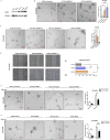
Effect of PKN2 on the proangiogenic effect of colon cancer cells in vitro. (A) The expression of PKN2 in various colon cancer cells. (B) Tube formation by EA.hy926 cells treated with conditioned medium from control shRNA or shPKN2 HT29 cells was assessed. (C) Tube formation by EA.hy926 cells treated with conditioned medium from control, PKN2 knockdown, or PKN2 overexpression HCT116 cells was assessed. (D) EA.hy926 cells were treated as indicated in section B and a migration assay was conducted. Quantification was conducted by measuring the migrated distance. (E) HCT116 or HT29 cells (F) stably overexpressing PKN2 or a vector cultured aerobically and anaerobically for 12 h. The supernatant was used to treat EA.hy926 cells, and tube formation was subsequently evaluated (200×). *p < 0.05, **p < 0.01, ***p < 0.001.
FIGURE 3
FIGURE 3
PKN2 suppressed the expression and secretion of VEGFA and bFGF in colon cancer cells, leading to impaired angiogenesis. (A) RT‐qPCR of proangiogenesis factors in HCT116 and HT29 cells with a PKN2 vector. (B, C) Levels of VEGFA and bFGF in the supernatants from control, PKN2 overexpression, and PKN2 knockdown HCT8 cells and HCT116 cells. (D) and (E) HCT116 cells were stably infected as indicated. The transcription factor binding activities of VEGFA and bFGF (E) were detected. Relative fold‐change in luciferase activity is shown. (F) and (G) Conditioned medium from PKN2 knockdown or control HT‐29 and SW480 (G) cells with human VEGFA, bFGF antibodies, or control isotype was used for the tube formation assay for EA.hy926 cells (200×). *p < 0.05, **p < 0.01, ***p < 0.001.
FIGURE 4
FIGURE 4
Effect of PKN2 on colon cancer growth and angiogenesis in mice. (A) The tumor diameter in nude mice following subcutaneously injection of overexpressed PKN2 and control HCT116 cells. (B) Representative photos of the subcutaneous tumors. (C) The tumor weight in both groups of nude mice. (D) Confocal assay of CD31 (red) and PKN2 (green) in tumor tissues (200× and 400×). The arrows indicate microvessels. (E) The IOD of CD31 staining. (F) and (G) VEGFA and bFGF levels in tumor tissue samples were determined by ELISA. *p < 0.05, **p < 0.01, ***p < 0.001.
FIGURE 5
FIGURE 5
PKN2 suppressed the transcription of VEGFA and bFGF by inhibiting the binding of HIF‐1α to their promoters. (A–C) HCT8 cells (A), HCT116 cells (B), and SW480 cells (C) stably transfected with PKN2‐WT or control vector were cultured aerobically or anaerobically. HIF‐1α expression in whole cells and nuclei was evaluated by Western blot, using GAPDH and Lamin B1 as loading controls. (D) Schematic of predicted HIF‐1α binding sites on the −1000 to + 1 bp promoter regions of VEGFA and bFGF from JASPAR and TRANSFAC databases. (E–H) ChIP assays of HIF‐1α on the promoters of VEGFA and bFGF in HCT116 cells and HCT8 cells stably transduced with PKN2‐WT or control vector. **p < 0.01.
FIGURE 6
FIGURE 6
PKN2 interacts with HIF‐1α and promotes its ubiquitination and degradation. (A, B) HCT116 cell lysates were immunoprecipitated with anti‐ PKN2 antibody and coprecipitated HIF‐1α was detected by Western blot. (C) Confocal microscopy of HCT116 cells stained with anti‐PKN2 and anti‐HIF‐1α antibodies. (D) HCT116 cells that stably expressed PKN2‐flag or vector control were analyzed for HIF‐1α and pHIF‐1α expression by Western blot. (E) The expression of HIF‐1α and pHIF‐1α in control, PKN2 overexpression, and PKN2 knockdown SW480 cells were measured by Western blot. (F) HIF‐1α and pHIF‐1α expression in PKN2‐WT, PKN2‐K686R, and control HCT116 cells. (G) HCT116 cells stably expressing 3 μg or 6 μg PKN2‐flag or vector control were treated with 5 μM MG132 for 6 h. Cell lysates were immunoprecipitated with an anti‐HIF‐1α antibody and blotted for ubiquitin. (H) HCT116 cells transfected with indicated constructs and treated with 10 μg/mL CHX for the indicated time. The expression of HIF‐1α was detected by Western blotting, using GAPDH as an internal reference.

References

    1. Siegel R. L., Miller K. D., Fedewa S. A., et al., “Colorectal Cancer Statistics, 2017,” CA: A Cancer Journal for Clinicians 67, no. 3 (2017): 177–193. - PubMed
    1. Mody K., Baldeo C., and Bekaii‐Saab T., “Antiangiogenic Therapy in Colorectal Cancer,” Cancer Journal 24, no. 4 (2018): 165–170. - PubMed
    1. Vempati P., Popel A. S., and Mac Gabhann F., “Extracellular Regulation of VEGF: Isoforms, Proteolysis, and Vascular Patterning,” Cytokine & Growth Factor Reviews 25, no. 1 (2014): 1–19. - PMC - PubMed
    1. Katoh M., “FGFR Inhibitors: Effects on Cancer Cells, Tumor Microenvironment and Whole‐Body Homeostasis (Review),” International Journal of Molecular Medicine 38, no. 1 (2016): 3–15. - PMC - PubMed
    1. Jain R. K., “Antiangiogenesis Strategies Revisited: From Starving Tumors to Alleviating Hypoxia,” Cancer Cell 26, no. 5 (2014): 605–622. - PMC - PubMed

MeSH terms

Substances

LinkOut - more resources