. 2025 Jun 24;44(6):115848.
doi: 10.1016/j.celrep.2025.115848. Epub 2025 Jun 13.

Transient glycan shield reduction induces CD4-binding site broadly neutralizing antibodies in SHIV-infected macaques

Affiliations

Transient glycan shield reduction induces CD4-binding site broadly neutralizing antibodies in SHIV-infected macaques

Daniel J Morris et al. Cell Rep. .

Abstract

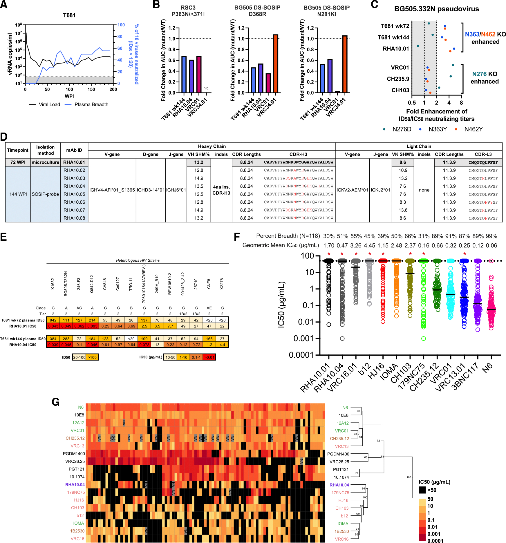
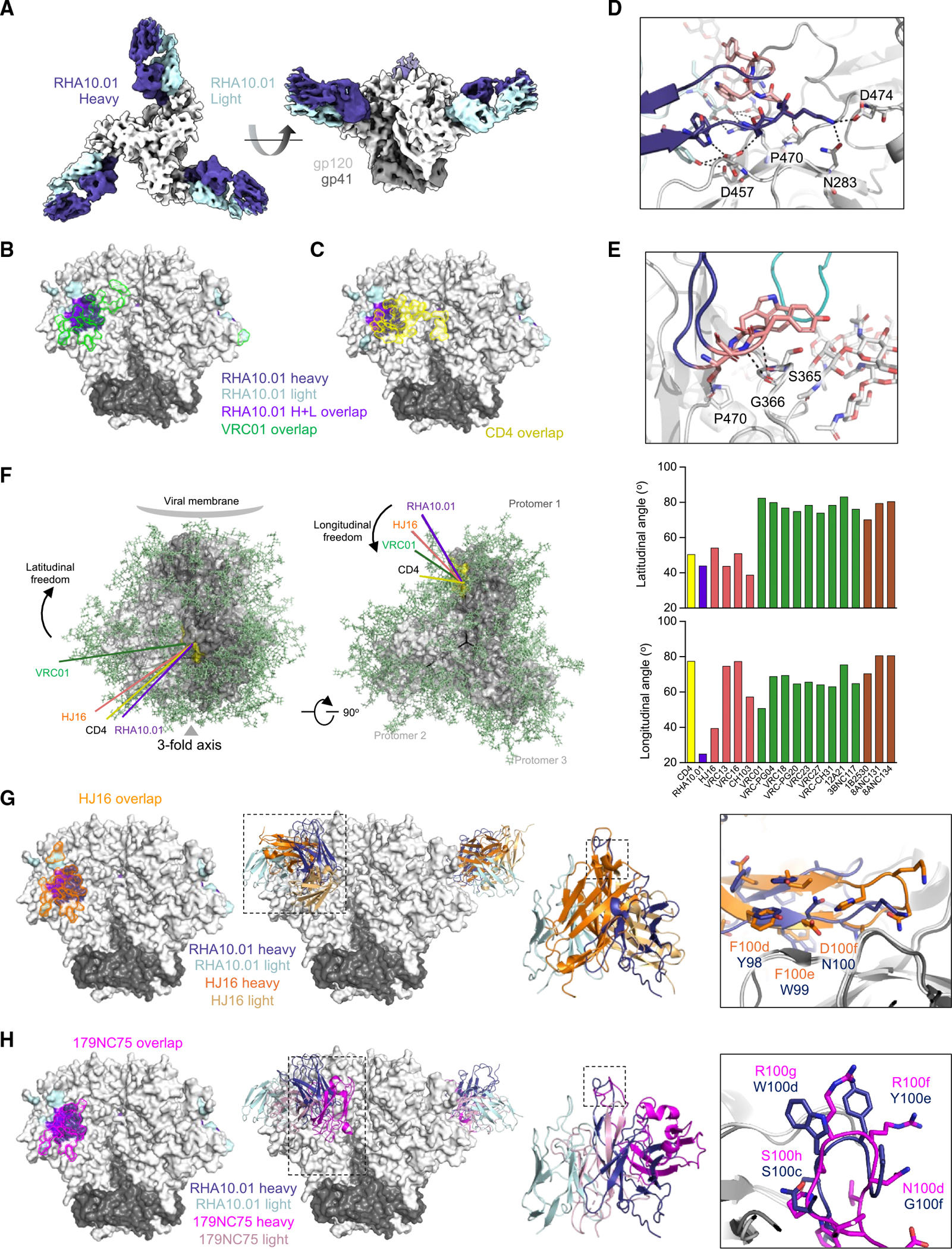
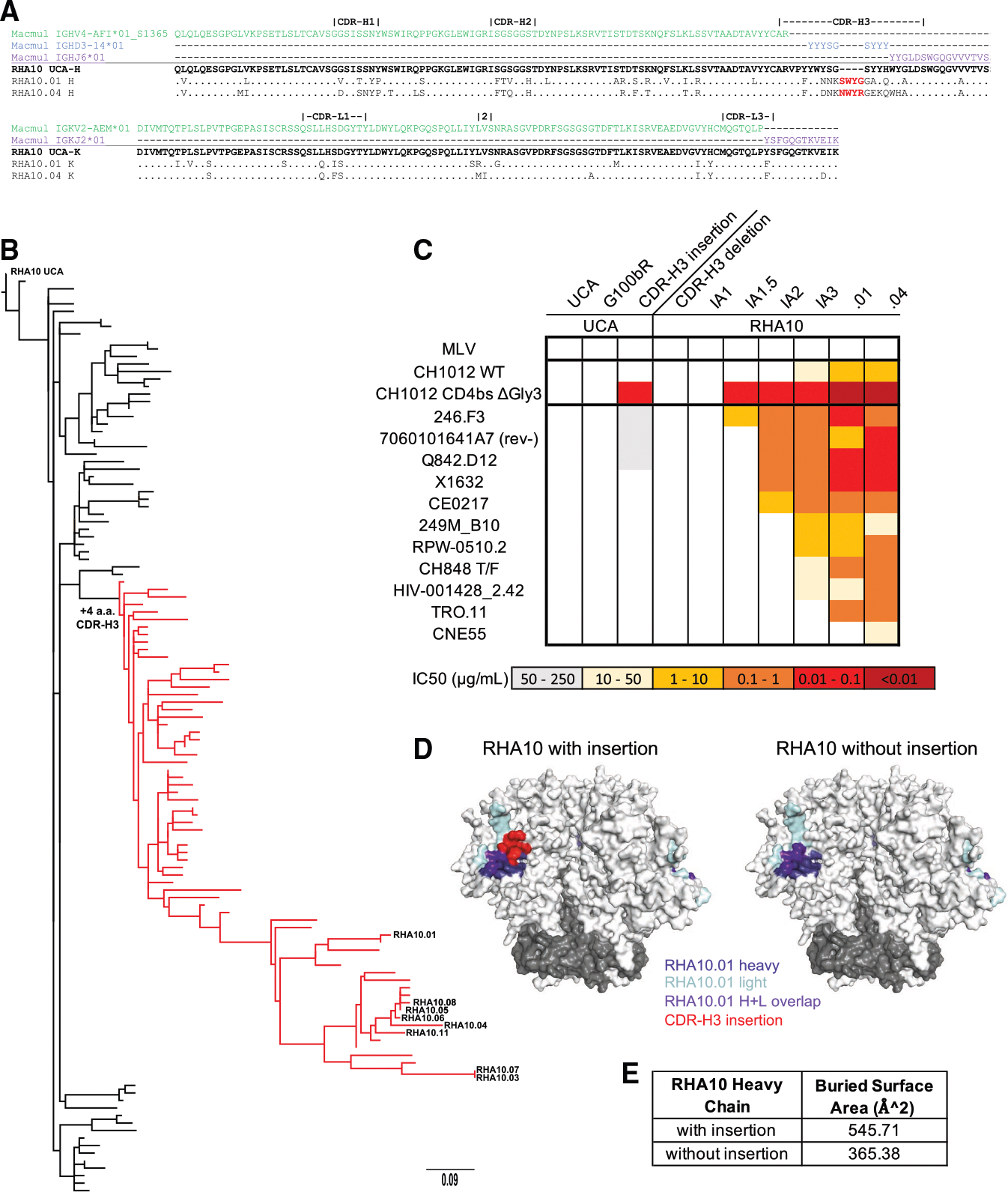
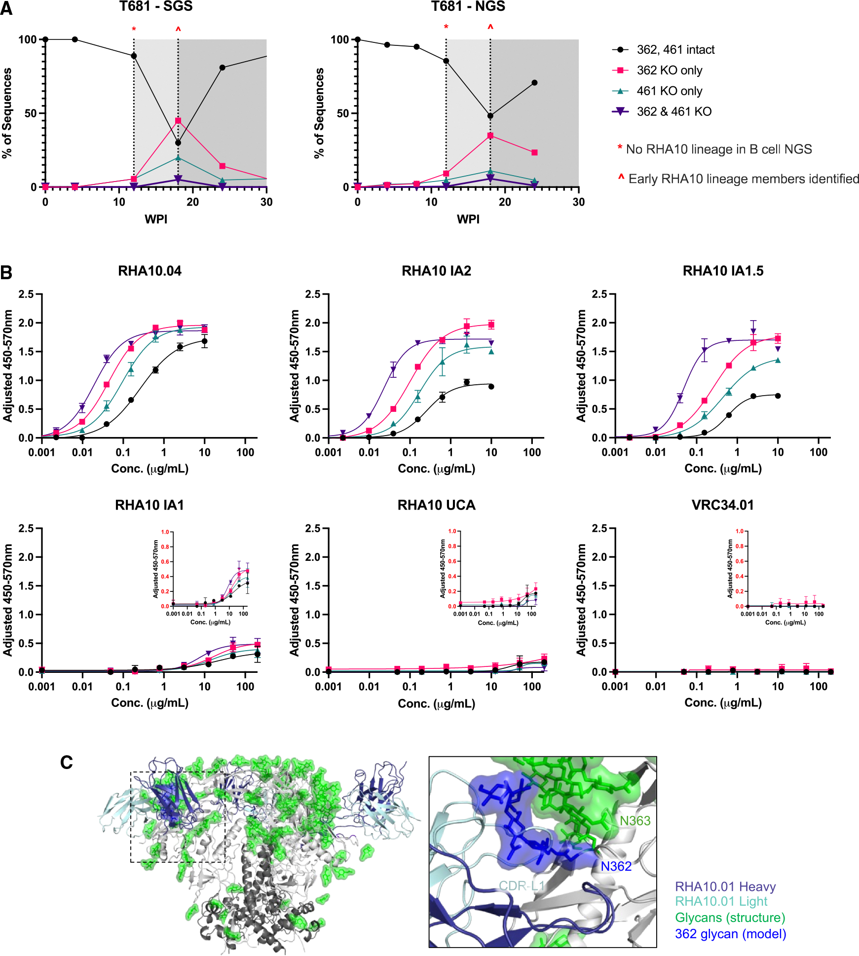
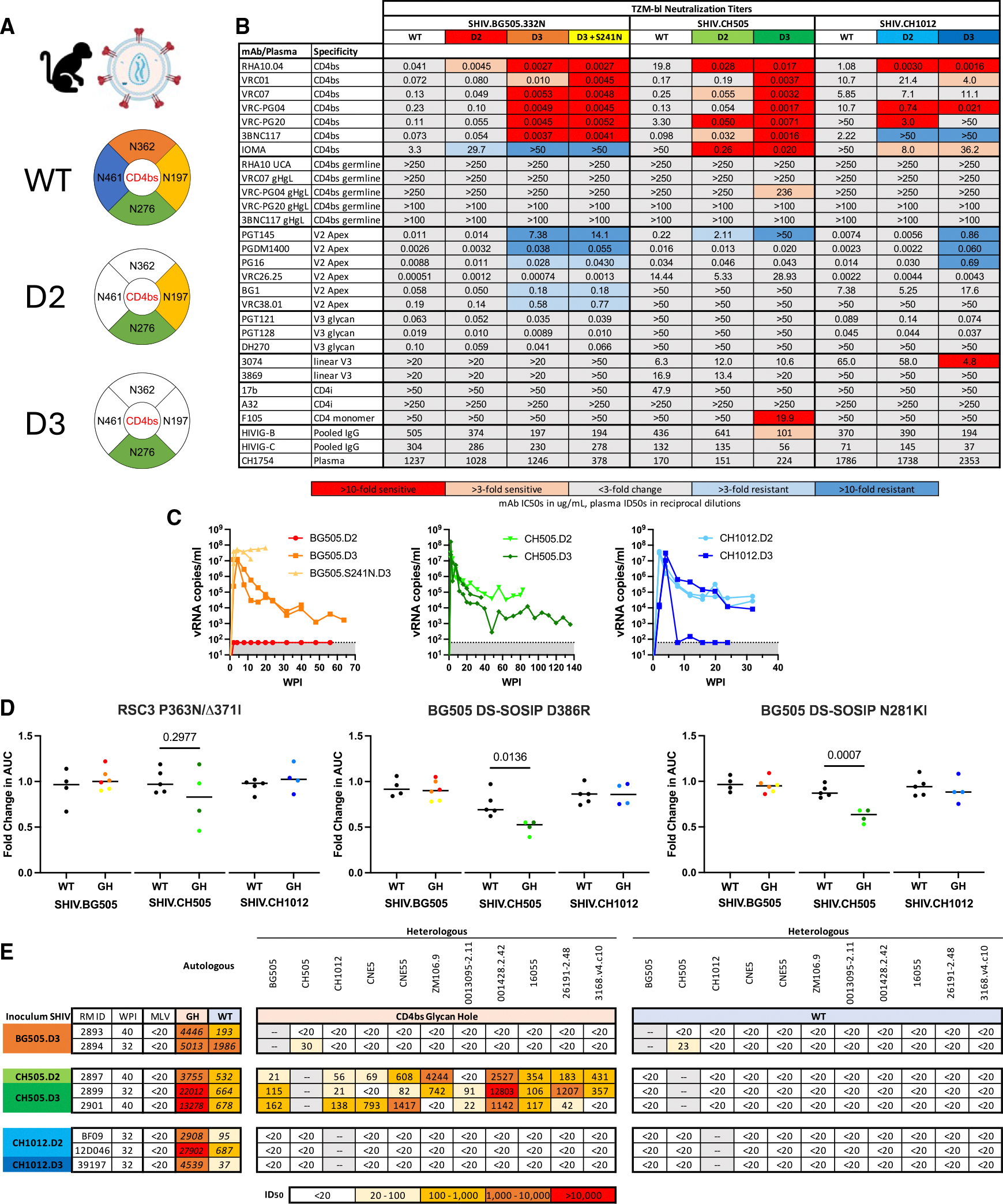
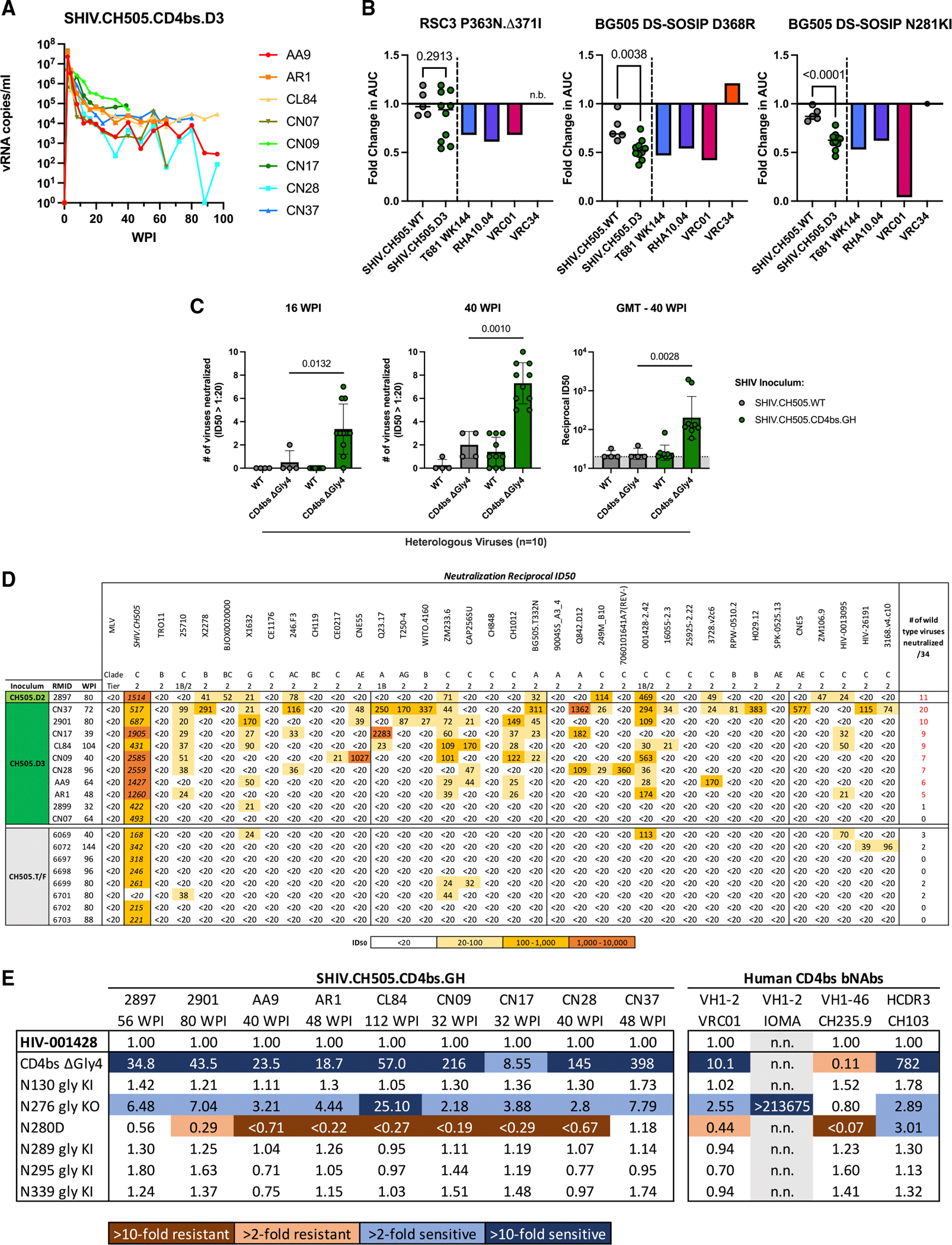
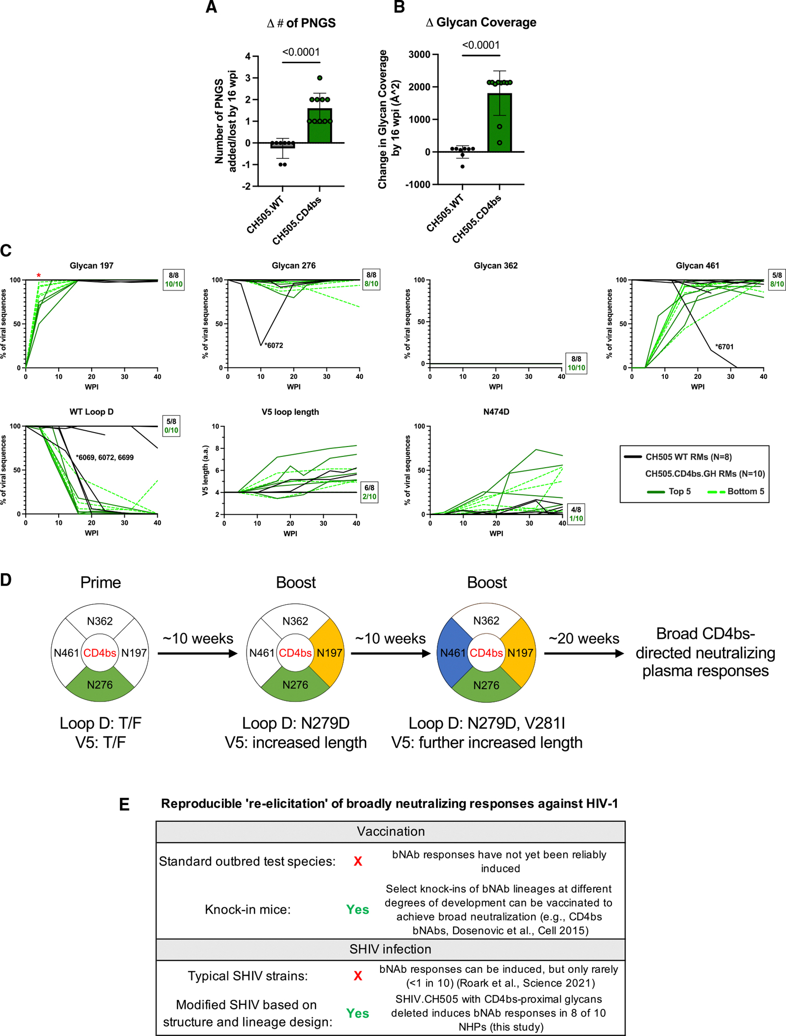
Broadly neutralizing antibodies (bNAbs) targeting the HIV-1 CD4-binding site (CD4bs) occur infrequently in macaques and humans and have not been reproducibly elicited in any outbred animal model. To address this challenge, we first isolated RHA10, an infection-induced rhesus bNAb with 51% breadth. The cryoelectron microscopy (cryo-EM) structure of RHA10 with the HIV-1 envelope (Env) resembled prototypic human CD4bs bNAbs with CDR-H3-dominated binding. Env-antibody co-evolution revealed transient elimination of two Env CD4bs-proximal glycans near the time of RHA10-lineage initiation, and these glycan-deficient Envs bound preferentially to early RHA10 intermediates, suggesting that glycan deletions in infecting SHIVs could induce CD4bs bNAbs. To test this hypothesis, we constructed SHIV.CH505 variants with CD4bs-proximal glycan deletions. Infection of 11 macaques resulted in accelerated CD4bs bNAb responses in 9 compared with 1 of 115 control macaques. Glycan hole-based immunofocusing coupled to Env-Ab co-evolution can consistently induce broad CD4bs responses in macaques and serve as a model for HIV vaccine design.

Keywords: CD4-binding site; CP: Immunology; HIV-1; broadly neutralizing antibody; co-evolution; glycan deletion; simian-human immunodeficiency virus (SHIV); structure-guided vaccine design.


Conflict of interest statement

Declaration of interests The authors declare no competing interests.

Figures

Figure 1.
Figure 1.. RHA10 is a rhesus CD4bs bNAb with 51% neutralization breadth that is N276 glycan agnostic
(A) Longitudinal plasma vRNA load (lower limit of quantification: 62 RNA copies/mL) and percent heterologous plasma neutralization breadth of an 18-strain panel in macaque T681. (B) Fold change in binding AUC between wild-type (WT) and CD4bs KO mutant RSC3 cores or BG505 DS-SOSIPs. n.b., no binding to WT protein. (C) Fold change in neutralization titers of T681 plasma or mAbs against BG505.332N pseudovirus glycan KO mutants. Enhancement is defined as a greater than 2-fold increase in ID50/IC50 titers. (D) Immunogenetics summary of the RHA10 lineage, with RHA10.01 highlighted as a reference with further SHM in the V(D)J junction shown in red. (E) Recapitulation of T681 plasma neutralization breadth and potency by respective RHA10 lineage members. (F) Neutralization breadth and potency of RHA10 members and human CD4bs bNAbs tested on a 118-virus panel; percent breadth and geometric mean IC50 are listed above each mAb. Median IC50 titers are represented as black bars. Human CD4bs bNAb neutralization data were obtained from HIV LANL CATNAP. (G) Heatmap and hierarchical clustering of large pseudovirus panel neutralization data. Only bootstrap values above 50% are shown. CD4bs bNAbs are colored by class: RHA10.04 (purple), CDR-H3 dominated (salmon), VH1-2 restricted (green), and VH1-46 restricted (brown). See also Figures S1 and S2 and Table S1.
Figure 2.
Figure 2.. RHA10 binds the CD4bs primarily through its long CDR-H3
(A) Cryo-EM structure of RHA10.01 bound to BG505 DS-SOSIP, colored by chain. (B) RHA10.01 binding footprint overlaid by VRC01 binding footprint (green). (C) RHA10.01 binding footprint overlaid by CD4 footprint (yellow). (D) RHA10.01 CDR-H3 stem Env contact details. CDR-H3 insertion, pink. (E) RHA10.01 CDR-H3 tip Env contact details. CDR-H3 insertion, pink. (F) Angle of approach analysis. CDR-H3-dominated class, salmon; VH1-2 class, green; VH1-46 class, brown. (G) RHA10.01 and HJ16 structural comparison by footprint, ribbon diagram overlap, and CDR-H3 geometries. (H) RHA10.01 and 179NC75 structural comparison by footprint, ribbon diagram overlap, and CDR-H3 geometries. See also Figure S3 and Table S2.
Figure 3.
Figure 3.. RHA10 developed a CDR-H3 insertion during maturation, crucial for broad neutralization
(A) Aa alignments of RHA10.01 and RHA10.04 heavy- and light-chain genes to inferred germline genes. Dots represent identity to the unmutated common ancestor (UCA). 2, CDR-L2 of 3 aa. (B) Phylogenetic tree of RHA10-lineage heavy chains rooted to the RHA10 UCA. Members containing the CDR-H3 insertion are highlighted in red. (C) Neutralization titers of RHA10-lineage evolutionary members and recombinant mutants against autologous and heterologous viral strains. CH1012 CD4bs ΔGly3 lacks the N197, N362, and N461 glycans. (D) Binding footprint comparison of RHA10.01 modeled with and without the CDR-H3 insertion. (E) Buried surface area of the RHA10 heavy chain, calculated by PDBePISA. See also Figure S4 and Table S3.
Figure 4.
Figure 4.. Transient deletion of glycans surrounding the CD4bs preceded RHA10 elicitation
(A) Percentage of single-genome viral sequences (SGS; left) or next-generation sequences (NGS; right) with mutated PNGS at residue(s) 362 and/or 461. Dotted vertical lines represent milestones in RHA10 lineage development highlighted in the text. wpi, weeks post infection. (B) ELISA curves of CH1012 gp120s binding by RHA10 UCA, inferred intermediates (IAs), RHA10.04, and negative control VRC34.01. Error bars represent standard deviation from the mean. (C) RHA10.01 structure from Figure 2 with glycans highlighted. RHA10.01 CDR-L1 clashing predicted with N362 glycan present in CH1012 (blue) is modeled next to N363 glycan present in BG505 (green). The N362 glycan model is based on gp120 alignment with PDB: 5FYK. See also Figure S5 and Table S4.
Figure 5.
Figure 5.. CD4bs glycan-deficient SHIVs improved CD4bs antigenicity, maintained infectivity, and elicited improved CD4bs targeting
(A) CD4bs glycan KO design schematic, created in BioRender. D2 designs lack the N362 and N461 glycans. D3 designs lack the N197, N362, and N461 glycans. (B) Neutralization titers against CD4bs glycan-deficient SHIV designs, with IC50 fold change highlighted. (C) Viral load kinetics of CD4bs.GH SHIV-infected RMs; lower limit of quantification = 62 vRNA copies/mL. (D) Fold change in binding AUC of rhesus plasma between WT and mutant RSC3 and BG505 DS-SOSIP proteins. Unpaired t tests were used for comparing WT and GH groups. (E) Plasma ID50 values of autologous and heterologous viruses with or without the CD4bs glycans from CD4bs.GH SHIV-infected RMs. See also Figure S6 and Table S5.
Figure 6.
Figure 6.. SHIV.CH505.CD4bs.GH elicited Abs capable of broadly recognizing the underlying CD4bs backbone
(A) Viral load kinetics of eight additional SHIV.CH505.D3-infected macaques. (B) Fold change in binding AUC of rhesus plasma between WT and mutant RSC3 and BG505 DS-SOSIP proteins. Unpaired t tests were used for comparing WT and GH groups. (C) Neutralization (number and titer) of heterologous viruses with or without the CD4bs glycans by SHIV.CH505.WT- and SHIV.CH505.CD4bs.GH-infected macaques. Error bars represent standard deviation from the mean. Significance was determined by Mann-Whitney tests. GMT, geometric mean titer. (D) Heterologous plasma ID50 titers in SHIV.CH505.CD4bs.GH-infected macaques at peak breadth and in SHIV.CH505.WT infected macaques at euthanasia. (E) Neutralization ID50/IC50 fold change of SHIV.CH505.CD4bs.GH macaques with breadth and human CD4bs bNAbs against strain HIV-001428 and point mutations, colored by direction and magnitude. CD4bs ΔGly4: lacks N197, N276, N363, and N462 glycans, gly KI: restores a common M-group glycan, gly KO: removes a common M-group glycan. n.n., not neutralized below 50 μg/mL. See also Table S6.
Figure 7.
Figure 7.. Sequential glycan reconstitution and CD4bs epitope viral evolution in SHIV.CH505.CD4bs.GH-infected macaques
(A) Comparison of the number of PNGS added by 16 wpi between WT and CH505.CD4bs.GH-infected macaques. Significance was determined by Mann-Whitney test. (B) Comparison of total glycan coverage of 16 wpi consensus sequences between WT and CH505.CD4bs.GH-infected macaques. Significance was determined by Mann-Whitney test. (C) SGS feature prevalence; lines represent individual macaques. Boxed numbers represent the number of overlapping macaque lines at 40 wpi; i.e., all 8 WT. SHIV.CH505 and all 10 SHIV.CH505.CD4bs.GH macaques lack a 362 PNGS in every Env sequence at 40 wpi. Solid green lines, top 5 breadth SHIV.CH505. CD4bs.GH RMs; dotted green lines, bottom 5 SHIV.CH505.CD4bs.GH RMs. Mann-Whitney test was applied to compare heterologous breadth in the top 5 and bottom 5 RMs; *p < 0.01. (D) Proposed immunization based on longitudinal sequencing in SHIV.CH505.D3 RMs. (E) Overview of CD4bs bNAb elicitation by vaccination or SHIV infection. See also Figure S7.

Update of

Similar articles

References

    1. Haynes BF, Burton DR, and Mascola JR (2019). Multiple roles for HIV broadly neutralizing antibodies. Sci. Transl. Med. 11, eaaz2686. 10.1126/scitranslmed.aaz2686. - DOI - PMC - PubMed
    1. Keele BF, Giorgi EE, Salazar-Gonzalez JF, Decker JM, Pham KT, Salazar MG, Sun C, Grayson T, Wang S, Li H, et al. (2008). Identification and characterization of transmitted and early founder virus envelopes in primary HIV-1 infection. Proc. Natl. Acad. Sci. 105, 7552–7557. 10.1073/pnas.0802203105. - DOI - PMC - PubMed
    1. Li H, Wang S, Kong R, Ding W, Lee F-H, Parker Z, Kim E, Learn GH, Hahn P, Policicchio B, et al. (2016). Envelope residue 375 substitutions in simian–human immunodeficiency viruses enhance CD4 binding and replication in rhesus macaques. Proc. Natl. Acad. Sci. 113, E3413–E3422. 10.1073/pnas.1606636113. - DOI - PMC - PubMed
    1. Roark RS, Li H, Williams WB, Chug H, Mason R, Gorman J, Wang S, Lee F, Rando J, Bonsignori M, et al. (2020). Recapitulation of HIV-1 Env-antibody coevolution in macaques leading to neutralization breadth. Science 2638, 1–32. - PMC - PubMed
    1. Zhou T, Lynch RM, Chen L, Acharya P, Wu X, Doria-Rose NA, Joyce MG, Lingwood D, Soto C, Bailer RT, et al. (2015). Structural repertoire of HIV-1-Neutralizing antibodies targeting the CD4 supersite in 14 donors. Cell 161, 1280–1292. - PMC - PubMed

MeSH terms

LinkOut - more resources