

Protocol for phenotyping and isolation of dendritic cell subsets from blood and lymphoid organs of non-human primates and humans by flow cytometry
- PMID: 40516056
- PMCID: PMC12205786
- DOI: 10.1016/j.xpro.2025.103897
Protocol for phenotyping and isolation of dendritic cell subsets from blood and lymphoid organs of non-human primates and humans by flow cytometry
Abstract
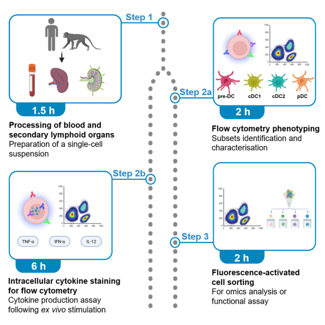
Dendritic cells (DCs) encompass several subsets that are essential for shaping immune responses. Here, we present a protocol for ex vivo functional analysis and purification of DC subsets from blood and secondary lymphoid organs using flow cytometry. We describe the steps for isolating mononuclear cells from blood, lymph nodes, and spleen. We then detail the procedures for phenotypic characterization of DC subsets, intracellular staining to assess their cytokine production, and fluorescence-activated cell sorting (FACS) to isolate individual DC populations. For complete details on the use and execution of this protocol, please refer to Gardet et al.1.
Keywords: Cell Biology; Cell isolation; Cell-based Assays; Flow Cytometry; Immunology; Molecular Biology; Single Cell.
Copyright © 2025 The Authors. Published by Elsevier Inc. All rights reserved.
Conflict of interest statement
Declaration of interests The authors declare no competing interests.
Figures


Similar articles
-
Protocol for in vivo lineage tracing of regeneration-associated macrophages from injured skeletal muscle of adult mice.STAR Protoc. 2025 Jun 20;6(2):103844. doi: 10.1016/j.xpro.2025.103844. Epub 2025 May 24. STAR Protoc. 2025. PMID: 40413749 Free PMC article.
-
Protocol to isolate and quantify large aging neutrophil-derived vesicles.STAR Protoc. 2025 Jun 20;6(2):103886. doi: 10.1016/j.xpro.2025.103886. Epub 2025 Jun 10. STAR Protoc. 2025. PMID: 40503932 Free PMC article.
-
Protocol for in vitro generation of innate lymphoid cells from human embryonic tissues.STAR Protoc. 2025 Mar 21;6(1):103525. doi: 10.1016/j.xpro.2024.103525. Epub 2024 Dec 20. STAR Protoc. 2025. PMID: 39708328 Free PMC article.
-
Neuraminidase inhibitors for preventing and treating influenza in healthy adults and children.Cochrane Database Syst Rev. 2012 Jan 18;1:CD008965. doi: 10.1002/14651858.CD008965.pub3. Cochrane Database Syst Rev. 2012. Update in: Cochrane Database Syst Rev. 2014 Apr 10;(4):CD008965. doi: 10.1002/14651858.CD008965.pub4. PMID: 22258996 Updated.
-
Factors that influence parents' and informal caregivers' views and practices regarding routine childhood vaccination: a qualitative evidence synthesis.Cochrane Database Syst Rev. 2021 Oct 27;10(10):CD013265. doi: 10.1002/14651858.CD013265.pub2. Cochrane Database Syst Rev. 2021. PMID: 34706066 Free PMC article.
References
-
- Gardet M., Haigh O., Meurisse F., Coindre S., Dimant N., Desjardins D., Bourgeois C., Goujard C., Vaslin B., Relouzat F., et al. Identification of macaque dendritic cell precursors in blood and tissue reveals their dysregulation in early SIV infection. Cell Rep. 2024;43 doi: 10.1016/j.celrep.2024.113994. - DOI - PubMed
-
- Guilliams M., Dutertre C.A., Scott C.L., McGovern N., Sichien D., Chakarov S., Van Gassen S., Chen J., Poidinger M., De Prijck S., et al. Unsupervised High-Dimensional Analysis Aligns Dendritic Cells across Tissues and Species. Immunity. 2016;45:669–684. doi: 10.1016/j.immuni.2016.08.015. - DOI - PMC - PubMed
LinkOut - more resources
Full Text Sources
