. 2025 Jun 14;16(1):449.
doi: 10.1038/s41419-025-07757-z.

USP10 promotes the progression and attenuates gemcitabine chemotherapy sensitivity via stabilizing PLK1 in PDAC

Affiliations

USP10 promotes the progression and attenuates gemcitabine chemotherapy sensitivity via stabilizing PLK1 in PDAC

Xuezhi Du et al. Cell Death Dis. .

Abstract

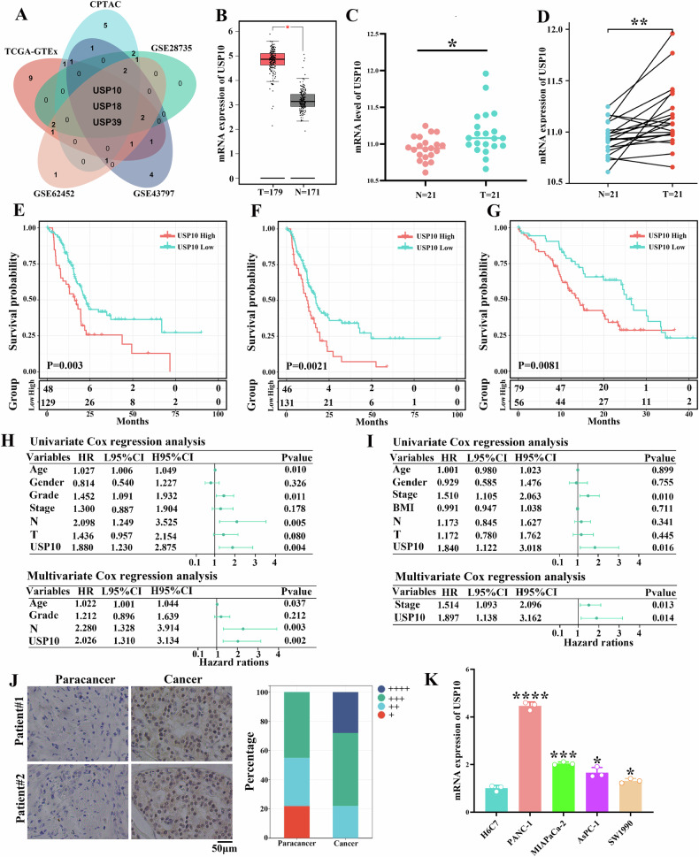
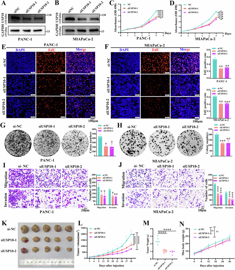
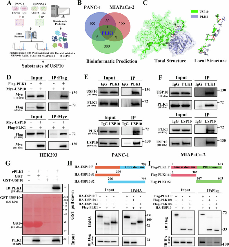
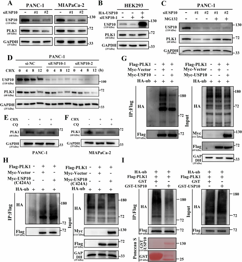
Pancreatic ductal adenocarcinoma (PDAC) is one of the most malignant tumors with limited treatment options, and chemotherapy resistance contributes to poor prognosis. An increasing number of studies have shown that ubiquitin specific peptidases (USPs), a subtype of deubiquitinases, can affect tumor progression by regulating the stability or biological function of substrate proteins. Thus, USPs are becoming attractive targets for cancer treatment. In this study, we investigated the role of USPs in PDAC. This study illustrated significant upregulation of USP10 expression in PDAC, which was found to be correlated with unfavorable prognosis. Further evaluation showed that USP10 exhibited the ability to facilitate PDAC progression in vitro and in vivo. The assays of immunoprecipitation-mass spectrometry, CO-IP, and GST pull-down suggested that USP10 directly interacted with PLK1. Deubiquitination assays indicated that USP10 could reduce the ubiquitination of PLK1 and increase protein stability. Moreover, USP10 may promote autophagy in PDAC cells through PLK1 and further attenuate the response of PDAC cells to gemcitabine (GEM). Finally, we demonstrated that the inhibition of USP10 combined with GEM synergistically inhibited the progression of PDAC in vitro and in vivo. In summary, we revealed that USP10, as a tumor promoter, promoted the progression and attenuated GEM chemotherapy sensitivity via stabilizing PLK1 in PDAC, providing a potential target for the treatment of PDAC.


Conflict of interest statement

Competing interests: The authors declared no competing interests.

Figures

Fig. 1
Fig. 1. USP10 is upregulated in PDAC samples and associated with unfavorable prognosis.
A A Venn diagram showing that USP10, USP18, and USP39 are differentially expressed in five public databases. B The difference of USP10 expression level between PDAC tissues and normal pancreatic tissues in TCGA-GTEx cohort. C The difference of USP10 expression level between PDAC tissues and paracancer tissues in CPTAC database. D The expression of USP10 in paired PDAC tissues and paracancer tissues. The overall survival (OS) (E) and progression free survival F in TCGA cohort showed a negative correlation with the expression of USP10. G The expression of USP10 was negatively correlated to the OS in CPTAC cohort. Univariate and multivariate Cox regression analysis between USP10 and OS in TCGA cohort (H) and CPTAC cohort (I). J Representative images of USP10 IHC staining in PDAC tissues and paracancer tissues. K The expression level of USP10 in four PDAC cells was detected via RT-qPCR. *P < 0.05, **P < 0.01, ***P < 0.001, ****P < 0.0001.
Fig. 2
Fig. 2. Biological functions of USP10 in PANC-1 and MIAPaCa-2 cells.
A, B The verification of USP10 siRNAs knockdown efficiency. C, D The effect of USP10 knockdown on proliferation detected through CCK-8 assays. E, F The change of proliferation after USP10 knockdown detected via EdU assays. G, H The influence of USP10 knockdown on the colony formation ability. I, J Transwell assays revealed decreased migration and invasion after USP10 knockdown. K Image of tumors in xenograft models following siRNA treatment. L, M Curves of tumor growth and weight for animal study. N Mice body weight among the groups. Data are presented as mean ± sd. from three biologically independent samples. **P < 0.01, ***P < 0.001, ****P < 0.0001.
Fig. 3
Fig. 3. USP10 directly interacts with PLK1.
A Flow chart of the USP10 substrate screening process. B A Venn diagram showing that PLK1 is the only substrate of USP10. C The interaction between USP10 and PLK1 was confirmed through molecular docking analysis. D Exogenous CO-IP assay of USP10 and PLK1 in HEK293 cells. E, F Endogenous interaction of USP10 and PLK1 detected via Co-IP assay in PDAC cells. G The direct interaction between USP10 and PLK1 was validated using a GST pull-down assay. H HEK293 cells were transfected with the respective plasmids, and full length and fragments of USP10 were used to pull down full-length PLK1. I HEK293 cells were transfected with the respective plasmids, and full length and fragments of PLK1 were used to pull down full-length USP10.
Fig. 4
Fig. 4. USP10 maintains PLK1 protein stability by triggering deubiquitination of PLK1.
A The change of USP10 and PLK1 protein after transfection with USP10 siRNAs in PDAC cells. B The protein level of USP10 and PLK1 were detected via western blot after transfection with HA-USP10 plasmids. C PANC-1 cells were transfected with si-NC, siUSP10-1, and siUSP10-2 and treated with 20 μM MG132 for 24 h, and the protein level of USP10 and PLK1 were detected through western blot. D PANC-1 cells were transfected with different siRNAs and treated with 10 μg/ml CHX for 0 h, 4 h, 8 h, and 12 h. The change of USP10 and PLK1 proteins were detected. The PANC1 E and MIAPaCa-2 F cells were treated with protein synthesis inhibitor CHX and autophagy inhibitor CQ, and the protein level of PLK1 was detected. G Myc-USP10 and other plasmids were transfected into HEK293 cells and treated with 20 μM MG132 for 6 h. The ubiquitination level of PLK1 was detected via IP assay. H Myc-USP10 (C424A) and other plasmids were transfected into HEK293 cells and treated with 20 μM MG132 for 6 h. The ubiquitination level of PLK1 was detected via IP assay. I The ubiquitinated Flag-PLK1 was purified from HEK293, and GST-USP10 was purified from E. coli BL21 (DE3). Next, the two proteins were incubated in the deubiquitination buffer at 37 °C for 2 h. The ubiquitination level of Flag-PLK1 was detected through western blot.
Fig. 5
Fig. 5. Biological functions of PLK1 in PANC-1 and MIAPaCa-2 cells.
A, B Confirmation of PLK1 knockdown efficiency. C, D CCK-8 assay was used to assess the impact of PLK1 knockdown on the proliferation. E, F EdU assay was employed to assess the impact of PLK1 knockdown on the proliferation. G, H Influence of PLK1 knockdown on the colony formation ability. I, J The change in migration ability after PLK1 knockdown confirmed through wound healing assay. K, L Influence of PLK1 knockdown on the migration and invasion ability, detected using Transwell assay. Data are presented as mean ± sd. from three biologically independent samples. *P < 0.05, **P < 0.01, ***P < 0.001, ****P < 0.0001.
Fig. 6
Fig. 6. USP10 affects the autophagy of PDAC cells.
A, B The si-NC, siUSP10-1, and siUSP10-2 were transfected into PDAC cells. When autophagy needed to be activated, the cells were treated with EBSS medium for 8 h. Subsequently, the autophagy-related proteins were detected through western blot. C, D PDAC cells were infected with the lentivirus Mcherry-EGFP-LC3B. Next, USP10 was knocked down, and the cells were treated with EBSS medium for 8 h before being observed under a confocal microscope. E The PDAC cells were treated with EBSS medium for 8 h following USP10 knockdown and were observed using transmission electron microscope. Data are presented as mean ± sd. from 3 biologically independent samples. *P < 0.05, **P < 0.01, ***P < 0.001, ****P < 0.0001.
Fig. 7
Fig. 7. USP10 affects the autophagy of PDAC cells partially through PLK1.
A, B The si-NC, siUSP10-1, siUSP10-2, and the overexpression plasmid of PLK1 were transfected into PDAC cells as required. The EBSS medium was used to activate autophagy. The autophagy-related proteins were detected. C, D PDAC cells were infected with the lentivirus Mcherry-EGFP-LC3B. Next, si-USP10 and the overexpression plasmid of PLK1 were transfected into PDAC cells as required, and the cells were treated with EBSS medium for 8 h before being observed under a confocal microscope. E si-USP10 and the overexpression plasmid of PLK1 were transfected into PDAC cells according to the requirement, and the cells were treated with EBSS medium for 8 h before being observed under a transmission electron microscope. *P < 0.05, **P < 0.01, ***P < 0.001, ****P < 0.0001.
Fig. 8
Fig. 8. USP10 affects the chemotherapy sensitivity of PDAC cells to gemcitabine (GEM) partially through autophagy.
A, B After USP10 knockdown, the IC50 of GEM for PANC-1 and MIAPaCa-2 cells was determined by treating the cells with different concentrations of GEM. C, D The PANC-1 and MIAPaCa-2 cells were treated with GEM after USP10 knockdown. The proliferation was detected through CCK-8 assay after the cells were treated with 100 nM Rapa for 48 h, as required. E, F The PANC-1 and MIAPaCa-2 cells were treated with GEM after USP10 knockdown. TUNEL assay was used to detect the cell apoptosis after the cells were treated with 100 nM Rapa for 48 h, as required. *P < 0.05, **P < 0.01, ***P < 0.001, ****P < 0.0001.
Fig. 9
Fig. 9. Inhibition of USP10 combined with gemcitabine (GEM) can synergistically inhibit the progress of PDAC.
A, B The synergistic inhibitory effect of USP10 knockdown combined with GEM on the cell proliferation detected through CCK-8 assay. CF The TUNEL assay demonstrated that USP10 knockdown combined with GEM had a synergistic stimulative effect, significantly enhancing cell apoptosis. G, H PANC-1 cells were used to construct xenograft tumor models. USP10 knockdown or GEM inhibited the growth of the tumors in vivo, whereas USP10 knockdown combined with GEM had synergistic inhibitory effect on tumor growth. I Mice body weight among the groups. Data are presented as mean ± sd. from three biologically independent samples. **P < 0.01, ***P < 0.001, ****P < 0.0001.

Similar articles

References

    1. Li O, Li L, Sheng Y, Ke K, Wu J, Mou Y, et al. Biological characteristics of pancreatic ductal adenocarcinoma: initiation to malignancy, intracellular to extracellular. Cancer Lett. 2023;574:216391. - PubMed
    1. Sung H, Ferlay J, Siegel RL, Laversanne M, Soerjomataram I, Jemal A, et al. Global cancer statistics 2020: GLOBOCAN estimates of incidence and mortality worldwide for 36 cancers in 185 countries. CA Cancer J Clin. 2021;71:209–49. - PubMed
    1. Bray F, Laversanne M, Sung H, Ferlay J, Siegel RL, Soerjomataram I, et al. Global cancer statistics 2022: GLOBOCAN estimates of incidence and mortality worldwide for 36 cancers in 185 countries. CA Cancer J Clin. 2024;74:229–63. - PubMed
    1. Mizrahi JD, Surana R, Valle JW, Shroff RT. Pancreatic cancer. Lancet. 2020;395:2008–20. - PubMed
    1. Wood LD, Canto MI, Jaffee EM, Simeone DM. Pancreatic cancer: pathogenesis, screening, diagnosis, and treatment. Gastroenterology. 2022;163:386–402.e1. - PMC - PubMed

MeSH terms