. 2025 Jun 14;11(1):109.
doi: 10.1038/s41522-025-00743-5.

M.marinum lacking epsH shows increased biofilm formation in vitro and boosted antibiotic tolerance in zebrafish

Affiliations

M.marinum lacking epsH shows increased biofilm formation in vitro and boosted antibiotic tolerance in zebrafish

Saara Lehmusvaara et al. NPJ Biofilms Microbiomes. .

Abstract

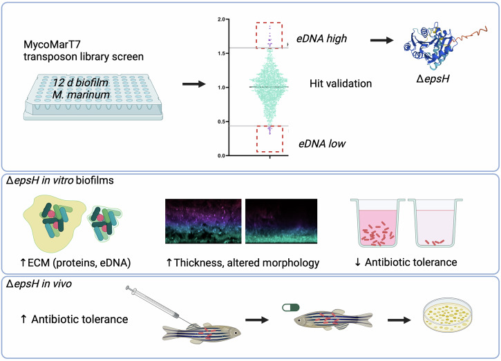
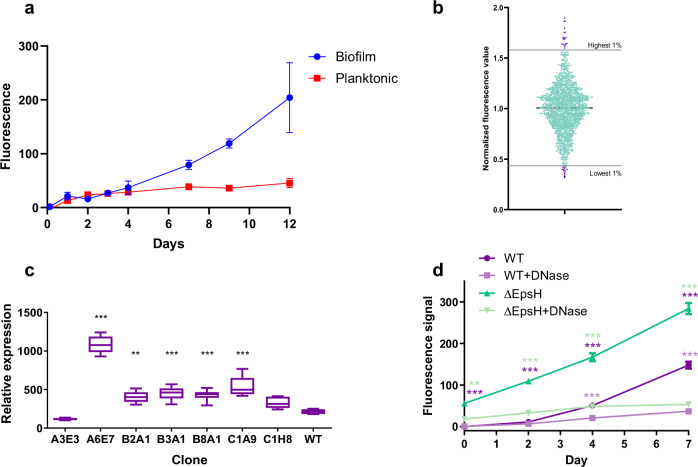
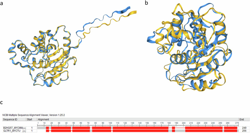
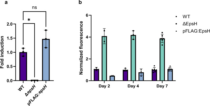
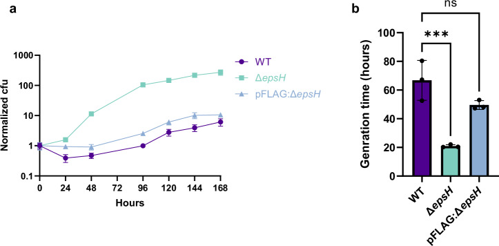
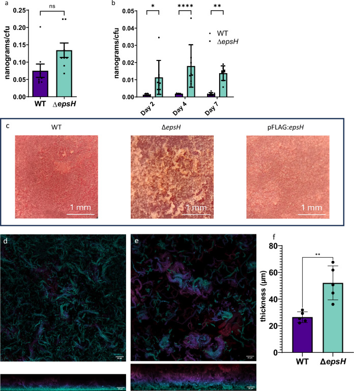
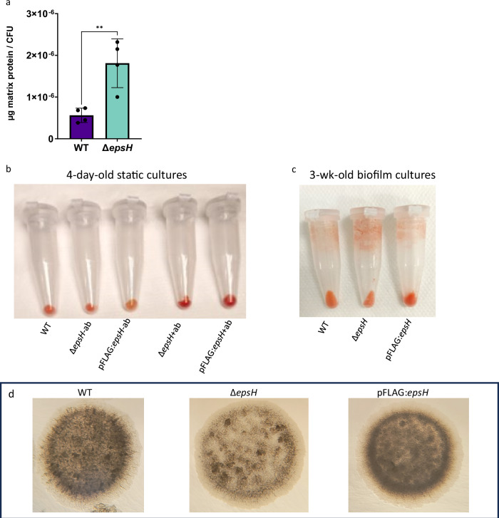
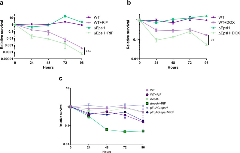
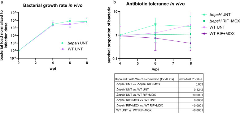
Recent discoveries have indicated that biofilm communities may play a role in natural drug tolerance of Mycobacterium tuberculosis. A transposon-based mutation library of a closely related species, Mycobacterium marinum, was used to identify clones in which the relative amount of extracellular DNA (eDNA), an important component of the extracellular matrix of biofilms, is altered. The disruption of a putative glycosyl transferase gene QDR78 11175, epsH, caused a substantial increase of the eDNA content of biofilms, and increased the growth rate and the biomass/cell in biofilm-forming conditions compared to wild-type. The increased abundance of biomass was mainly due to the elevated levels of eDNA and proteins in the extracellular matrix. The growth of the ΔepsH strain in the zebrafish was normal, but the mutant developed greater antibiotic tolerance in the adult zebrafish model. These results suggest that the extracellular matrix of biofilms increases antibiotic tolerance of mycobacteria during infection.


Conflict of interest statement

Competing interests: The authors declare no competing interests.

Figures

Fig. 1
Fig. 1. A summarizing figure of the work.
A transposon library screen of Mycobacterium marinum identified a biofilm mutant that lacked the gene encoding for a putative glycosyl transferase called epsH. The mutant had increased production of extracellular matrix in which extracellular DNA and proteins were increased. The drug tolerance of the mutant was decreased in biofilms cultured in vitro but increased in vivo in the adult zebra fish.
Fig. 2
Fig. 2. MycoMarT7 transposon library screen reveals mutants with altered eDNA levels in biofilm-forming culture conditions.
a GelRed-based measurement of eDNA levels in biofilm and planktonic M. marinum cultures revealed higher eDNA content in the biofilm. b Normalized fluorescence values of single clones grown in biofilm forming conditions after GelRed staining. Values were normalized to the average value of the plate. The clones having highest and lowest 1% of fluorescence were selected for further analysis. c Secondary screen of top and bottom 1% of eDNA abundancy clones by AccuBlue staining confirms the altered eDNA levels. One-way ANOVA followed by Dunnett's multiple comparison test. d The fluorescence signal of DNA binding dye AccuBlue increases by time in biofilm forming conditions and is significantly higher in epsH mutant strain than in WT. DNase treatment significantly reduces the fluorescence signal. The colour of the asterisk indicates the condition which the statistical analysis is compared to. Two-way ANOVA followed by Bonferroni’s multiple comparison test. P-values: * < 0.05, ** < 0.01, *** < 0.001.
Fig. 3
Fig. 3. Biofilm screen identifies a high eDNA clone lacking an EpsH glycosyltransferase.
Based on sequence homology and BLASTP analysis, M. ulcerans also has EpsH in its genome. Figure a shows the structural overlap of the structures of M. ulcerans EpsH and M. marinum EpsH structures as predicted by AlphaFold 2.0. The overlapping image was created with FoldSeek. OrthoInspector 3.0 identified Rv2957 as the closest homologous protein in Mtb. Figure b shows the structural overlap of the structures of Rv2957 and M. marinum EpsH structures as predicted by AlphaFold 2.0. The overlapping image was created with FoldSeek. c The sequence alignment shows the conserved areas between M. marinum EpsH and Rv2957 in red.
Fig. 4
Fig. 4. ΔepsH has an altered phenotype in eDNA deposition under biofilm-forming conditions.
a Quantitative PCR analysis of epsH expression in WT, ΔepsH and pFLAG:epsH strain shows restored epsH expression in the complemented strain. One-way ANOVA followed by Šídák's multiple comparisons test, error bar: mean ± SD. Three biological replicates were performed and shown as individual points. b Accublue staining of the biofilms shows that the complementation of the epsH gene reduces the level of eDNA close to the WT. Fluorescence values were normalized to OD600. Error bar: mean ± SD.
Fig. 5
Fig. 5. epsH deletion affects the growth in biofilm-forming conditions.
a CFU counts indicate faster cell proliferation of ΔepsH in biofilm-forming conditions compared to the WT. b ΔepsH demonstrated significantly shorter generation time in comparison to WT. One-way ANOVA followed by Dunnett’s multiple comparisons test, error bar: mean ± SD. ***p = 0.0009.
Fig. 6
Fig. 6. epsH deletion increases biomass per cell and affects colony morphology and biofilm structure.
a epsH deletion does not significantly influence the biomass in planktonic condition after 7 days of culture. Unpaired t test, error bar: mean ± SEM. b epsH deletion increases the total biomass per CFU under biofilm-forming conditions. Two-way ANOVA followed by Šídák's multiple comparisons test. *p = 0.0131, **p = 0.0023, ****p < 0.0001, error bar: mean ± SEM. c 2-week-old WT, ΔepsH and pFLAG:epsH biofilm on Congo red agar. 2-week-old WT and pFLAG:epsH bacterial lawns on agar have smoother surface compared to the rough morphology of the ΔepsH strain. The images were taken through a stereo microscope using 5x magnification. Confocal microscope images of 7-day-old tdTomato expressing d WT and e ΔepsH biofilms show differences in biofilm structure and thickness. Images were taken with Nikon A1R+ confocal laser scanning microscope equipped with a Nikon Apo LWD 40x WI λS DIC N2 objective. Image processing was carried out with Fiji-ImageJ software. Color represents z-distance, with warmer colors indicating higher positions and cooler colors showing deeper regions of the biofilm. f ΔepsH biofilms are significantly thicker in comparison to WT biofilms (N = 5). Biofilm thickness was calculated from confocal z-stacks acquired with a 0.325 µm step size. Slices with a mean intensity below 15 were excluded from the top and bottom, and the remaining slices were multiplied by the step size to determine total thickness. Unpaired t test with Welch's correction, p = 0.0085, error bar: mean ± SD.
Fig. 7
Fig. 7. epsH deletion increases protein levels, but not cellulose, in biofilm-forming conditions and affects colony opacity.
a epsH deletion results in elevated total protein levels in the ECM. Unpaired t test, p = 0.0064, error bar: mean ± SD. b Congo Red staining of 4-day old static cultures. ΔepsH and pFLAG:epsH were grown in the absence (−ab) and presence ( + ab) of strain specific antibiotics. Cultures were stained with 40 µg/ml Congo red for 2 h. c Congo Red staining of 3-week-old biofilm cultures. Cultures were stained with 40 µg/ml Congo red for 2 h. d ΔepsH strain forms less opaque colonies on agar. 6-day-old 5 µl inoculation spots.
Fig. 8
Fig. 8. epsH deletion reduces antibiotic tolerance in biofilm forming conditions.
1-week-old microaerophilic, detergent-free liquid M. marinum biofilm cultures were exposed to high concentrations of antibiotics and surviving bacteria were plated daily to create time-kill curves for tolerance measurements. a The ΔepsH strain was less tolerant to 400 µg/ml of rifampicin (RIF) and b 250 µg/ml doxycycline (DOX). c Complementation of the epsH-deficient strain with epsH-expressing plasmid reverted the tolerance to wildtype levels.
Fig. 9
Fig. 9. ΔepsH mutant strain demonstrates unaltered growth rate and increased drug tolerance in vivo.
a Adult zebrafish were infected per intraperitoneal injection with 35 cfu of wt or 44 cfu of ΔepsH M. marinum. Bacterial loads were quantified at 4-, 6-, and 8-weeks post-infection (N = 16-24 per time point per group) by plating and counting colonies. The medians and 95% CI are shown. There was no difference in growth rate between the strains. b Adult zebrafish infected as in part a were treated with 32 mg/kg of rifampicin (RIF) and 15 mg/kg moxifloxacin (MOX) per os between 4 and 8 weeks of infection (N = 16–24 per group per time-point). The bacterial loads were quantified as in a and normalized to the median load at the beginning of the treatment. The medians and 95% CI are shown. The ΔepsH strain demonstrated significantly increased tolerance to antibiotics compared to the WT.

Similar articles

References

    1. Global tuberculosis report 2024. Geneva: World Health Organization; 2024. Licence: CC BY-NC-SA 3.0 IGO.
    1. WHO Consolidated Guidelines on Tuberculosis. Module 4: Treatment - Drug-Susceptible Tuberculosis Treatment. (World Health Organization, Geneva, 2022). - PubMed
    1. Chakraborty, P., Bajeli, S., Kaushal, D., Radotra, B. D. & Kumar, A. Biofilm formation in the lung contributes to virulence and drug tolerance of Mycobacterium tuberculosis. Nat. Commun.12, 1606 (2021). - PMC - PubMed
    1. Flemming, H.-C. et al. Biofilms: an emergent form of bacterial life. Nat. Rev. Microbiol.14, 563–575 (2016). - PubMed
    1. Aldridge, B. B. et al. Asymmetry and aging of mycobacterial cells lead to variable growth and antibiotic susceptibility. Science335, 100–104 (2012). - PMC - PubMed

MeSH terms

LinkOut - more resources