. 2025 Jun 14;23(1):444.
doi: 10.1186/s12951-025-03466-z.

Coptis chinensis-derived extracellular vesicle-like nanoparticles delivered miRNA-5106 suppresses NETs by restoring zinc homeostasis to alleviate colitis

Affiliations

Coptis chinensis-derived extracellular vesicle-like nanoparticles delivered miRNA-5106 suppresses NETs by restoring zinc homeostasis to alleviate colitis

Yi Yang et al. J Nanobiotechnology. .

Abstract

Background: Inflammatory bowel disease (IBD) is a chronic disorder marked by persistent inflammation and damage to the intestinal mucosa. Despite significant advances in treatment, there remains an unmet need for more effective and safer therapeutic strategies.

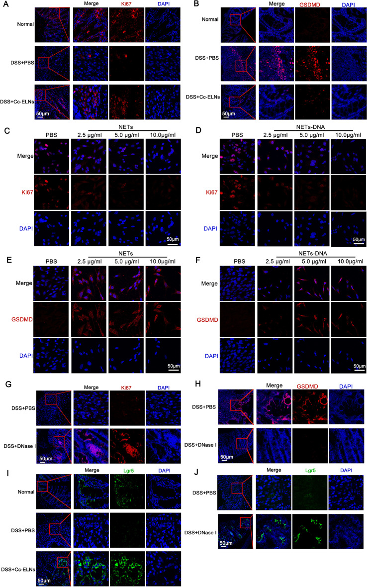
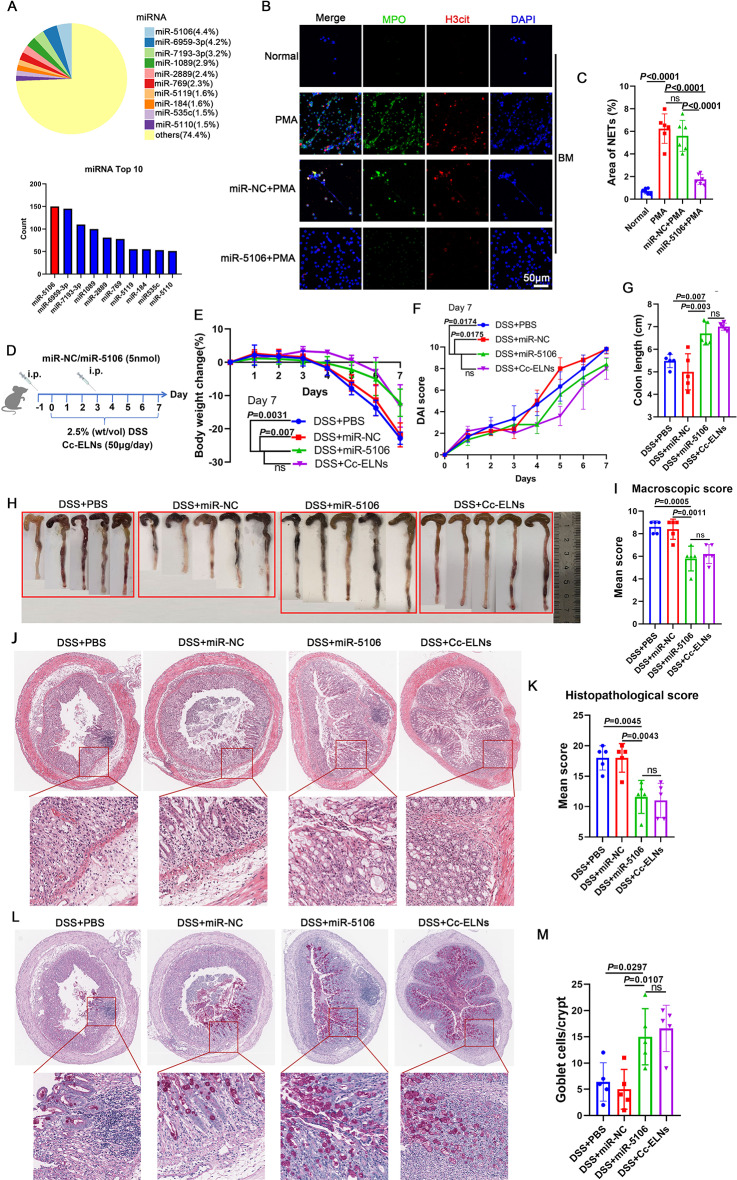
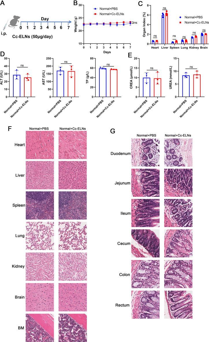
Results: In this study, we isolated and characterized extracellular vesicle-like nanoparticles (ELNs) derived from Coptis chinensis (Cc-ELNs) and evaluated their therapeutic potential in IBD. Intraperitoneal administration of Cc-ELNs in dextran sulfate sodium (DSS)-induced colitis mice demonstrated selective targeting of inflamed intestinal regions. Cc-ELNs significantly alleviated colitis by reducing neutrophil recruitment and inhibiting the formation of neutrophil extracellular traps (NETs). Furthermore, by suppressing NET formation, Cc-ELNs mitigated pyroptosis in intestinal epithelial cells (IECs) and promoted the proliferation of both IECs and intestinal stem cells (ISCs). Mechanistically, Cc-ELNs delivered miR-5106, which downregulated Slc39a2 expression, thereby restoring zinc homeostasis in neutrophils and reducing NET formation.

Conclusions: These findings establish Cc-ELNs as a novel, natural, and effective therapeutic candidate for IBD, highlighting the potential of plant-derived nanoparticle-based therapies.

Keywords: Coptis chinensis; Extracellular vesicle-like nanoparticles; Inflammatory bowel disease; Neutrophil; Neutrophil extracellular trap.


Conflict of interest statement

Declarations. Ethics approval and consent to participate: Ethics approval Animal protocols were approved by the Animal Care and Use Committee of Guangzhou Medical University (approval number: G2023-726) and complied with the National Institutes of Health Guidelines for the Care and Use of Laboratory Animals in China. Consent for publication: All authors of this study agreed to publish. Competing interests: The authors declare no competing interests.

Figures

Fig. 1
Fig. 1
Isolation and characterization of extracellular vesicle-like nanoparticles (Cc-ELNs) derived from Coptis chinensis and their protective effect against DSS-induced colitis in mice. (A) Cc-ELNs were isolated from fresh Coptis chinensis juice through a multi-step differential centrifugation process. (B) Transmission electron microscopy (TEM) image showing the cup-shaped morphology of Cc-ELNs. (C, D) Nanoflow cytometry analysis was used to determine the size distribution (C) and concentration (D) of Cc-ELNs. (E) DiR-labeled Cc-ELNs were injected intraperitoneally into mice, and their distribution was observed in the intestinal tissues (jejunum, ileum, cecum, and colon) after 24 h post-injection. (F) Mice with DSS-induced colitis were treated with intraperitoneal injections of Cc-ELNs, with berberine (BBR) serving as a positive control. (G) Time course of body weight changes in the experimental groups, normalized to Day 0 body weight. (H) Disease Activity Index (DAI) scores based on symptoms such as diarrhea, bleeding, and body weight loss. (I) Measurement of colon length from mice in different treatment groups. (J) Macroscopic appearance of the colon, as represented by a colon with mean colon length and typical injury findings. (K) Macroscopic colon scores indicating the severity of colon injury. (L) Histopathological examination of colon tissues stained with H&E. (M) Histopathological scores for the colon tissue samples determined through H&E staining. (N, O) Alcian blue-periodic acid-Schiff (AB-PAS) staining to detect goblet cell depletion (N), with goblet cell counts presented (O). n = 5–12; Statistical significance was determined using one-way ANOVA followed by Tukey’s multiple comparison test
Fig. 2
Fig. 2
Cc-ELNs inhibit neutrophil recruitment in DSS-induced mice. (A) Immunofluorescence analysis of neutrophil counts in the colon tissue of mice. (B) DiR-labeled Cc-ELNs were intraperitoneally injected into mice, and the biodistribution of Cc-ELNs was assessed in the femur, heart, liver, spleen, lungs, kidneys, brain, and gut using ex vivo imaging. (CF) Percentages of neutrophil counts in the spleen and bone marrow (BM) were analyzed by flow cytometry (C, E), and the statistical analysis results are shown (D, F). n = 5–7; Statistical significance was determined using one-way ANOVA followed by Tukey’s multiple comparison test
Fig. 3
Fig. 3
Cc-ELNs inhibit NET formation in DSS-induced colitis mice. (A) Immunofluorescence analysis was used to evaluate NET formation in colon tissues. Co-localization of H3cit and MPO was used as a marker for NET detection. (BD) Neutrophils isolated from the BM and spleen of mice were cultured in vitro for 24 h. Immunofluorescence analysis and scanning electron microscopy (SEM) were used to assess NET formation. (E) Neutrophils were incubated with PKH26-labeled Cc-ELNs, and internalization of Cc-ELNs was visualized through laser scanning confocal microscopy. (FH) Neutrophils from BM and spleens of normal mice were treated with Cc-ELNs and BBR, followed by PMA-induced NET formation in vitro. NETs were assessed by immunofluorescence and SEM. n = 6; Statistical significance was determined using one-way ANOVA followed by Tukey’s multiple comparison test
Fig. 4
Fig. 4
Cc-ELNs alleviate intestinal epithelial injury by inhibiting NET formation. (A) Ki67 staining was performed on colon tissues to evaluate cell proliferation levels. (B) Immunofluorescence analysis was used to detect gasdermin D (GSDMD), a marker of pyroptosis, in the colon tissues. (CF) Intestinal epithelial cells (IECs) were treated with NETs and NETs-derived DNA, and the levels of Ki67 and GSDMD were assessed by immunofluorescence analysis. (G, H) Colitis mice were treated with DNase I to inhibit colonic NET formation, and Ki67 and GSDMD expression levels in the colon tissue were evaluated. (I) Lgr5+ cells in the intestinal mucosa were detected by immunofluorescence to assess intestinal stem cells (ISCs). (J) DNase I was used to inhibit colonic NET formation in colitis mice, and ISCs in the intestinal mucosa were detected using immunofluorescence analysis
Fig. 5
Fig. 5
Cc-ELNs protect mice against DSS-induced colitis by delivering miR-5106 in high abundance. (A) Expression profile of miRNAs in Cc-ELNs was analyzed using small RNA sequencing, with the top 10 most highly expressed miRNAs presented. (B, C) Neutrophils treated with PMA and miR-5106 were analyzed for NET formation using immunofluorescence microscopy. (D) Mice with DSS-induced colitis were treated through intraperitoneal injection with miR-NC, miR-5106, or Cc-ELNs. (E) Body weight loss during the colitis progression was recorded, with Day 0 serving as the baseline (set at 0%). (F) Changes in the DAI were assessed. (G) On Day 7, mice were euthanized, and their colon tissues were excised and measured for length. (H) Macroscopic appearance of the colons was recorded. (I) Average macroscopic colon scores were calculated for each group. (J) Histopathological changes in colon tissues were examined using H&E staining. (K) Histopathological scoring was conducted for the colon tissue samples. (L, M) AB-PAS staining was used to assess goblet cell depletion in the colon (L), and quantification of goblet cells shown (M). n = 5–6; Statistical significance was determined using one-way ANOVA followed by Tukey’s multiple comparison test
Fig. 6
Fig. 6
MiR-5106 delivery by Cc-ELNs inhibits neutrophil infiltration and NET formation in colitis mice. (A) Immunofluorescence analysis was performed to assess neutrophil counts in colon tissues. (B, C) Percentage of neutrophils in the spleen was determined using flow cytometry (B), and the results of statistical analysis are shown (C). (D) Immunofluorescence was used to evaluate NET formation in the colon tissues of colitis mice. (EH) Neutrophils isolated from the BM and spleen of mice were cultured in vitro for 24 h. SEM and immunofluorescence analyses were performed to assess NET formation. n = 5–6; Statistical significance was determined using unpaired two-sample t-test
Fig. 7
Fig. 7
miR-5106 inhibits NET formation by targeting Slc39a2 to restore intracellular zinc homeostasis. (A) Slc39a2 mRNA expression was analyzed using qRT-PCR. (B) Wild-type and mutated m-Slc39a2-3′-untranslated region (UTR) was cloned into pmirGLO, and the predicted binding site of miR-5106 in the 3′-UTR of the Slc39a2 gene. (C) HEK293T cells were transfected with the Slc39a2 UTR reporter plasmid along with miR-5106 mimic or miR-NC mimic. Dual-luciferase reporter assays were performed to measure the impact of miR-5106 on Slc39a2 expression. (DG) Immunofluorescence analyses were performed to detect Slc39a2 expression in intestinal epithelium and neutrophils in the colon. (H) Neutrophils were treated with ZnCl2, and NET formation was evaluated using immunofluorescence assay. (I) Neutrophils were treated with miR-NC, miR-NC + ZnCl2, and miR-5106 + ZnCl2, and the mitochondrial situation was evaluated using the Mito Tracker Red fluorescent probe. n = 5–6; Statistical significance was determined using one-way ANOVA followed by Tukey’s multiple comparison test (A) and unpaired two-sample t-test (C, H)
Fig. 8
Fig. 8
Safety assays for Cc-ELNs. (A) Schematic of intraperitoneal Cc-ELNs administration. (B) Body weight variations in mice. (C) Organ indices (organ weight/body weight×100%). (D, E) Serological indices of liver (D) and kidney function (E). (F) Representative images of H&E-stained heart, liver, spleen, lung, kidney, brain, and BM sections. (G) H&E staining of intestinal segments (duodenum, jejunum, ileum, cecum, colon, and rectum). n = 3; Statistical significance was determined using an unpaired two-sample t-test
Fig. 9
Fig. 9
Schematic diagram of the experimental study. Cc-ELNs effectively alleviated colitis by reducing neutrophil recruitment and inhibiting NET formation. Cc-ELNs inhibited pyroptosis in IECs and promoted the proliferation of IECs and ISCs by reducing NETs. Mechanistically, miR-5106 delivery by Cc-ELNs inhibited NET formation by targeting Slc39a2 to restore intracellular zinc homeostasis

References

    1. Massironi S, Viganò C, Palermo A, et al. Inflammation and malnutrition in inflammatory bowel disease. Lancet Gastroenterol Hepatol. 2023;8:579–90. - PubMed
    1. Rubin DT, Ananthakrishnan AN, Siegel CA, et al. ACG clinical guideline: ulcerative colitis in adults. Am J Gastroenterol. 2019;114:384–413. - PubMed
    1. Maloy KJ, Powrie F. Intestinal homeostasis and its breakdown in inflammatory bowel disease. Nature. 2011;474:298–306. - PubMed
    1. Zhou G, Yu L, Fang L, et al. CD177 + neutrophils as functionally activated neutrophils negatively regulate IBD. Gut. 2018;67:1052–63. - PubMed
    1. Mantovani A, Cassatella MA, Costantini C, Jaillon S. Neutrophils in the activation and regulation of innate and adaptive immunity. Nat Rev Immunol. 2011;11:519–31. - PubMed

MeSH terms

LinkOut - more resources