. 2025 Jun 14;56(1):118.
doi: 10.1186/s13567-025-01554-4.

Genetic characterization and pathogenicity analysis of three porcine epidemic diarrhea virus strains isolated from North China

Affiliations

Genetic characterization and pathogenicity analysis of three porcine epidemic diarrhea virus strains isolated from North China

Ying Liu et al. Vet Res. .

Abstract

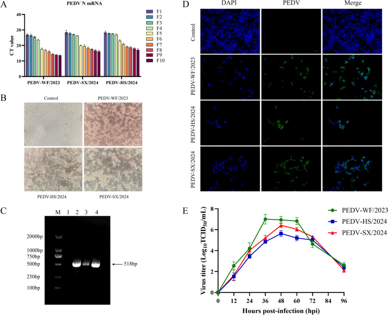
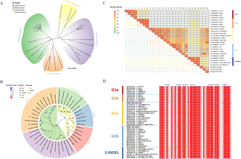
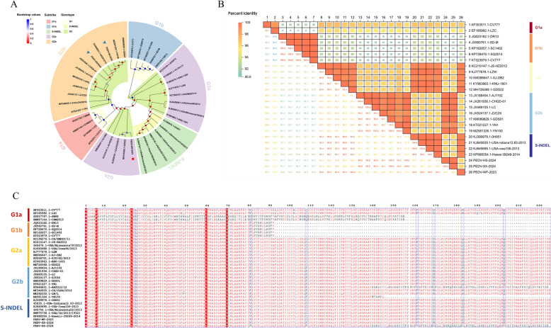
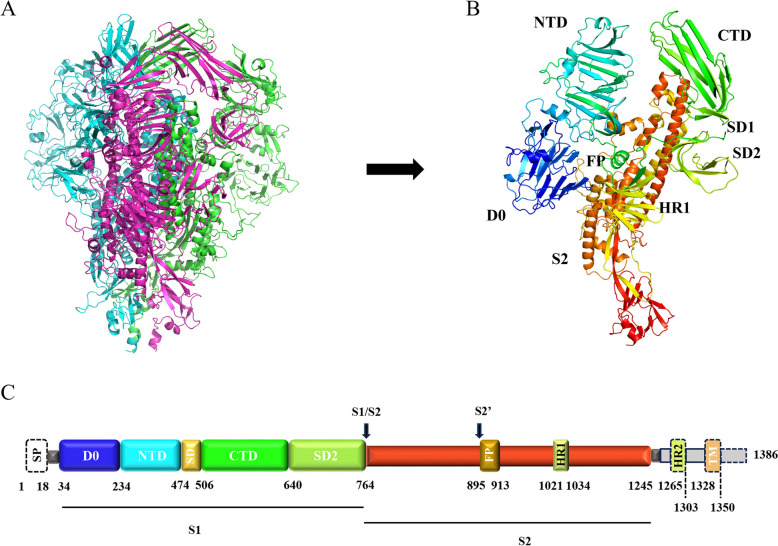
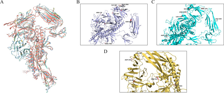
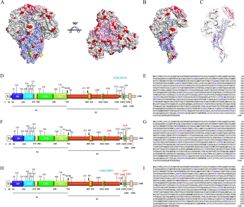
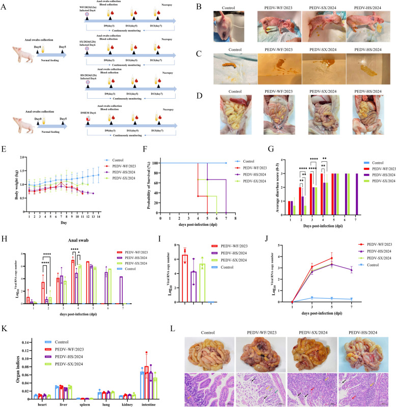
Porcine epidemic diarrhea (PED) is a highly contagious intestinal disease owing to porcine epidemic diarrhea virus (PEDV) infection. It is extremely detrimental to newborn piglets and has caused huge economic losses to the global pig industry. In this study, three PEDV strains of G2a PEDV-WF/2023, G2b PEDV-SX/2024 and PEDV-HS/2024 were successfully isolated from small intestine tissue samples with the analysis of their molecular structure characteristics, genetic characteristics and pathogenicity. Notably, these three PEDV strains had multiple unique aa mutations and extensive N-glycosylation in the D0 region, S1-NTD, COE epitope and SS6, respectively. Therefore, their structures were different compared to CV777 and PT-P5 strains. Furthermore, all the three PEDV strains caused severe clinical symptoms in 1-day-old piglets after infection. Among them, G2a PEDV-WF/2023 was the most detrimental to piglets, with highly levels of viral RNA in vivo. In contrast, PEDV-HS/2024 showed relatively weak pathogenicity to piglets, but it also caused the death of piglets. It might be attributed to the occurrence of individual mutations consistent with the amino acid sequence of G1b subtype in PEDV-HS/2024 strain. Findings in this study allow us to confirm that the G2a PEDV-WF/2023 strain is currently one of the most harmful epidemic strains to piglets. This study may benefit our understanding of the molecular structure characteristics, evolution trend and transmission dynamics of the epidemic strains in China. Moreover, it may provide potential reference for formulating more targeted PEDV vaccines, preventing and controlling this infection, and further curbing the cross-species spread of PEDV.

Keywords: PEDV; S protein; evolution trend; molecular structure characterization; pathogenic analysis.


Conflict of interest statement

Declarations. Ethics approval and consent to participate: The Ethics Committee at Hebei Agricultural University granted permission for the animal study, identified by the approval number 2021069. The research adhered to both institutional guidelines and local regulations. Competing interests: The authors declare that they have no competing interests.

Figures

Figure 1
Figure 1
Isolation and characterization of the PEDV-WF/2023, PEDV-SX/2024 and PEDV-HS/2024 strains. A RT-qPCR detection method was used to identify the mRNA expression level of N gene of each virus strain from F1 to F10 generations. B Cytopathic effects of each isolate under light microscopy. C The RT-PCR detection method was used to detect the infection of Vero cells by each isolated strain at the F5 generation for the PEDV-M gene. (M) 2000 Marker; (1) Negative Control. (2) PEDV-WF/2023 strain; (3) PEDV-HS/2024 strain; (4) PEDV-SX/2024 strain. D Indirect immunofluorescence identification of each isolated virus strain. E Detection of growth kinetics of each isolated virus strain.
Figure 2
Figure 2
Phylogenetic tree and genetic characteristics analysis based on S gene. A Coronavirus phylogenetic tree. Different genera are marked with different colors: Alpha-coronavirus (green), Beta-coronavirus (purple), Gamma-coronavirus (yellow) and Delta-coronavirus (orange). B Phylogenetic tree of PEDV strains. Different genotypes and subtypes are marked with different colors: G1 (yellow), S-INDEL (grass green), G2 (green) and G1a (red), G1b (blue), S-INDEL (green), G2a (purple), G2b (orange). Phylogenetic trees were constructed with MAGE 11 software using the neighbor-joining method. Bootstrap analysis was set up in 1000 replicates, the color of the dots at the nodes represents the bootstrap values. C Heat map of Homology analysis. Different colors represent levels of homology. The change from orange to blue represents the homology from high to low. Among them, the larger the square, the higher the homology, and the smaller the square, the lower the homology. D Based on S gene amino acid sequence alignment. Identical amino acid positions are represented by a red background, and mutated amino acids are represented by black fonts. The red square represents the PEDV-WF/2023 strain obtained in this study, and the blue triangle represents the PEDV-SX/2024 and PEDV-HS/2024 strains obtained in this study.
Figure 3
Figure 3
Phylogenetic tree and genetic characteristics analysis based on ORF3 gene. A Phylogenetic tree of PEDV strains. Different genotypes and subtypes are marked with different colors: G1 (yellow), S-INDEL (grass green), G2 (green) and G1a (red), G1b (blue), S-INDEL (green), G2a (purple), G2b (orange). Phylogenetic trees were constructed with MAGE 11 software using the neighbor-joining method. Bootstrap analysis was set up in 1000 replicates, the color of the dots at the nodes represents the bootstrap values. B Heat map of Homology analysis. Different colors represent levels of homology. The change from orange to blue represents the homology from high to low. Among them, the larger the square, the higher the homology, and the smaller the square, the lower the homology. C Based on ORF3 gene amino acid sequence alignment. Identical amino acid positions are represented by a red background, and mutated amino acids are represented by black fonts. The red square represents the PEDV-WF/2023 strain obtained in this study, and the blue triangle represents the PEDV-SX/2024 and PEDV-HS/2024 strains obtained in this study.
Figure 4
Figure 4
Schematic structure of PEDV S protein. A Trimer prefusion structure of PEDV S protein. Different colors represent different monomers. B Monomer structure of PEDV S protein. Different structural domains are represented by different colors. C Genome structure diagram of PEDV S protein. The different colored boxes represent the different domains of the S protein. The same domains in B and C are colored in the same scheme.
Figure 5
Figure 5
Structural analysis of PEDV S protein. A Comparative analysis of S protein monomer structure between PEDV-WF/2023, PEDV-SX/2024 and PEDV-HS/2024 strains and CV777 and PT-P5 strains. The S protein of PEDV-WF/2023, PEDV-SX/2024, PEDV-HS/2024, CV777 and PT-P5 strains cartoon modeling is shown as purple, light blue, yellow, red and blue, respectively. B Visualization of PEDV-WF/2023 amino acid mutations. C Visualization of PEDV-SX/2024 amino acid mutations. D Visualization of PEDV-HS/2024 amino acid mutations. Each strain in A.B.C.D is colored in the same scheme.
Figure 6
Figure 6
The N-glycosylation structure maps of S protein in three PEDV strains obtained in this study. A Atomic model of S protein trimer. B S protein monomer atomic model. C. Cartoon model of S protein monomer. N-glycosylation sites are shown in red. The lost N-glycosylation sites are indicated in yellow. The pink represents the receptor binding domain (RBD). The S1 and S2 subunits are shown in gray and purple, respectively. D. Distribution of N-glycosylation sites in the genome of PEDV-WF/2023 strain. E. Prediction results of N-glycosylation of PEDV-WF/2023 strain. F Distribution of N-glycosylation sites in the genome of PEDV-SX/2024 strain. G Prediction of N-glycosylation of PEDV-SX/2024 strain. H Distribution of N-glycosylation sites in genome of PEDV-HS/2024 strain. I PEDV-HS/2024 strain N-glycosylation prediction results. D, F, H in blue represent lost N-glycosylation sites. E, G, I Represent the results of specific tripeptide sequence outputs in blue and N-glycosylation modifications in red.
Figure 7
Figure 7
Pathogenicity test of piglets with PEDV-WF/2023, PEDV-SX/2024 and PEDV-HS/2024 strains. A Experimental challenge program for piglets. B Representative clinical signs of piglets in each group. C Fecal morphology evaluation of piglets in each group. D Gross lesions of the small intestine of piglets in each group. E Weight monitoring of piglets. F Survival rate of piglets. G.Piglet fecal scores. H Detection of viral RNA in anal swabs of piglets by RT-qPCR. *, P < 0.05; **, P < 0.01; ***, P < 0.001; ****, P < 0.0001. I. Viral load in jejunal tissues of piglets in different groups. J Viral load in blood of piglets in different groups. K Organ index of piglets in each group. L Histopathological examination and H&E staining of jejunum of piglets in PEDV challenge group or control group. Black arrow: mucosal epithelial cells necrosis, shedding; Red arrow: blood vessel congestion; Yellow arrow: focal aggregation of lymphocytes; Green arrow: intestinal gland; Light green arrow: connective tissue of the lamina propria of the intestinal villi; Scale bar, 100 μm.

References

    1. Yu K, Liu X, Lu Y, Long M, Bai J, Qin Q, Su X, He G, Mi X, Yang C, Wang R, Wang H, Chen Y, Wei Z, Huang W, Ouyang K (2023) Biological characteristics and pathogenicity analysis of a low virulence G2a porcine epidemic diarrhea virus. Microbiol Spectr 11:e0453522 - PMC - PubMed
    1. Cui J, Li F, Shi ZL (2019) Origin and evolution of pathogenic coronaviruses. Nat Rev Microbiol 17:181–192 - PMC - PubMed
    1. Have P, Moving V, Svansson V, Uttenthal A, Bloch B (1992) Coronavirus infection in mink (Mustela vison). Serological evidence of infection with a coronavirus related to transmissible gastroenteritis virus and porcine epidemic diarrhea virus. Vet Microbiol 31:1–10 - PMC - PubMed
    1. Peng Q, Fan B, Song X, He W, Wang C, Zhao Y, Guo W, Zhang X, Liu S, Gao J, Li K, Zhang B, Zhou J, Li Y, Guo R, Li B (2023) Genetic signatures associated with the virulence of porcine epidemic diarrhea virus AH2012/12. J Virol 97:e0106323 - PMC - PubMed
    1. Pensaert MB, de Bouck P (1978) A new coronavirus-like particle associated with diarrhea in swine. Arch Virol 58:243–247 - PMC - PubMed

MeSH terms

LinkOut - more resources