. 2025 Jun 12:85:103729.
doi: 10.1016/j.redox.2025.103729. Online ahead of print.

Fructose intake enhances lipoteichoic acid-mediated immune response in monocytes of healthy humans

Affiliations

Fructose intake enhances lipoteichoic acid-mediated immune response in monocytes of healthy humans

Raphaela Staltner et al. Redox Biol. .

Abstract

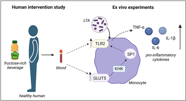
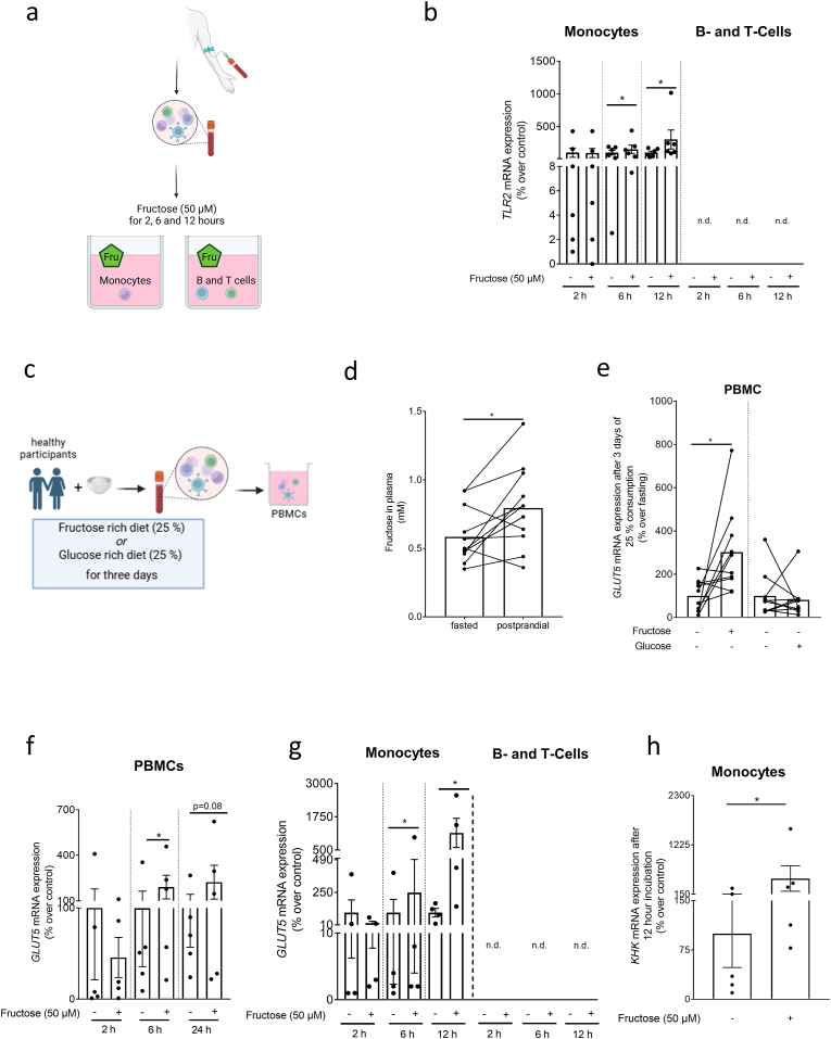
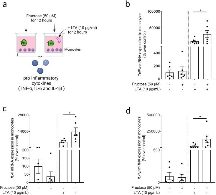
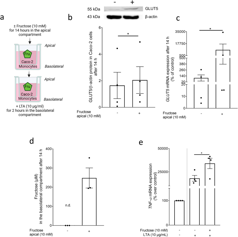
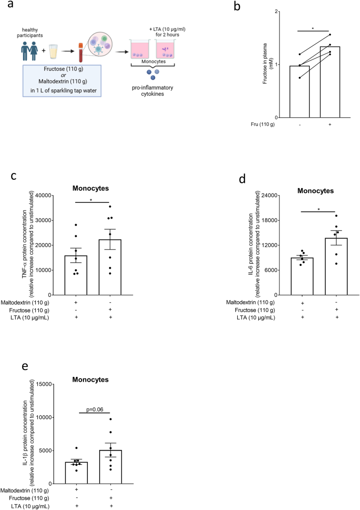
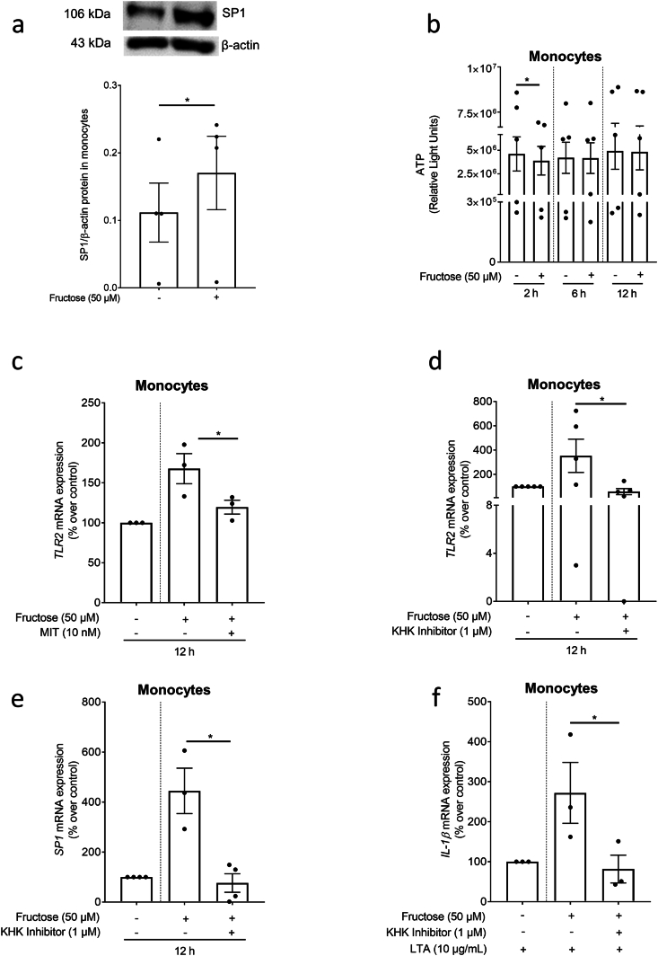
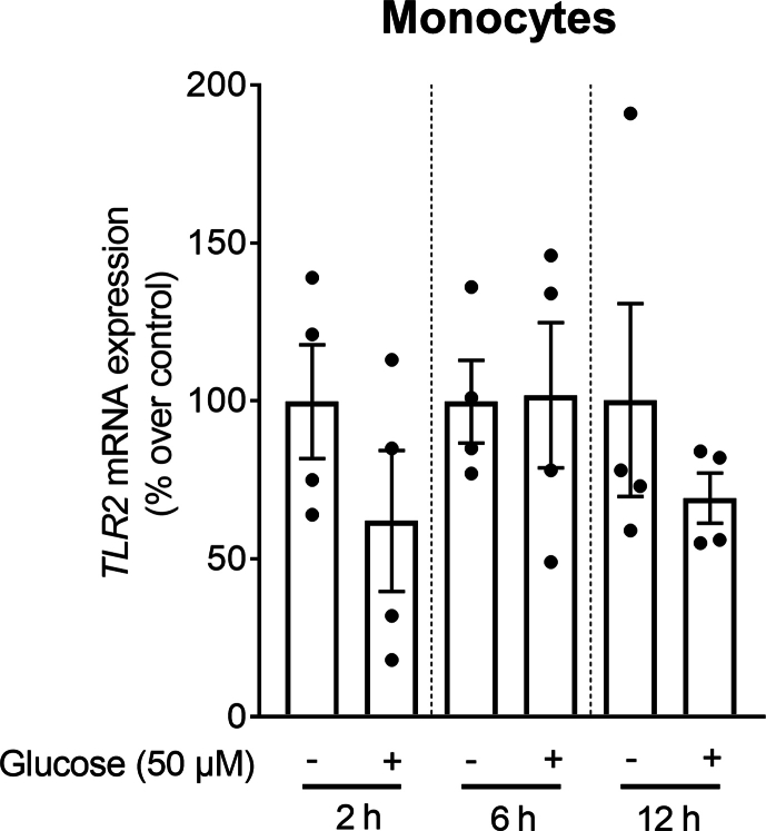
Metabolic diseases like type 2 diabetes are afflicted with higher rates of infections and longer, more complicated infection course as well as higher fatality rates. The impact of nutrition and specific nutrients like free fructose herein has not yet been fully understood. Here, we performed dietary intervention studies in healthy individuals and performed ex vivo experiments in isolated blood immune cells to assess the effects of dietary fructose intake on Gram-positive bacterial toxin induced immune responses. Acute and extended intake of fructose but not glucose was related with an induction of Toll like receptor 2 mRNA expression in monocytes and enhanced the LTA-dependent release of proinflammatory cytokines from monocytes. Blocking fructose metabolism and transcription factor SP1 attenuated the fructose-related induction of Toll like receptor 2 mRNA expression and augmentation of proinflammatory cytokine release further suggesting that fructose-dependent metabolic alterations are critical in enhancing immune responsiveness of humans after fructose consumption.

Keywords: Gram-positive bacteria; Ketohexokinase; Lipoteichoic acid; SP1; Sugar; Toll like receptor 2.


Conflict of interest statement

Declaration of competing interest All authors declare no conflict of interest.

Figures

Image 1
Graphical abstract
Fig. 1
Fig. 1
Expression of TLR2, GLUT5, and KHK mRNA in PBMCs, monocytes, and B- as well as T-cells isolated from healthy donors or healthy humans after the intake of fructose. (a) Schematic drawing of experimental set-up of experiments shown in (b) and (f, g, h). (b) TLR2 mRNA expression in monocytes and B- and T-lymphocytes isolated from buffy coats and ex vivo challenged with fructose (50 μM) for 2, 6 and 12 h. (c) Study design of the human intervention study (study A) with an acute intake of fructose (40 g) and a fructose rich diet for 3 days (25 % E of total caloric intake). (d) Fructose levels in plasma before and 3 h after a light breakfast containing 40 g fructose as well as (e) GLUT5 mRNA expression in isolated PBMCs after three days of a fructose or glucose rich diet (25 % E of total caloric intake) of healthy participants. (f) GLUT5 mRNA expression in PBMCs from healthy human donors challenged ex vivo with fructose (50 μM) for 2, 6 and 24 h. (g) GLUT5 mRNA expression in monocytes and B- and T-cells of healthy human donor's ex vivo challenged with fructose (50 μM) for 2, 6 and 12 h. (h) KHK mRNA expression in monocytes isolated from buffy coats and challenged with fructose (50 μM) for 12 h ∗p≤0.05. Fig. 1 (a); n = 6; Fig. (d, e): n = 10; Fig. (f): n = 5; Fig. (g): n = 4; Figure (h): n = 5. Fig. 1 (a) and (c) were created with biorender.com. TLR2: Toll like receptor 2; GLUT5: glucose transporter 5; PBMC: peripheral blood mononuclear cells: KHK: ketohexokinase.
Fig. 2
Fig. 2
Effect of fructose on the LTA-dependent induction of proinflammatory cytokine expression in monocytes obtained from healthy donors. (a) Graphical illustration of experimental set-up. (b) TNFα, (c) IL-6 and (d) IL-1β mRNA expression in monocytes pre-incubated for 12 h with 50 μM fructose and challenged with 10 μg/ml LTA for 2 h n = 6. ∗p≤0.05. Fig. 2 (a) was created with biorender.com. LTA: lipoteichoic acid; TNFα: tumor necrosis factor; IL: interleukin.
Fig. 3
Fig. 3
Effect of fructose on fructose transport and the LTA-dependent induction of TNFα mRNA expression in a co-culture model of differentiated Caco-2 cells and human primary monocytes. (a) Experimental set-up. (b) GLUT5 protein concentration in Caco-2 cells and (c) GLUT5 mRNA expression in monocytes 14 h after challenging Caco-2 cells with 0–10 mM fructose (d) fructose concentration in the basolateral compartment as well as (e) TNFα mRNA expression in monocytes after the 14 h challenge of Caco-2 cells with fructose (10 mM) and 2 h incubation of monocytes with LTA (10 μg/ml). n = 3–4. ∗p≤0.05.Fig. 3 (a) was created with biorender.com. GLUT5: glucose transporter 5; LTA: lipoteichoic acid; TNFα: tumor necrosis factor.
Fig. 4
Fig. 4
Effect of an acute intake of an isocaloric fructose- or maltodextrin-rich beverage on fructose levels in blood and LTA-induced cytokine release in monocytes. (a) Study design of the human intervention study (study B) and ex vivo experiments. (b) Plasma fructose levels 2 h after the intake of 110 g fructose. Relative increase (compared to unstimulated) of (c) TNFα, (d) IL-6, and (e) IL-1β protein concentration in monocytes isolated after the intake of maltodextrin or fructose challenged ex vivo with and without 10 μg/ml LTA for 2 h n = 4–7. ∗p≤0.05.Fig. 4 (a) was created with biorender.com.; LTA: lipoteichoic acid; TNFα: tumor necrosis factor; IL: interleukin.
Fig. 5
Fig. 5
Effect of fructose on SP1 protein and ATP levels in primary monocytes and on SP1 and KHK-1-dependent regulation of TLR2 expression in primary monocytes. Effect of the incubation of monocytes with fructose (0–50 μM) for 12 h on (a) SP1 protein concentration. (b) ATP units in monocytes after 2, 6 and 12 h of incubation with fructose. TLR2 mRNA expression in monocytes incubated with fructose (c) ± MIT (10 nM) and (d) ± KHK inhibitor 1. (e) SP1 mRNA expression of monocytes incubated with fructose±KHK inhibitor 1 (1 μM) and (f) IL- mRNA expression after additional stimulation with LTA (10 μg/ml) for 2 h n = 3–5. ∗p≤0.05. MIT: mithramycin; SP1: transcription factor 1; TLR2: Toll like receptor 2; KHK: ketohexokinase.
figs1
figs1

Similar articles

References

    1. Organization W.H. The top 10 causes of death. 2024. https://www.who.int/news-room/fact-sheets/detail/the-top-10-causes-of-death
    1. Collaborators G.B.D.A.R. Global mortality associated with 33 bacterial pathogens in 2019: a systematic analysis for the Global Burden of Disease Study 2019. Lancet. 2022;400:2221–2248. - PMC - PubMed
    1. Boyce M.R., Katz R., Standley C.J. Risk factors for infectious diseases in urban environments of sub-saharan Africa: a systematic review and critical appraisal of evidence. Trop. Med. Infect. Dis. 2019;4 - PMC - PubMed
    1. Krznaric J., Vince A. The role of non-alcoholic fatty liver disease in infections. Life (Basel) 2022;12 - PMC - PubMed
    1. Wu Q., Liu Y., Ma Y.B., Liu K., Chen S.H. Incidence and prevalence of pulmonary tuberculosis among patients with type 2 diabetes mellitus: a systematic review and meta-analysis. Ann. Med. 2022;54:1657–1666. - PMC - PubMed

LinkOut - more resources