

Optimizing malignancy prediction: A comparative analysis of transfer learning techniques on EBUS images
- PMID: 40517683
- PMCID: PMC12206062
- DOI: 10.1016/j.clinsp.2025.100703
Optimizing malignancy prediction: A comparative analysis of transfer learning techniques on EBUS images
Abstract
Background: Improving diagnostic accuracy in EBUS image analysis using machine learning is a current challenge. This study aimed to identify the most effective transfer learning model for predicting lymph node malignancy.
Methods: EBUS images collected between 2020-2023 were retrospectively analyzed. Demographic data, sampled lymph nodes, and pathology results were retrospectively collected from the files. Eight pre-trained CNN models (VGG, ResNet, InceptionNet, Xception, MobileNet, DenseNet, NasNet, EfficientNet) were evaluated.
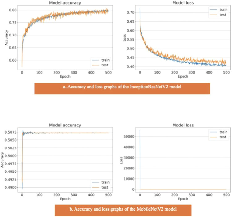
Results: The study shows that the VGG19, EfficientNetV2L and DenseNet201 models have the highest performance in malignancy prediction, achieving areas under the curve of 0.96, 0.96 and 0.95 respectively, with consistent training and testing accuracy, indicating successful models without overfitting. In contrast, the ResNet152V2, Xception, and NasNet models show lower performance with areas under the curve of 0.88, 0.85, and 0.84 respectively, indicating overfitting due to discrepancies between training and test data. The MobileNetV2 model, with an area under the curve of 0.50, fails to discriminate between benign and malignant cases, resulting in an accuracy of only 0.51.
Conclusions: The application of transfer learning to the analysis of EBUS images offers significant potential for improving diagnostic accuracy in thoracic medicine, particularly in lung cancer.
Keywords: EBUS; InceptionNet; Lung cancer; Machine learning; ResNet; Transfer learning; VGG.
Copyright © 2025. Published by Elsevier España, S.L.U.
Conflict of interest statement
Declaration of competing interest The authors declare that they have no known competing financial interests or personal relationships that could have appeared to influence the work reported in this paper.
Figures


References
-
- Vilmann P., Clementsen P.F., Colella S., Siemsen M., De Leyn P., Dumonceau J.M., et al. Combined endobronchial and esophageal endosonography for the diagnosis and staging of lung cancer: european Society of Gastrointestinal Endoscopy (ESGE) Guideline, in cooperation with the European Respiratory Society (ERS) and the European Society of Thoracic Surgeons (ESTS) Endoscopy. 2015;47(6):c1. - PubMed
-
- Lin C.K., Wu S.H., Chua Y.W., Fan H.J., Cheng Y.C. TransEBUS: the interpretation of endobronchial ultrasound image using hybrid transformer for differentiating malignant and benign mediastinal lesions. J Formos Med Assoc. 2025;124(1):28–37. - PubMed
LinkOut - more resources
Full Text Sources
