Review
. 2025 Aug;14(21):e2500918.
doi: 10.1002/adhm.202500918. Epub 2025 Jun 16.

Integration of Bioengineered Tools in Assisted Reproductive Technologies

Affiliations
Review

Integration of Bioengineered Tools in Assisted Reproductive Technologies

Aslı Ak et al. Adv Healthc Mater. 2025 Aug.

Abstract

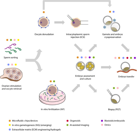
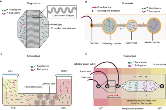
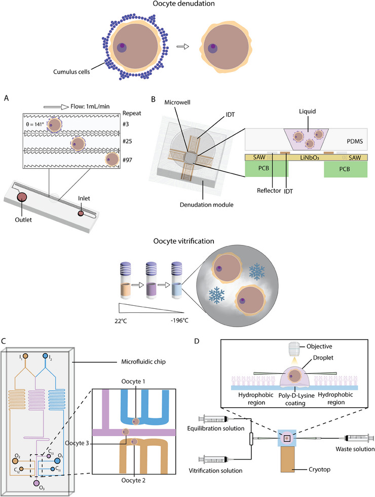
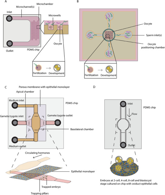
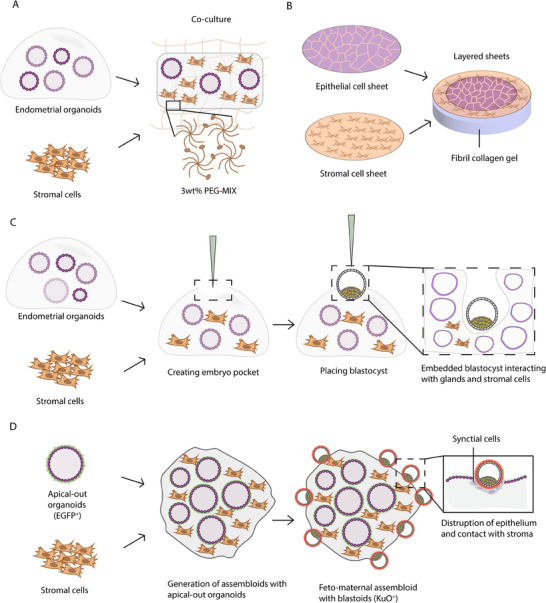
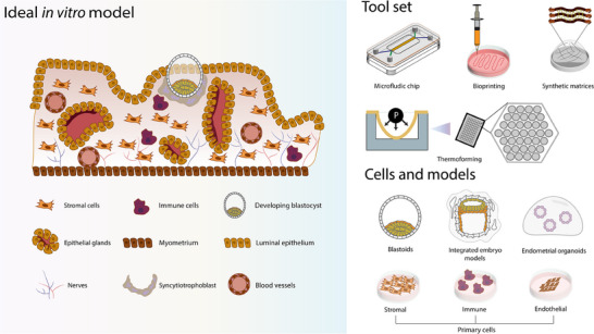
Infertility affects ≈17.5% of the adult population worldwide, posing significant clinical, emotional, and socioeconomic challenges. Recent advances at the intersection of reproductive medicine and bioengineering offer promising avenues to enhance assisted reproductive technologies (ART). This review synthesizes emerging microengineered and stem cell-based platforms designed to improve key ART stages from gamete handling and fertilization to embryo culture and implantation. State-of-the-art microfluidic systems that refine sperm selection by leveraging directional behavioral responses, enhancing motility, and preserving DNA integrity is discussed. Moreover, devices for oocyte denudation and cryopreservation have been developed to mitigate cellular stress associated with conventional processing techniques. While microengineered platforms demonstrate promise in sperm sorting and reducing stress on gametes, their broader application in ART, particularly for oocyte handling and embryo culture, requires further development. The review further addresses stem cell-based embryo models and bioengineered endometrial platforms, which aim to recapitulate the dynamic in vivo microenvironments needed for successful fertilization, embryo development, and implantation. Despite encouraging preliminary results, challenges such as scalability, reproducibility, clinical validation, and ethical considerations remain. By identifying these gaps and proposing future directions, this review considers integrating microengineering and bioengineering approaches to streamline ART procedures, ultimately enabling more personalized and effective reproductive therapies.

Keywords: assisted reproductive technologies; bioengineered platforms; fertilization; implantation; in vitro models; stem cell‐based embryo models.


Conflict of interest statement

The authors declare no conflict of interest.

Figures

Figure 1
Figure 1
Overview of assisted reproductive technologies (ART) and the integration of emerging bioengineering and computational tools.
Figure 2
Figure 2
Microengineered platforms for sperm selection based on taxis. A) Microfluidic chip design based on boundary‐following behavior of sperm. This study developed chips with 85 parallel microchannels with 150 µm curvature for selecting good quality sperm. Adapted from.[ 60 ] B) Microfluidic chip design based on rheotaxis behavior of sperm. This simple setup made use of flow to select good quality sperm based on their ability to swim against. Adapted from.[ 47 ] C) Microfluidic chip design based on chemotaxis (and thermotaxis). In this setup, cumulus cells of oocytes were placed in the tube for releasing chemoattractant molecules and guiding sperm towards themselves. Adapted from.[ 58 ] D) Microfluidic chip design based on thermotaxis (and rheotaxis). In this setup, a temperature gradient was applied for creating an enhancing recovery rate zone. The good quality sperm moved toward a more ideal temperature in the gradient, which was further sorted with flow. Adapted from.[ 51 ]
Figure 3
Figure 3
Microfluidic approaches for oocyte denudation from cumulus‐oocyte complexes and for precise oocyte vitrification. A) An oocyte denudation device consisting of 150 constriction‐expansion units with varying geometry (width) and surface (smooth or jagged‐teeth). Tubing attached to syringe pump was pre‐filled with culture medium and was inserted in the inlet to create flow. In the insert, denudation of an oocyte was depicted following the repeated units. Naked oocytes were collected from the outlet. Adapted from.[ 86 ] B) Oocyte denudation module consisting of microwell and IDTs for generating SAW. The PDMS module was placed on top of printed circuit board (PCB). Side view showing the arrangement of IDTs around the edge of the microwell on top of the LiNbO3 substrate and how the traveling SAW interacts with the liquid and oocytes inside the well. Adapted from.[ 87 ] C) Microfluidic perfusion chip for studying permeability of multiple oocytes at the same time. A serpentine channel was designed to precisely mix the solution and create varying concentrations, which was used to study how permeability depends on concentration. I1 = inlet 1, I2 = inlet 2, Ci1 = cell inlet 1, Ci2 = cell inlet 2, Ci3 = cell inlet 3, O1 = outlet 1, O2 = outlet 2, O3 = outlet 3. Magnified view showing three oocytes in different channels with different CPA concentrations. Adapted from.[ 88 ] D) Microfluidic chip for efficient loading of cryoprotectants for vitrification. The chip was attached to the cryotop (a film strip for vitrification medium volume minimization) and to three different syringes for loading both equilibration and vitrification solutions through the micropipettes on both side and one for waste. The chip consisted of hydrophobic, and poly‐D‐Lysine coated surfaces for stabilizing the droplet containing oocyte. Adapted from.[ 89 ]
Figure 4
Figure 4
Microfluidic platforms for in‐chip fertilization and blastocyst culturing as an alternative to static culture in IVF labs. A) Microfluidic design for in vitro fertilization. This chip had an integrated oocyte‐positioning system for motile sperm selection and fertilization. Switching from fertilization medium to development medium allowed embryos to develop into blastocyst stage. Adapted from.[ 163 ] B) A simpler microfluidic design for oocyte trapping. In this setup, the micro‐chamber consisted of microwells allowed the oocyte trapping for fertilization. Flow was introduced for medium substitution and debris cleaning. Adapted from.[ 164 ] C) Microfluidic chip designed for trapping gamete/zygotes by the trapping pillars on the apical chamber. The porous membrane above the apical chamber seeded with oviduct epithelial monolayer provided molecules for embryo development. On the top, the basolateral chamber provided flow which led to hormone circulation. Adapted from.[ 167 ] D) A simpler microfluidic chip design for embryo culture. Oviduct epithelial cells were seeded in the chamber prior to embryo culture. Tubes were attached for automated medium changing. Embryos from different stages were cultured on the chip, with and without the epithelial cells. Adapted from.[ 168 ]
Figure 5
Figure 5
Advanced endometrial culture methods: from mimicking the endometrium to simulating implantation processes. A) Co‐culture of endometrial organoids with stromal cells in a tailored PEG‐MIX hydrogel for promoting the growth of both cell types. Adapted from.[ 260 ] B) Creating epithelial and stromal cell sheets for co‐culture in fibril collagen gel to study the cell–cell interactions, as well as testing the effect of hormones. Adapted from.[ 265 ] C) Co‐culture of endometrial organoids, stromal cells and blastocysts that were placed in “embryo pockets.” The insert highlights the interaction between the blastocyst and co‐culture in the gel. Adapted from.[ 266 ] D) Generation of assembloids with apical‐out EGFP+ endometrial organoids and stromal cells. Once the assembloids were generated, KuO+ blastoids were co‐cultured and showed implantation‐like behaviors, including disrupting the epithelium and contacting with stromal cells. EGFP = Green fluorescent protein. KuO = Kusabira Orange. Adapted from.[ 267 ]
Figure 6
Figure 6
An ideal in vitro model of endometrium for embryo implantation with available tool sets, models, and cells.

References

    1. Inhorn M. C., Patrizio P., Hum. Reprod. Update 2015, 21, 411. - PubMed
    1. Njagi P., Groot W., Arsenijevic J., Dyer S., Mburu G., Kiarie J., Hum. Reprod. Open 2023, 2023, hoad007. - PMC - PubMed
    1. Carson S. A., Kallen A. N., JAMA, J. Am. Med. Assoc. 2021, 326, 65. - PMC - PubMed
    1. Hanson B., Johnstone E., Dorais J., Silver B., Peterson C. M., Hotaling J., J. Assist. Reprod. Genet. 2017, 34, 167. - PMC - PubMed
    1. Vander Borght M., Wyns C., Clin. Biochem. 2018, 62, 2. - PubMed