

Rare case report: Liver metastasis from cervical adenocarcinoma presenting with cystic mass and obstructive jaundice
- PMID: 40519293
- PMCID: PMC12162578
- DOI: 10.3389/fonc.2025.1558946
Rare case report: Liver metastasis from cervical adenocarcinoma presenting with cystic mass and obstructive jaundice
Abstract
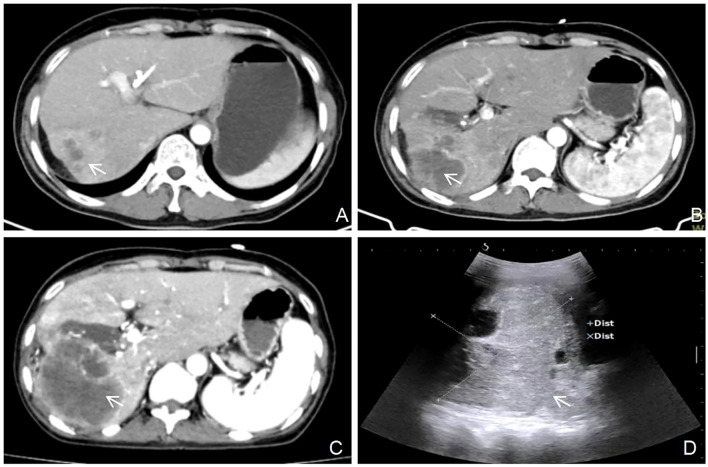
Liver metastases can originate from primary tumors in various organs; however, metastasis from cervical cancer to the liver is rare. Cervical cancer patients with distant metastases have a poor prognosis and reduced survival rates. This report describes a case of a cystic liver mass with obstructive jaundice, observed four years after resection of cervical adenocarcinoma. The lesion lacked typical imaging characteristics of hepatic metastases and was initially suspected to be a biliary neoplasm. A contrast-enhanced, ultrasound-guided needle biopsy was performed to confirm the diagnosis. Histopathological analysis confirmed adenocarcinoma of the liver, and immunohistochemical staining suggested a uterine or adnexal origin. Considering the patient's surgical history, the final diagnosis was liver metastasis originating from cervical adenocarcinoma. This report reviews relevant literature to discuss the clinical features, diagnostic challenges, and therapeutic strategies for liver metastasis of cervical cancer.
Keywords: case report; cervical cancer; contrast-enhanced ultrasound; hepatic cystic neoplasms; liver metastases.
Copyright © 2025 Li, Wei and Luo.
Conflict of interest statement
The authors declare that the research was conducted in the absence of any commercial or financial relationships that could be construed as a potential conflict of interest.
Figures

Similar articles
-
Obstructive Jaundice Induced by Hilar Mucinous Cystic Neoplasm of the Liver: A Rare Case Report and Literature Review.Curr Oncol. 2025 Feb 23;32(3):126. doi: 10.3390/curroncol32030126. Curr Oncol. 2025. PMID: 40136330 Free PMC article. Review.
-
Metastatic Biliary Stricture with "Beaded" Appearance from Cervical Adenocarcinoma.Euroasian J Hepatogastroenterol. 2021 Jul-Dec;11(2):97-99. doi: 10.5005/jp-journals-10018-1350. Euroasian J Hepatogastroenterol. 2021. PMID: 34786364 Free PMC article.
-
A rare case of cardiac metastasis from uterine cervical adenocarcinoma.J Cardiol Cases. 2020 Dec 13;24(1):30-33. doi: 10.1016/j.jccase.2020.12.002. eCollection 2021 Jul. J Cardiol Cases. 2020. PMID: 34257758 Free PMC article.
-
Solid pseudopapillary neoplasm of the pancreas causing obstructive jaundice: Case report of a rare entity.Int J Surg Case Rep. 2025 Jan;126:110635. doi: 10.1016/j.ijscr.2024.110635. Epub 2024 Nov 21. Int J Surg Case Rep. 2025. PMID: 39612899 Free PMC article.
-
Cervical metastasis of gingival carcinoma misdiagnosed as branchiogenic carcinoma, a rare entity - report of a case and review of literature.BMC Oral Health. 2017 Nov 28;17(1):139. doi: 10.1186/s12903-017-0435-9. BMC Oral Health. 2017. PMID: 29183323 Free PMC article. Review.
References
-
- Lin A, Ma S, Dehdashti F, Markovina S, Schwarz J, Siegel B, et al. Detection of distant metastatic disease by positron emission tomography with 18F-fluorodeoxyglucose (FDG-PET) at initial staging of cervical carcinoma. Int J Gynecol Cancer. (2019) 29:487–91. doi: 10.1136/ijgc-2018-000108 - DOI - PMC - PubMed
Publication types
LinkOut - more resources
Full Text Sources