. 2025 May 19;33(2):101493.
doi: 10.1016/j.omtm.2025.101493. eCollection 2025 Jun 12.

Preclinical toxicological assessment of an α-galactosylceramide-adjuvanted mRNA cancer vaccine in Wistar Han rats and domestic pigs

Affiliations

Preclinical toxicological assessment of an α-galactosylceramide-adjuvanted mRNA cancer vaccine in Wistar Han rats and domestic pigs

Sofie Meulewaeter et al. Mol Ther Methods Clin Dev. .

Abstract

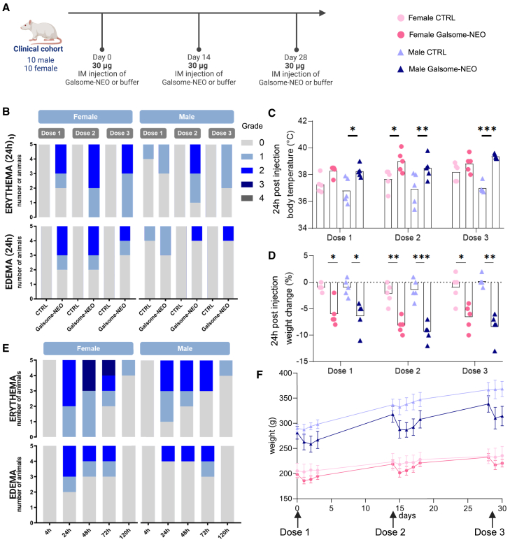
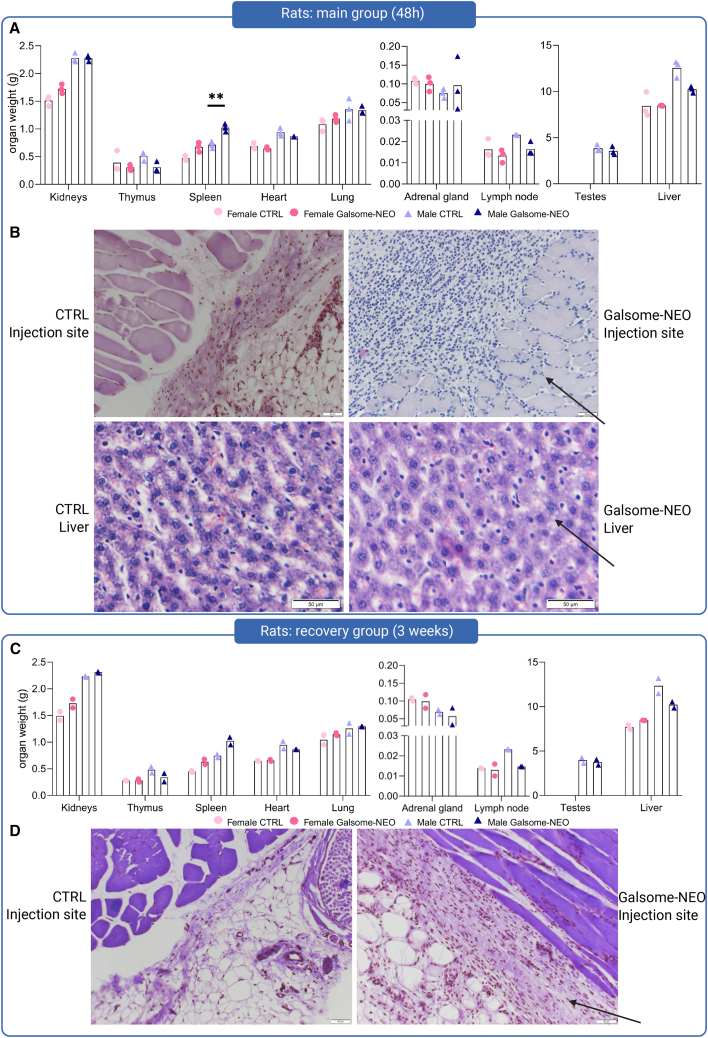
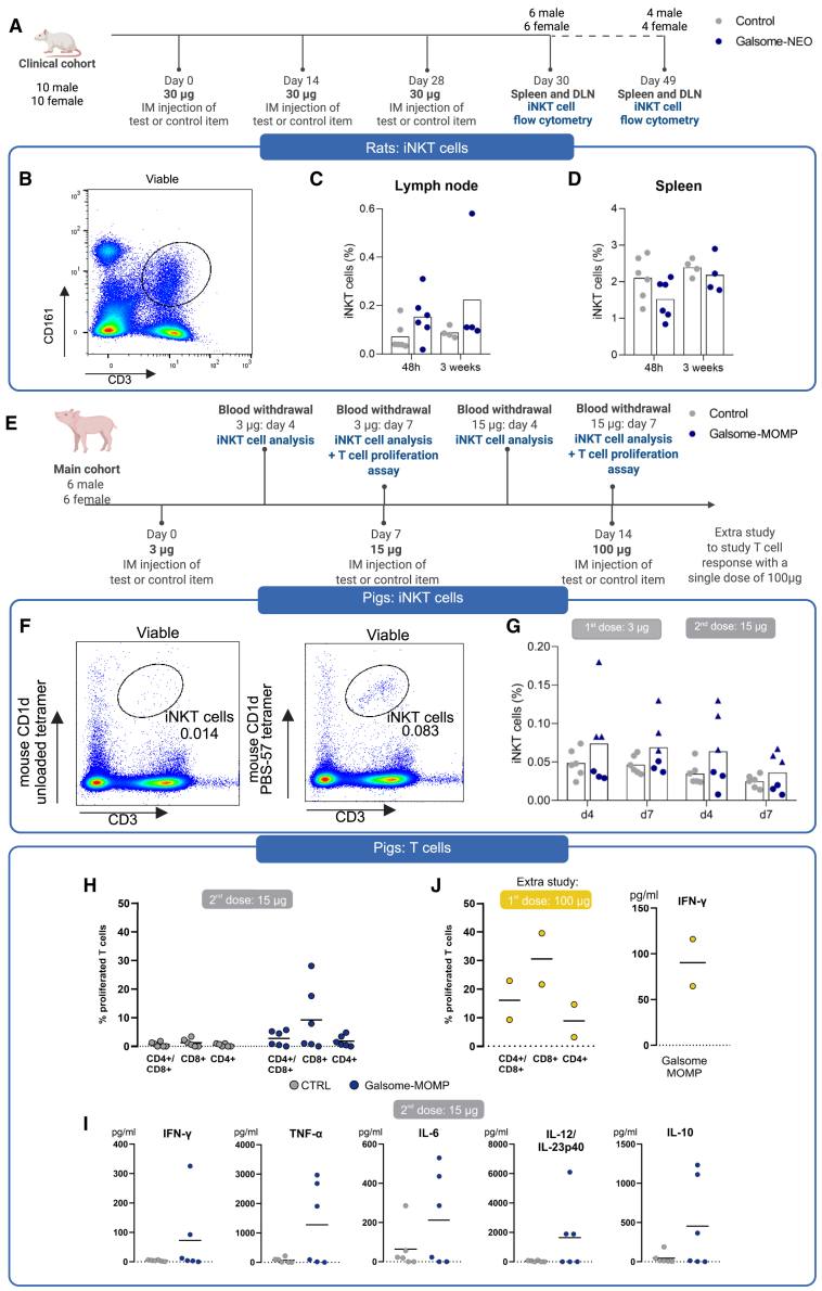
Galsome-NEO is a glycolipid-adjuvanted mRNA lipid nanoparticle (LNP) cancer vaccine encoding neo-epitopes for evaluation in a phase 1 study in patients with non-small cell lung cancer. To assess the safety of Galsome-NEO, a repeated-dose toxicity study was conducted in Wistar Han rats involving three intramuscular doses of 30 μg mRNA. A dose-escalation study in piglets tested three doses of 3, 15, and 100 μg mRNA. Rats showed a pronounced pro-inflammatory response, evidenced by cytokine secretion and an acute phase reaction. Clinical findings included temporary local reactions (maximum grade 3), elevated temperatures, and weight loss. In pigs, all doses were well tolerated. Blood analysis showed elevated alkaline phosphatase and decreased thrombocytes in rats, while pigs had reduced reticulocyte counts. Histology revealed hepatocyte vacuolation in rats and immune infiltration at injection sites in both species. In rats, blood and histology alterations resolved 3 weeks post dosing, except for immune infiltration in the connective tissue at injection sites in two females. Galsomes with mRNA encoding the Chlamydia trachomatis major outer membrane protein induced T cell responses in pigs. Natural killer T cell activation was observed in both species. These findings align with the safety data for the COVID-19 mRNA vaccine, Comirnaty, and demonstrate Galsomes' potential in large animals.

Keywords: NKT cell; Wistar Han rats; lipid nanoparticle; mRNA cancer vaccine; pigs; toxicology study; α-galactosylceramide.


Conflict of interest statement

R.V., I.L., and S.C.D.S. are contributors to patent applications no. WO2020058239A1: Therapeutic nanoparticles and methods of use thereof and no. WO2023209103: Prevention and treatment of infections with intracellular bacteria, together with I.A.

Figures

None
Graphical abstract
Figure 1
Figure 1
Study design of preclinical toxicity studies performed in Wistar Han Rats and domestic piglets Schematic overview of the dosing regimen and days of sacrifice for the repeated-dose study in Wistar Han rats (A) and dose-escalation study in domestic piglets (B). In a separate study, 1 male and 1 female piglet (5 weeks) received a single dose i.m. of 100 μg MOMP-encoding mRNA packaged in Galsomes. Blood was collected in heparin tubes 7 days after injection, and PBMCs were isolated and restimulated with MOMP peptide to assess MOMP-specific T cell proliferation. Created with Biorender.com.
Figure 2
Figure 2
Clinical evaluation of Wistar Han rats vaccinated with three repeated doses of 30 μg Galsome-NEO (A) Ten male and 10 female 10-week-old Wistar Han rats were randomly allocated to the clinical cohort and received intramuscularly (i.m.) in the biceps femoris three repeated doses of either 30 μg patient neo-epitope mRNA encapsulated in Galsomes (Galsome-NEO rats) or an equal volume of 9% sucrose Tris 20 mM buffer (CTRL rats). The total dose was divided over both hind legs. (B–D) Clinical parameters in male and female rats 24 h after i.m. injection with buffer (CTRL) or 30 μg Galsome-NEO. (B) Local reactions (erythema and edema) were evaluated using the Draize scoring system and received a grade 0–4 depending on the severity of the local reactions. Both injection sites were evaluated, and the highest score obtained for each rat is shown in the graph. (C) Rectal temperature 24 h post injection. (D) Body weight change (%) 24 h after injection relative to the weight of the same animal 0 h after injection. (C and D) Symbols represent individual data points and mean is indicated by a bar, statistical analyses on datasets were performed by using two-way ANOVA (∗p < 0.05, ∗∗p < 0.01, ∗∗∗p < 0.001). (E) Local reactions (erythema and edema) in male and female rats 4, 24, 48, 72, and 120 h after i.m. injection with Galsome-NEO. (F) Absolute daily body weights (g) of male and female rats treated with 9% sucrose Tris 20 mM buffer (CTRL) or Galsome-NEO (mean ± SD). Images created with Biorender.com.
Figure 3
Figure 3
Clinical evaluation of domestic piglets vaccinated with three increasing doses of Galsome-MOMP (A) Six male and 6 female 7-week-old piglets received intramuscularly (i.m.) in the left dorsal longitudinal muscle increasing doses of 3, 15, or 100 μg C. trachomatis major outer membrane protein (MOMP) encoding mRNA encapsulated in Galsomes (Galsome-MOMP piglets) or an equal volume of 9% sucrose Tris 20 mM buffer (CTRL piglets). (B–D) Clinical parameters in male and female piglets 24 h after i.m. injection with buffer (CTRL) or increasing doses of Galsome-MOMP. (B) Local reactions (erythema and edema) were evaluated using the Draize scoring system and received a grade 0–4 depending on the severity of the local reactions. (C) Body temperature is measured by scanning a subcutaneously implanted thermal microchip. (D) Body weight change (%) 24 h after injection relative to the weight of the same animal 0 h after injection. (C and D) Individual data are shown and bars indicate mean value, statistical analyses on datasets were performed by using two-way ANOVA but did not detect any significance. (E) Local reactions at the injection site in piglets 4, 24, 48, 72, and 96 h after i.m. injection with Galsome-MOMP. (F) Absolute daily body weights (kg) of individual male and female piglets (individual data points) treated with 9% sucrose Tris 20 mM buffer (CTRL) or Galsome-MOMP, full line represents mean weights. Images created with Biorender.com.
Figure 4
Figure 4
Cytokine response in rats and pigs (A) A separate cytokine cohort of 6 male and 6 female rats was included in the study to evaluate cytokine secretion in rats shortly (6 h) after injection. The rats received an i.m. injection of either 30 μg of patient neo-epitope mRNA encapsulated in Galsomes (Galsome-NEO) or an equal volume of 9% sucrose Tris 20 mM buffer (control). (B–E) Plasma cytokines (TNF-α, IL-10, IFN-γ, CXCL1, MCP-1, GM-CSF, IL-18, IL-12p70, IL-1β, IL-17A, IL-33, IL-1α, and IL-6) were measured using a LEGENDplex assay. Elevated cytokine levels after NEO-Galsome injection are shown for CXCL1 (B), IL-6 (C), MCP-1 (D), and IFN-γ (E). (F) In pigs, blood was collected from the main cohort (6 male and 6 female pigs) shortly (5 h) after injection. The piglets received an i.m. injection of 3, 15, or 100 μg MOMP mRNA encapsulated in Galsomes (Galsome) or an equal volume of 9% sucrose Tris 20 mM buffer (control). (G) Serum cytokines (IFN-α, IFN-γ, IL-1β, IL-10, IL-12/IL-23p40, IL-4, IL-6, IL-8, and TNF-α) were measured using a ProcartaPlex assay. Among these, only IL-6 showed a notable elevation (p = 0.06). Individual data points and means are shown. Statistical analyses on datasets were performed using the Mann-Whitney test, and data points below the limit of detection (LoD) were set at the LoD value for analysis (∗p < 0.05, ∗∗p < 0.01, ∗∗∗p < 0.001). Images created with Biorender.com.
Figure 5
Figure 5
Postmortem organ evaluation 48 h and 3 weeks after the final dose in rats reveals morphological alterations in the liver and at the injection site (A and C) Absolute organ weights (g) of kidneys, thymus, testes, spleen, heart, lung, liver, adrenal gland, and lymph node of rats in the main group (n = 6) and recovery group (n = 4). Organs were excised and weighed 48 h (A) or 3 weeks (C) after the last dose of a series of three repeated doses from rats of the main group and recovery group, respectively. Symbols represent individual data points (n = 3/2) and mean values are shown. Multiple unpaired t test was performed without correction (∗p < 0.05, ∗∗p < 0.01, ∗∗∗p < 0.001). (B) Representative images of H&E-stained liver tissue sections and tissue from the injection site from rats of the main group. Inflammatory cells located between and around muscle cells of Galsome-NEO-treated rats are indicated by arrows. Liver tissue sections show vacuoles in hepatocytes (indicated by arrows) of Galsome-NEO-treated rats. (D) H&E-stained tissue sections from the injection site 3 weeks after the final injection (recovery group) show macrophages (indicated by arrows) in the connective tissue surrounding muscle cells of female Galsome-NEO-treated rats.
Figure 6
Figure 6
Organ weights of selected organs in pigs 48 h after the final dose (A) Absolute organ weights (g) of kidneys, testes, spleen, heart, lung, liver, and lymph node of piglets (6 females and 6 males). Organs were excised and weighed 48 h after the final dose of repeated injections of Galsome-MOMP or buffer (control). Symbols represent individual data points (n = 3) with mean values shown. Multiple unpaired t test was performed without correction and no significant differences were found. (B) Representative images of H&E-stained sections of liver tissue and tissue from the injection site in pigs 48 h after the final dose. Inflammatory cells located between muscle cells and around muscle cells of Galsome-MOMP-treated pigs are indicated by arrows. No clear vacuoles were observed in the liver tissue of pigs.
Figure 7
Figure 7
Immune responses to Galsome vaccination in rats and piglets (A) Wistar Han rats in the clinical cohort were sacrificed 48 h (main group) or 3 weeks (recovery group) after the final injection of three successive injections of either 30 μg Galsome-NEO (Galsome-NEO) or buffer (control). Spleen and inguinal lymph node were isolated and processed for flow cytometry analysis of iNKT cells. (B) Gating strategy for identifying iNKT cells in rats. Viable cells positive for CD161 (NK1.1 receptor) and CD3 were classified as iNKT cells (CD161+/CD3+). Fraction of iNKT cells within the viable cell population in the (C) spleen and (D) lymph node of control animals (3 doses of buffer) and Galsome-NEO (3 doses of 30 μg TMGNEO mRNA Galsomes) 48 h (n = 6) and 3 weeks (n = 4) after the final dose. (E) Heparinized blood was drawn from piglets (6 females/6 males) 4 and 7 days after the first and second dose of buffer (control) or MOMP-encoding mRNA packed in Galsomes (Galsome-MOMP). The first dose contained 3 μg mRNA and the second dose 15 μg mRNA. (F) Four and 7 days after the first and second injection, whole blood was stained to detect iNKT cells. The gating strategy is shown, with cells positive for CD3 and mouse CD1d PBS-57 tetramer identified as iNKT cells. An unloaded CD1d tetramer was included as a control to ensure specific binding. (G) Fraction of iNKT cells within the viable cell population in the blood of piglets 4 and 7 days after the first (3 μg) and second (15 μg) dose. Three piglets consistently showed elevated iNKT cell numbers and are indicated with triangle symbols. (H and I) PBMCs were isolated 7 days after the second dose (3 and 15 μg, respectively) or after a single dose of 100 μg in 2 piglets (6 weeks, 1 female and 1 male), and stained with CellTrace Violet. Cells were cultured for 4 days either in the presence of an MOMP-derived peptide (25 aa containing CD4+ and CD8+ epitopes) or in cell culture medium only. Proliferating cells were defined by decreased CellTrace Violet signal with the number of proliferating cells in the unstimulated condition (autoproliferation) subtracted. (J) In addition, cell culture supernatant of peptide-pulsed PBMCs was taken and analyzed for the presence of cytokines (IFN-α, IFN-γ, IL-1β, IL-10, IL-12/IL-23p40, IL-4, IL-6, IL-8, and TNF-α). Symbols represent individual data points and mean values are shown. Statistical analysis for immune cells was performed using the multiple t test with Holm-Sidak correction and Mann-Whitney for cytokines. No significant differences were found (p = 0.05). Images created with Biorender.com.

Similar articles

References

    1. Sung H., Ferlay J., Siegel R.L., Laversanne M., Soerjomataram I., Jemal A., Bray F. Global Cancer Statistics 2020: GLOBOCAN Estimates of Incidence and Mortality Worldwide for 36 Cancers in 185 Countries. CA Cancer J. Clin. 2021;71:209–249. doi: 10.3322/CAAC.21660. - DOI - PubMed
    1. Lahiri A., Maji A., Potdar P.D., Singh N., Parikh P., Bisht B., Mukherjee A., Paul M.K. Lung cancer immunotherapy: progress, pitfalls, and promises. Mol. Cancer. 2023;22:1–37. doi: 10.1186/S12943-023-01740-Y. - DOI - PMC - PubMed
    1. Adotévi O., Vernerey D., Jacoulet P., Meurisse A., Laheurte C., Almotlak H., Jacquin M., Kaulek V., Boullerot L., Malfroy M., Safety, et al. Immunogenicity, and 1-Year Efficacy of Universal Cancer Peptide-Based Vaccine in Patients with Refractory Advanced Non-Small-Cell Lung Cancer: A Phase Ib/Phase IIa De-Escalation Study. J. Clin. Oncol. 2023;41:373–384. doi: 10.1200/JCO.22.00096. - DOI - PubMed
    1. Besse B., Remon J., Felip E., Garcia Campelo R., Cobo M., Mascaux C., Madroszyk A., Cappuzzo F., Hilgers W., Romano G., et al. Randomized open-label controlled study of cancer vaccine OSE2101 versus chemotherapy in HLA-A2-positive patients with advanced non-small-cell lung cancer with resistance to immunotherapy: ATALANTE-1. Ann. Oncol. 2023;34:920–933. doi: 10.1016/j.annonc.2023.07.006. - DOI - PubMed
    1. Rodriguez P.C., Popa X., Martínez O., Mendoza S., Santiesteban E., Crespo T., Amador R.M., Fleytas R., Acosta S.C., Otero Y., et al. A Phase III Clinical Trial of the Epidermal Growth Factor Vaccine CIMAvax-EGF as Switch Maintenance Therapy in Advanced Non-Small Cell Lung Cancer Patients. Clin. Cancer Res. 2016;22:3782–3790. doi: 10.1158/1078-0432.CCR-15-0855. - DOI - PubMed

LinkOut - more resources