Spontaneous Spinal Epidural Hematoma Mimicking Stroke in a Young Patient: A Case Report
- PMID: 40519452
- PMCID: PMC12162385
- DOI: 10.7759/cureus.83994
Spontaneous Spinal Epidural Hematoma Mimicking Stroke in a Young Patient: A Case Report
Abstract
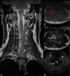
Spontaneous spinal epidural hematoma (SSEH) is a rare but serious neurological emergency characterized by the accumulation of blood in the epidural space without an identifiable cause. It typically presents with sudden-onset neck or back pain, followed by motor and sensory deficits. Although uncommon, SSEH can mimic ischemic stroke when presenting as hemiparesis, often leading to diagnostic delays. We report the case of a 35-year-old male patient who developed sudden-onset cervical pain, followed by right-sided hemiparesis and sensory deficits. Due to focal neurological signs, an acute ischemic stroke was initially suspected; however, brain CT was unremarkable. Given the inconclusive findings, a cervicothoracic MRI was performed, which revealed an epidural mass extending from C3 to T2, consistent with SSEH. Surgical decompression confirmed a subacute epidural hematoma with no identifiable source of bleeding. The patient achieved complete neurological recovery within one month of surgery. This case highlights the importance of including SSEH in the differential diagnosis of acute hemiparesis, particularly when preceded by severe neck or back pain and without cranial nerve involvement. It also emphasizes the need to consider spinal imaging when initial brain CT is inconclusive in patients presenting with acute neurological deficits. Early recognition of these clinical features is crucial, as prompt surgical intervention significantly improves neurological outcomes and reduces the risk of long-term disability.
Keywords: case report; cervical pain; hemiparesis; spinal cord compression; stroke mimic.
Copyright © 2025, Castelo-Pablos et al.
Conflict of interest statement
Human subjects: Consent for treatment and open access publication was obtained or waived by all participants in this study. Conflicts of interest: In compliance with the ICMJE uniform disclosure form, all authors declare the following: Payment/services info: All authors have declared that no financial support was received from any organization for the submitted work. Financial relationships: All authors have declared that they have no financial relationships at present or within the previous three years with any organizations that might have an interest in the submitted work. Other relationships: All authors have declared that there are no other relationships or activities that could appear to have influenced the submitted work.
Figures


Similar articles
-
Spontaneous spinal epidural hematoma mimicking transient ischemic attack: A case report.Medicine (Baltimore). 2017 Dec;96(49):e9007. doi: 10.1097/MD.0000000000009007. Medicine (Baltimore). 2017. PMID: 29245281 Free PMC article.
-
Paradoxical contralateral hemiparesis in spontaneous spinal epidural hematoma: a case report.BMC Neurol. 2023 Apr 1;23(1):138. doi: 10.1186/s12883-023-03179-6. BMC Neurol. 2023. PMID: 37005562 Free PMC article.
-
[Two cases of spontaneous cervical epidural hematoma mimicking cerebral infarction].No Shinkei Geka. 2014 Feb;42(2):143-8. No Shinkei Geka. 2014. PMID: 24501188 Review. Japanese.
-
Acute Cervical Spontaneous Spinal Epidural Hematoma Presenting with Minimal Neurological Deficits: A Case Report.Anesth Pain Med. 2016 Aug 27;6(5):e40067. doi: 10.5812/aapm.40067. eCollection 2016 Oct. Anesth Pain Med. 2016. PMID: 27853682 Free PMC article.
-
Spontaneous cervical epidural haematoma mimicking stroke: a case report and literature review.Folia Neuropathol. 2022;60(2):261-265. doi: 10.5114/fn.2022.116940. Folia Neuropathol. 2022. PMID: 35950479 Review.
Cited by
-
Acute Spontaneous Spinal Epidural Hematoma in a Patient on a Direct Oral Anticoagulant (DOAC): A Case Report.Cureus. 2025 Jul 16;17(7):e88105. doi: 10.7759/cureus.88105. eCollection 2025 Jul. Cureus. 2025. PMID: 40821279 Free PMC article.
References
-
- Spinal epidural hematomas: personal experience and literature review of more than 1000 cases. Domenicucci M, Mancarella C, Santoro G, Dugoni DE, Ramieri A, Arezzo MF, Missori P. J Neurosurg Spine. 2017;27:198–208. - PubMed
-
- Spinal epidural hematoma as a stroke mimic. Inatomi Y, Nakajima M, Yonehara T. J Stroke Cerebrovasc Dis. 2020;29:105030. - PubMed
-
- Spontaneous spinal epidural hematoma treated with tissue plasminogen activator mimicking ischemic stroke. Lee CH, Kwun KH, Jung KH. https://www.sciencedirect.com/science/article/pii/S2214751919302543?via%... Interdiscip Neurosurg. 2020;19:100569.
-
- Spinal hematoma: a literature survey with meta-analysis of 613 patients. Kreppel D, Antoniadis G, Seeling W. Neurosurg Rev. 2003;26:1–49. - PubMed
Publication types
LinkOut - more resources
Full Text Sources
Miscellaneous
