

Therapeutic Effects of Fire Needling Acupuncture on Pain Relief and Cartilage Protection in MIA-Induced Knee Osteoarthritis Rats: The Role of Macrophage Polarization in Synovium and Angiogenesis in Subchondral Bone
- PMID: 40519654
- PMCID: PMC12165211
- DOI: 10.2147/JIR.S518829
Therapeutic Effects of Fire Needling Acupuncture on Pain Relief and Cartilage Protection in MIA-Induced Knee Osteoarthritis Rats: The Role of Macrophage Polarization in Synovium and Angiogenesis in Subchondral Bone
Abstract
Purpose: Knee osteoarthritis (KOA) is a prevalent degenerative disease impacting bone and joint health. Clinical studies indicate that fire needling acupuncture can alleviate joint stiffness, pain, and dysfunction in KOA. Previous research demonstrated its efficacy in reducing pain, mitigating cartilage damage, and regulating macrophage polarization, but its effects on subchondral bone remain unclear. This study aimed to evaluate the therapeutic effects of fire needling acupuncture on subchondral bone in KOA.
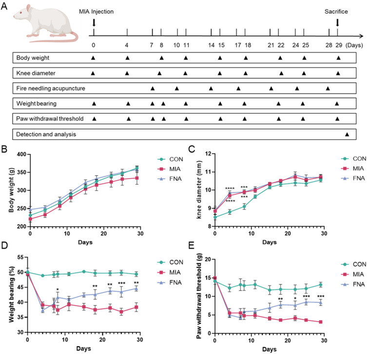
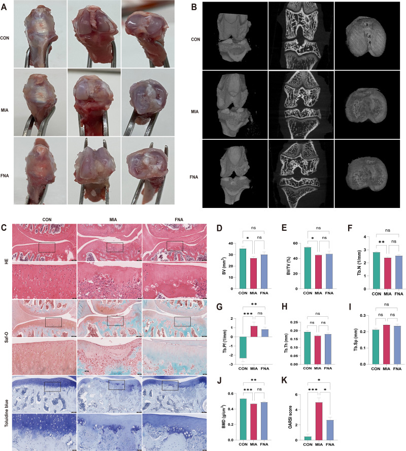
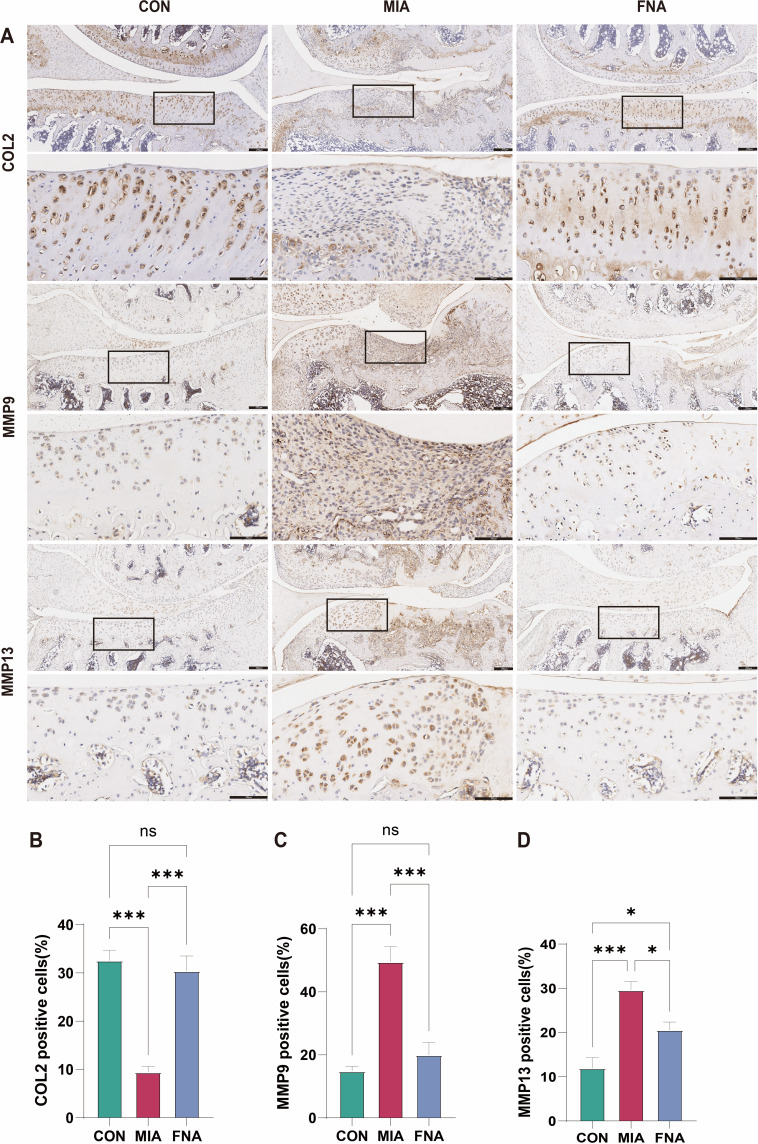
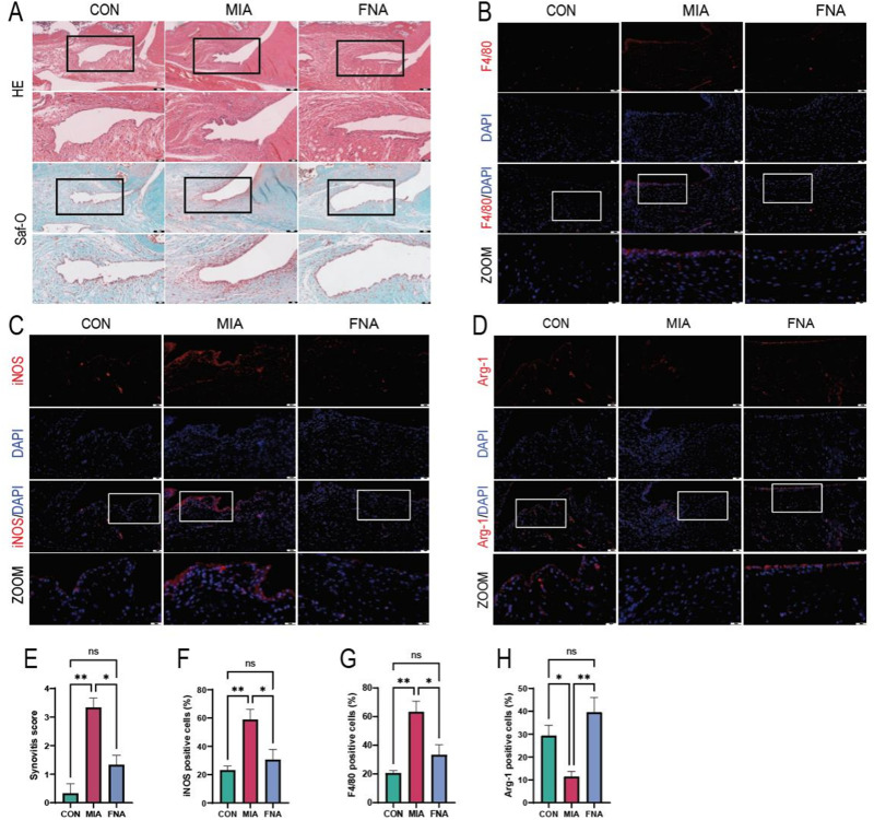
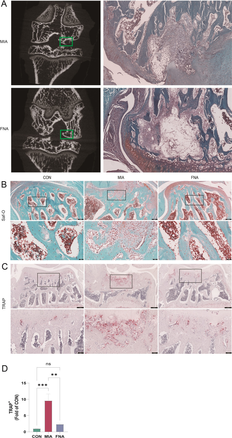
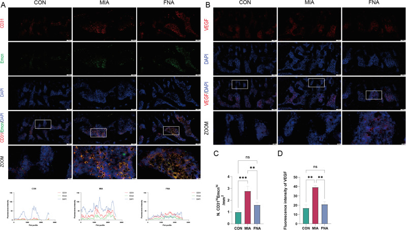
Methods: Sprague-Dawley rats were divided into three groups: control (CON), monosodium iodoacetate (MIA), and fire needling acupuncture (FNA) (n=6 per group). A KOA model was established using 0.3 mg/50 µL MIA, followed by acupuncture at acupoints SP10, ST34, ST35, EX-LE5, and ST36 twice weekly. Evaluations included body weight, joint diameter, weight distribution, and mechanical withdrawal threshold. Micro-CT imaging assessed tibial plateau bone mass changes. Histological evaluations used HE, Safranin O/Fast Green, and toluidine blue staining. Immunohistochemistry examined COL2, MMP9, and MMP13 expression, while macrophage polarization was analyzed using immunofluorescence for F4/80, iNOS, and Arg-1. TRAP staining assessed osteoclast activity, and immunofluorescence for CD31/Emcn and VEGF evaluated angiogenesis in subchondral bone.
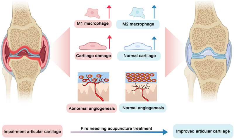
Results: Fire needling acupuncture significantly improved weight distribution and mechanical withdrawal thresholds, reduced synovial inflammation and abnormal changes in subchondral bone, and preserved cartilage integrity in MIA-induced KOA rats. Notably, F4/80 and iNOS expression levels decreased, while Arg-1 expression increased after treatment. Additionally, TRAP, CD31/Emcn, and VEGF expression in subchondral bone decreased following fire needling acupuncture.
Conclusion: Fire needling acupuncture mitigates pain behavior, synovial inflammation, cartilage degradation, and abnormal changes in subchondral bone in MIA-induced KOA rats. The therapeutic mechanism may involve modulation of synovial macrophage polarization and subchondral bone angiogenesis. Further research is warranted to elucidate the precise molecular pathways and the interaction between macrophage polarization and angiogenesis.
Keywords: cartilage degradation; fire needling acupuncture; knee osteoarthritis; macrophages polarization; subchondral bone angiogenesis.
© 2025 Wei et al.
Conflict of interest statement
The authors declare no potential conflicts of interest concerning this article’s research, authorship, and/or publication.
Figures
Similar articles
-
Fire Needling Acupuncture Suppresses Cartilage Damage by Mediating Macrophage Polarization in Mice with Knee Osteoarthritis.J Pain Res. 2022 Apr 13;15:1071-1082. doi: 10.2147/JPR.S360555. eCollection 2022. J Pain Res. 2022. PMID: 35444462 Free PMC article.
-
Fire needling acupuncture attenuates synovial inflammation and cartilage degeneration in knee osteoarthritis via SDF-1/CXCR4-mediated macrophage polarization.Clin Rheumatol. 2025 Aug 8. doi: 10.1007/s10067-025-07612-8. Online ahead of print. Clin Rheumatol. 2025. PMID: 40779147
-
[Warm needling reduces oxidative damage and inflammation of cartilage in knee osteoarthritis rats].Zhen Ci Yan Jiu. 2022 Apr 25;47(4):321-8. doi: 10.13702/j.1000-0607.20210308. Zhen Ci Yan Jiu. 2022. PMID: 35486011 Chinese.
-
[Complex network analysis on regularities of acupoint combinations and application characteristics of acupuncture and moxibustion in the treatment of knee osteoarthritis].Zhen Ci Yan Jiu. 2022 Jan 25;47(1):65-70. doi: 10.13702/j.1000-0607.20210080. Zhen Ci Yan Jiu. 2022. PMID: 35128873 Review. Chinese.
-
[Research progress of effect mechanism of acupotomy for knee osteoarthritis].Zhongguo Zhen Jiu. 2025 Jun 12;45(6):867-874. doi: 10.13703/j.0255-2930.20250106-k0003. Epub 2025 Mar 31. Zhongguo Zhen Jiu. 2025. PMID: 40518794 Review. Chinese.
References
-
- Alice C, Inès K, Nadine S, Sylvain M, Jérémie S. Osteoarthritis year in review 2024: epidemiology and therapy. Osteoarthr Cartilage. 2024;32(11):1397–1404. - PubMed
LinkOut - more resources
Full Text Sources
Research Materials
Miscellaneous
