Review
. 2025 May 29:13:100147.
doi: 10.1016/j.tcsw.2025.100147. eCollection 2025 Jun.

Sterols in plant biology - Advances in studying membrane dynamics

Affiliations
Review

Sterols in plant biology - Advances in studying membrane dynamics

Paul Vogel et al. Cell Surf. .

Abstract

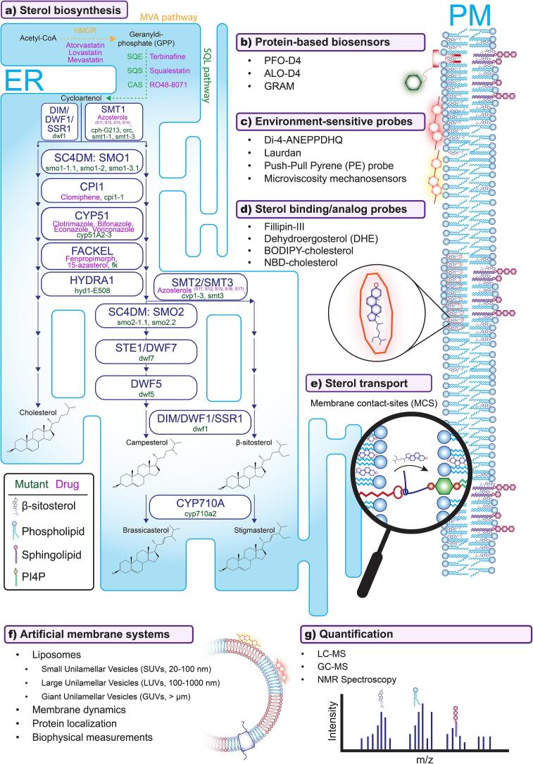
Plants sense their environment at the cell surface, i.e. the plasma membrane, where extracellular signals are perceived and transduced. Together with the cortical cytoskeleton and the cell wall, membrane lipids can influence these processes by acting on protein dynamics at the plasma membrane. Among these lipids, sterols regulate membrane fluidity and thus, protein functions. However, plant sterols are diverse in structure and particularly difficult to study due to technical limitations. Nevertheless, advances in sterol imaging, sterol-protein interaction studies, and sterol perturbation methods have resulted in a better understanding of their functions in plant development and physiology. Here we summarize the current knowledge and the latest breakthroughs, and discuss future challenges, in the field of plant sterol biology and cell surface organization.

Keywords: Lipid-order; Membrane-contact sites; Phytosterols; Plant membrane biology; Sterol conjugates; Sterol dynamics; Sterol transport; Sterols.


Conflict of interest statement

The authors declare that they have no known competing financial interests or personal relationships that could have appeared to influence the work reported in this paper.

Figures

Fig. 1
Fig. 1
General structure of the main sterol variants in plants. The tetracyclic steroid ring system forms a consistent backbone, with carbon atoms numbered according to the typical sterol numbering system (shown on backbone structure). The residue at the C3 position (red R groups) determines the type of sterol conjugate: Free Sterol (FS), Steryl Ester (SE), Steryl Glycoside (SG), or Acyl Steryl Glycoside (ASG). The alkyl side chain attached to C17 (green) defines the sterol species (β-sitosterol, stigmasterol, campesterol, or cholesterol). Key structural differences between sterol species are highlighted in blue. Adapted from (Ferrer et al., 2017). (For interpretation of the references to colour in this figure legend, the reader is referred to the web version of this article.)
Fig. 2
Fig. 2
Comprehensive methods for studying plant sterols. a) Sterol biosynthesis pathway highlighting key enzymes, mutants (green), and inhibitory drugs (orange). 3-hydroxy-3-methylglutaryl-CoA reductase (HMGR), geranyldi-phosphate (GPP), squalene epoxidase (SQE), cycloartenol synthase (CAS), STEROL METHYLTRANSFERASE1 (SMT1), STEROL C-4 DEMETHYLASE (SC4DM), STEROL 4-ALPHA-METHYL OXIDASE1 (SMO1), CYCLOPROPYL ISOMERASE1 (CPI1), CYTOCHROME P450 51 (CYP51), STEROL C-14 REDUCTASE (FACKEL), C-8,7 STEROL ISOMERASE (HYDRA1), STEROL 4-ALPHA-METHYL OXIDASE2 (SMO2), STEROL C-24 REDUCTASE/DWARF7 (STE1/DWF7), DWARF5 (DWF5), DIMINUTO/DWARF1/STEROL SIDE CHAIN REDUCTASE1 (DIM/DWF1/SSR1), and CYTOCHROME P450 710 A (CYP710A) b) Protein-based biosensors include Perfringolysin O Domain 4 (PFO-D4), Anthrolysin O Domain 4 (ALO-D4), and the Glycosyltransferases, Rab-like GTPase activators and Myotubularins (GRAM) domain. c) Environmental-sensitive probes include di-4-aminonaphthylethenylpyridinium (Di-4-ANEPPDHQ), push-pull pyrene (PE). Sterol binding probes shown are nitrobenzoxadiazole (NBD) and boron-dipyrromethene (BODIPY). d) Sterol binding and analog probes including Filipin-III, dehydroergosterol (DHE), BODIPY-cholesterol, and NBD-cholesterol for sterol visualization and tracking. e) Membrane contact sites (MCS) as potential mediators of sterol transport between cellular membranes and as such possibly novel targets for sterol perturbation. Depicted is the potential transport via the counter-transport principle utilizing OSBP (red) and VAPA (dark blue) proteins and involving PI4P (green) as described for animals. f) Artificial membrane systems including different classes of liposomes (SUVs, LUVs, GUVs) for studying membrane dynamics and protein localization. g) Quantification methods include liquid-chromatography mass spectrometry (LC-MS), gas-chromatography mass spectrometry (GS-MS), and nuclear magnetic resonance (NMR) spectroscopy. (For interpretation of the references to colour in this figure legend, the reader is referred to the web version of this article.)
Fig. 3
Fig. 3
Microscopic visualization of sterols in Arabidopsis. a) Filipin-III staining of the Arabidopsis root tip. Epidermal and cortical cells are labeled upon filipin-III staining when excited at 364 nm, visualizing 3-β-hydroxysterols in complex with filipin-III (left panel). Untreated cell show no fluorescence signal when imaged with the same settings (right panel). Adapted from (Boutté et al., 2011). b) The protein-based biosensor SP-sfGFP-D4L localizes to potential sterol-rich punctae in Arabidopsis cotyledons. The cholesterol-binding domain 4 (D4) of perfringolysin O (PFO) from Clostridium perfringens with a D44L mutation was fused to a secretion peptide (SP) and a superfolder green fluorescent protein (sfGFP) yielding a potential sterol biosensor: SP-sfGFP-D4L. Expressed in 8-day old Arabidopsis cotyledons results in localization of the biosensor to punctae that could resemble sterol-rich nanodomains (upper panel). Treatment with the sterol-depleting drug methyl-β-cyclodextrin (MβCD) causes punctae to diffuse. Adapted from (Ukawa et al., 2022). c) Quantitative visualization of membrane order using the environment-sensitive probe di-4-ANEPPDHQ in Arabidopsis root hair. Using excitation at 488 nm, emissions in two channels were recorded with windows of 500–580 nm and 620–750 nm (first and second panel from the left). The merged image of the two emission channels does not give immediate inside in the distribution of lipid order (third panel from the left). To spatially visualize the degree of membrane order, generalized polarization (GP) processing was performed to quantitatively infer the degree of membrane order from the ratio of the two emission channels. GP values are indicated by Hue-Saturation-Brightness (HSB) with values ranging from − 0.69 (lower order, blue) to 0.92 (higher order, pink) showing higher lipid order at the root tip (last three images on the right). Adapted from (Zhao et al., 2015). Scale bar = 10 μm. (For interpretation of the references to colour in this figure legend, the reader is referred to the web version of this article.)

Similar articles

Cited by

References

    1. Aboobucker S.I., Showman L.J., Lübberstedt T., Suza W.P. Maize Zmcyp710a8 mutant as a tool to decipher the function of stigmasterol in plant metabolism. Front. Plant Sci. 2021;12 doi: 10.3389/fpls.2021.732216. - DOI - PMC - PubMed
    1. Amaro M., Reina F., Hof M., Eggeling C., Sezgin E. Laurdan and Di-4-ANEPPDHQ probe different properties of the membrane. J. Phys. D. Appl. Phys. 2017;50 doi: 10.1088/1361-6463/aa5dbc. - DOI - PMC - PubMed
    1. Ariola F.S., Li Z., Cornejo C., Bittman R., Heikal A.A. Membrane fluidity and lipid order in ternary giant unilamellar vesicles using a new bodipy-cholesterol derivative. Biophys. J. 2009;96:2696–2708. doi: j.bpj.2008.12.3922. - PMC - PubMed
    1. Asami T., Min Y.K., Nagata N., Yamagishi K., Takatsuto S., Fujioka S., Murofushi N., Yamaguchi I. Characterization of Brassinazole, a Triazole-Type Brassinosteroid Biosynthesis Inhibitor. Plant Physiology. 2000;123(1):93–100. - PMC - PubMed
    1. Asami T., Masaharu Mizutani, Yukihisa Shimada, Hideki Goda, Nobutaka Kitahata, Katsuhiko Sekimata, Han S.-Y., et al. Triadimefon, a Fungicidal Triazole-Type P450 Inhibitor, Induces Brassinosteroid Deficiency-like Phenotypes in Plants and Binds to DWF4 Protein in the Brassinosteroid Biosynthesis Pathway. Biochemical Journal. 2003;369(Pt 1):71–76. - PMC - PubMed

LinkOut - more resources