

Distribution characteristics and prognostic value of TIM-1 in patients with lung adenocarcinoma
- PMID: 40519901
- PMCID: PMC12162315
- DOI: 10.3389/fimmu.2025.1602868
Distribution characteristics and prognostic value of TIM-1 in patients with lung adenocarcinoma
Abstract
Background: T-cell immunoglobulin and mucin domain-containing protein 1 (TIM-1) has been identified as a promoter of tumor cell viability, migration, and invasion. However, the precise role and distribution characteristics of TIM-1 within the tumor microenvironment (TME) remain critical areas of investigation.
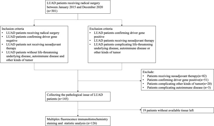
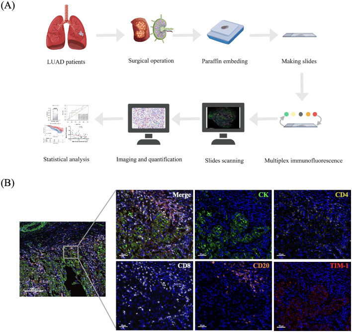
Methods: In this study, multiplex immunofluorescence (mIF) was performed on tissue slides from 126 patients with lung adenocarcinoma (LUAD) to investigate the distribution patterns of TIM-1 and the prognostic significance of three TIM-1 positive immune cell populations in both the primary tumor and tumor-draining lymph nodes (TDLN).
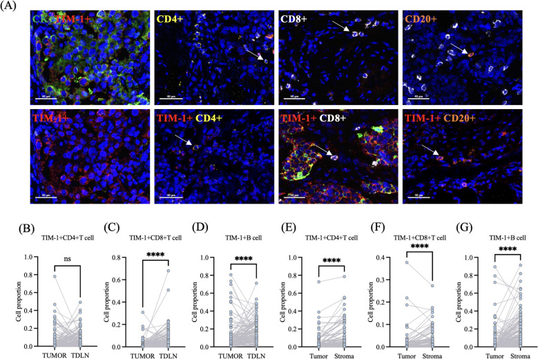
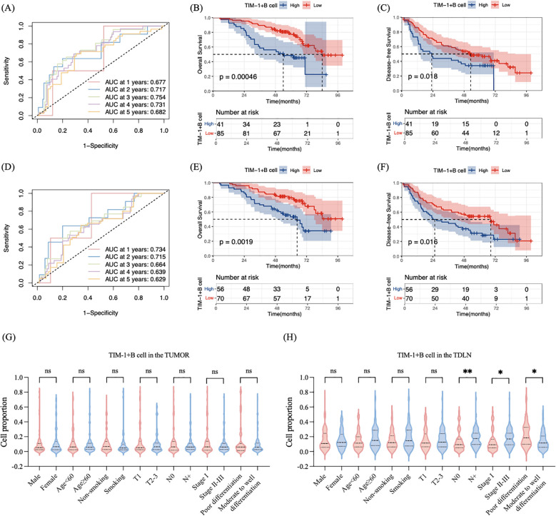
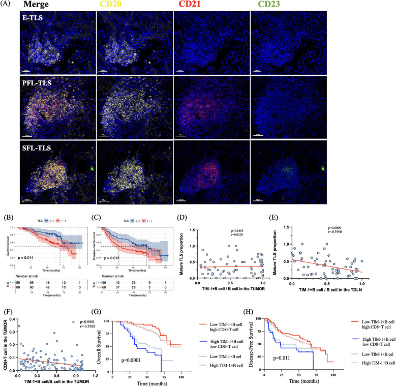
Results: Compared to the primary tumor, TIM-1+CD8+T cells and TIM-1+B cells exhibited significantly greater density in the TDLN (p<0.0001, p<0.0001 respectively). In the primary tumor, lower TIM-1+B cell density was associated with longer overall survival (OS) (mOS, 84 vs. 54 months; p<0.0001, HR=2.574) and disease-free survival (DFS) (mDFS, 53.0 vs. 23.1 months; p=0.018, HR=1.721). In the TDLN, lower TIM-1+B cell density was also correlated with longer OS (mOS, not reached vs. 64.7 months; p=0.0019, HR=2.3502) and DFS (mDFS, 68.5 vs. 28.9 months; p=0.016, HR=1.707). Higher TIM-1+B cell density in the TDLN was associated with a lower proportion of mature tertiary lymphoid structures (TLS) (p=0.0009, r=-0.3990) and increased density of TIM-1+B cells in the tumor was linked to reduced CD8+ T cell density (p=0.016, r=-0.2788).
Conclusions: Our findings confirm the immunosuppressive role of TIM-1+B cells in LUAD and suggest that TIM-1+B cells exert immune suppression by inhibiting TLS maturation and CD8+ T cell density. These findings highlight TIM-1+ B cells as a potential therapeutic target.
Keywords: TIM-1; lung adenocarcinoma; prognosis; tertiary lymphoid structure; tumor microenvironment; tumor-draining lymph nodes.
Copyright © 2025 Wen, Yun, Chen, Yin, Cui, Yu and Meng.
Conflict of interest statement
The authors declare that the research was conducted in the absence of any commercial or financial relationships that could be construed as a potential conflict of interest. The reviewer CM declared a shared affiliation with the authors to the handling editor at time of review.
Figures
Similar articles
-
Characterization of TIM-3 expression and its prognostic value in patients with surgically resected lung adenocarcinoma.Lung Cancer. 2018 Jul;121:18-24. doi: 10.1016/j.lungcan.2018.04.009. Epub 2018 Apr 19. Lung Cancer. 2018. PMID: 29858021
-
Prognostic effect of TCF1+ CD8+ T cell and TOX+ CD8+ T cell infiltration in lung adenocarcinoma.Cancer Sci. 2024 Jul;115(7):2184-2195. doi: 10.1111/cas.16177. Epub 2024 Apr 9. Cancer Sci. 2024. PMID: 38590234 Free PMC article.
-
Multi-omics profiling and experimental verification of tertiary lymphoid structure-related genes: molecular subgroups, immune infiltration, and prognostic implications in lung adenocarcinoma.Front Immunol. 2024 Sep 19;15:1453220. doi: 10.3389/fimmu.2024.1453220. eCollection 2024. Front Immunol. 2024. PMID: 39364403 Free PMC article.
-
Increased expression of TTC21A in lung adenocarcinoma infers favorable prognosis and high immune infiltrating level.Int Immunopharmacol. 2020 Jan;78:106077. doi: 10.1016/j.intimp.2019.106077. Epub 2019 Dec 5. Int Immunopharmacol. 2020. PMID: 31812070
-
Dendritic cells in tumor-associated tertiary lymphoid structures signal a Th1 cytotoxic immune contexture and license the positive prognostic value of infiltrating CD8+ T cells.Cancer Res. 2014 Feb 1;74(3):705-15. doi: 10.1158/0008-5472.CAN-13-1342. Epub 2013 Dec 23. Cancer Res. 2014. PMID: 24366885
References
MeSH terms
Substances
LinkOut - more resources
Full Text Sources
Medical
Research Materials
Miscellaneous