. 2025 May 30:16:1572757.
doi: 10.3389/fimmu.2025.1572757. eCollection 2025.

GPR65 is a novel immune biomarker and regulates the immune microenvironment in lung adenocarcinoma

Affiliations

GPR65 is a novel immune biomarker and regulates the immune microenvironment in lung adenocarcinoma

Hanxu Zhou et al. Front Immunol. .

Abstract

Background: The tumor microenvironment (TME) plays a crucial role in the progression of lung adenocarcinoma (LUAD), and it may serve as the best prognostic predictor of LUAD. GPR65 is an extracellular pH-sensing G protein-coupled receptor and a glycosphingolipid receptor, which is engaged in the functions of regulating tumor immunity. However, the prognostic value of GPR65 and its relevance to immune infiltration in LUAD are unknown.

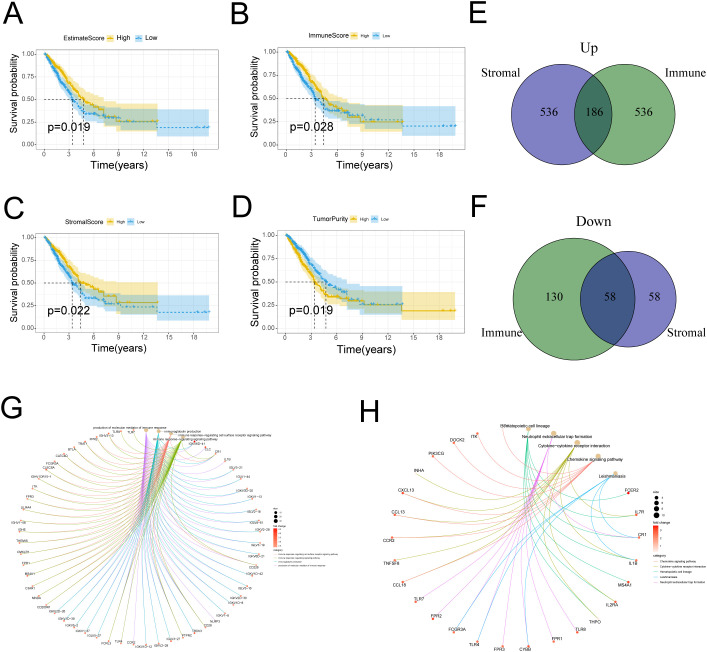
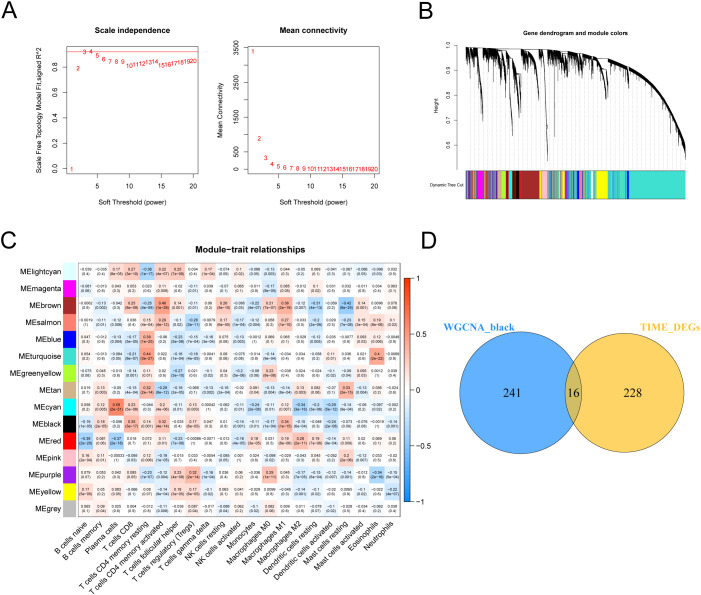
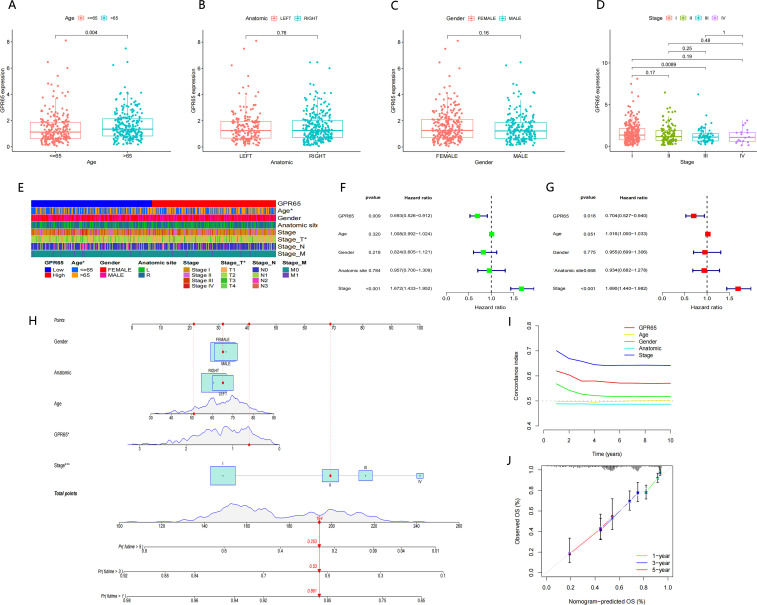
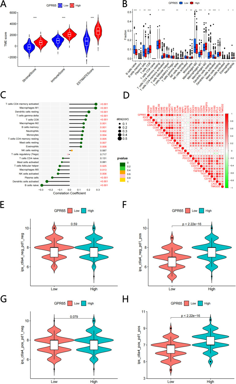
Methods: The proportion of immune, stromal and tumor cells in LUAD samples was assessed by ESTIMATE algorithm scores with RNA sequence data and clinical information from LUAD patients downloaded from The Cancer Genome Atlas (TCGA) database. We screened differential genes (DEGs) in the immune and stromal components, and then screened modular genes by the WGCNA algorithm, which were intersected with DEGs and incorporated into the LASSO-COX regression model. Additionally, nomogram containing GPR65 and clinical features were constructed for predicting patient prognosis. Then, the correlation between GPR65 and immune cell infiltration was assessed by CIBERSORT, and the impact of hub gene on immunotherapy was determined using correlation analysis between GPR65 and immune checkpoint molecules. Finally, we confirmed the expression of GPR65 in LUAD by Western Blot, Quantitative Real-time PCR and Immunohistochemistry.

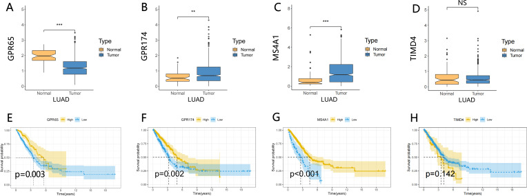
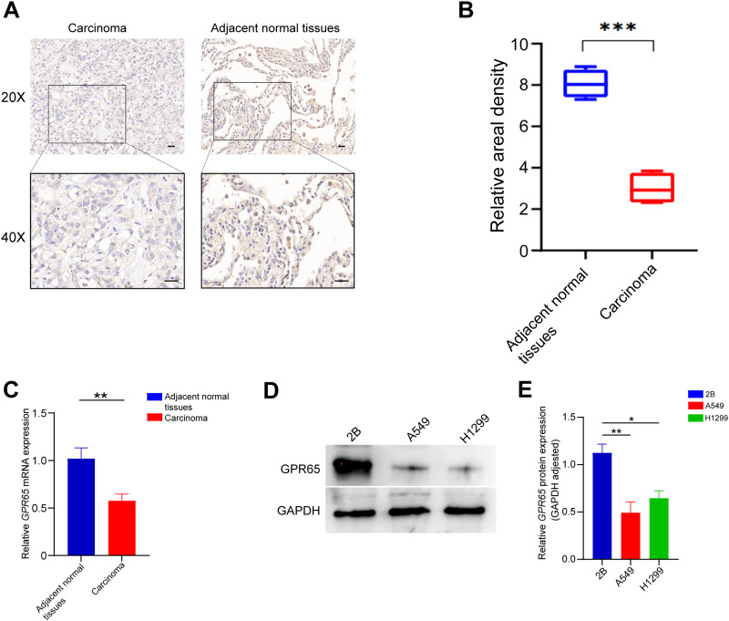
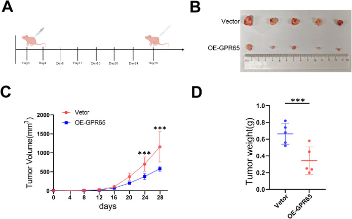
Results: In our study, we found that low expression of GPR65 was associated with poorer overall survival and primary treatment outcome in patients with LUAD. Moreover, GPR65 expression was found to be closely correlated with multiple tumor infiltrating immune cells (TIICs) and immune checkpoint molecules. Immunohistochemistry and Quantitative Real-time PCR results confirmed that the transcription levels and protein expression levels of GPR65 in LUAD tissues were significantly lower than in normal tissues. Western Blot results showed that the expression of GPR65 in human normal lung epithelial cell lines was significantly higher than the expression level in LUAD cell lines.

Conclusions: GPR65 may be an important immune biomarker in the prognosis and diagnosis of LUAD.

Keywords: Gpr65; biomarker; immune microenvironment; lung adenocarcinoma; prognosis.


Conflict of interest statement

The authors declare that the research was conducted in the absence of any commercial or financial relationships that could be construed as a potential conflict of interest.

Figures

Figure 1
Figure 1
Correlation of scores and tumor purity with the survival of patients with LUAD. (A) Kaplan–Meier survival analysis for LUAD patients grouped into high or low score ESTIMATEScore determined by the comparison with the median (P = 0.019 by log-rank test). (B) Kaplan–Meier survival curve for ImmuneScore with P = 0.028 by log-rank test. (C) Survival analysis with Kaplan–Meier method for LUAD patients grouped by StromalScore (P = 0.022 by log-rank test). (D) Kaplan–Meier survival analysis for TumorPurity with P = 0.019 by log-rank test. (E) Venn plot of commonly upregulated DEGs in the stromal and immune components (|log2 fold change (FC)|>1, adj P-value < 0.05). (F) Venn plot showing common down-regulated DEGs shared by ImmuneScore and StromalScore. (|log2 fold change (FC)|>1, adj P-value < 0.05). (G-H) Gene Ontology (GO) and Kyoto Encyclopedia of Genes and Genomes (KEGG) enrichment analysis of differentially expressed genes (DEGs).
Figure 2
Figure 2
WGCNA and DEGs identified GPR65 as a hub gene for LUAD. (A) Analysis of network topology for various soft-threshold powers. (B) Clustering dendrograms of genes, with dissimilarity based on topological overlap, together with assigned module colors. (C) Analysis of module-trait relationships of LUAD based on TCGA data. (D) Venn diagram of WGCNA and DEGs.
Figure 3
Figure 3
Integrated analysis of GPR65、GPR174、MS4A1 and TIMD4. (A–D) Different expression of GPR65、GPR174、MS4A1 and TIMD4 in the normal and cancer tissue; (E–H) Kaplan-Meier survival analysis between 4 gene expression levels and prognosis in LUAD patients.
Figure 4
Figure 4
Association between GPR65 expression and clinicopathological characteristics. Boxplots show that (A) Age is significantly associated with GPR65 expression, but other plots have no statistical difference with GPR65 (B–E) Strip chart showed that Age and Stage_T were significantly associated with GPR65. (F-G) Univariate and multivariate Cox regression analyses of GPR65 expression and four other clinicopathological parameters in the TCGA-LUAD cohort. (H) Nomogram of GPR65 and four other clinicopathological characteristics in the diagnosis of LUAD patients. (I) Time dependent C-index curves of the GPR65 and four other clinical traits. (J) Calibration graphs indicated that predicted 1,3, and 5 year survival rates were close to the actual survival rates.
Figure 5
Figure 5
GPR65 were associated with immune cell infltration and immune checkpoint. (A) Comparisons of the levels of three kinds of TME scores analysis in the high and low GPR65 groups(B) Diferential fractions of 22 immune cells in low and high-expressed groups. (C) Relationship between 22 immune cell types by CIBERSORT. (D) The correlation between 29 immune checkpoint-related genes and GPR65. (E-H) GPR65 high expressed were positively correlated with upregulated programmed cell death protein 1 (PD1) level and cytotoxic T lymphocyte-associated antigen-4 (CTLA4) plus PD1 level, whereas the other plots showed no statistical difference in patients with LUAD.
Figure 6
Figure 6
GPR65 is low expressed in both LUAD tissues and cell lines. (A-B) GPR65 was low expressed in LUAD tissues compared to adjacent normal tissues; (C) Low mRNA expression of GPR65 in LUAD tissues compared to adjacent normal tissues; (D-E) Low expression of GPR65 in LUAD cells compared to normal alveolar epithelial cells.
Figure 7
Figure 7
Up-regulation of GPR65 attenuates proliferation and invasiveness in vitro. (A) Clone formation assays performed with A549 and H1299 cells; (B-C) Representative images of indicated cells migration and invasion treated with medium from vetor lung cancer cells or si-GPR65. (D) Representative images of indicated cells invasion treated with medium from vetor lung cancer cells or OE-GPR65.
Figure 8
Figure 8
GPR65 inhibits tumors through the JAK2/STAT3 signaling pathway. (A) GSEA analysis of GPR65 in the TCGA database. (B-C) Immunoblot analysis of p-JAK2 and JAK2 as well as p-STAT3 and STAT3 expression in shCtr cells and siGPR65 cells with or without JAK2 inhibitor. gAPDH was used as an internal control. (D) Immunoblotting was performed to analyze the expression of p-JAK2 and JAK2 as well as p-STAT3 and STAT3 in overexpressing GPR65 cells. GAPDH was used as an internal control.
Figure 9
Figure 9
Overexpression of GPR65 inhibits tumorigenesis in vivo. (A) Schematic diagram of subcutaneous tumor formation in nude mice; (B) Representative tumors are shown; (C) Tumor growth curves depicting tumor volume over time; (D) Scatter plot of individual tumor weights at the experimental endpoint.

Similar articles

References

    1. Cheung CHY, Juan H-F. Quantitative proteomics in lung cancer. J Biomed Sci. (2017) 24:37. doi: 10.1186/s12929-017-0343-y - DOI - PMC - PubMed
    1. Hirsch FR, Scagliotti GV, Mulshine JL, Kwon R, Curran WJ, Wu YL, et al. Lung cancer: current therapies and new targeted treatments. Lancet. (2017) 389:299–311. doi: 10.1016/S0140-6736(16)30958-8 - DOI - PubMed
    1. Herbst RS, Morgensztern D, Boshoff C. The biology and management of non-small cell lung cancer. Nature. (2018) 553:446–54. doi: 10.1038/nature25183 - DOI - PubMed
    1. Succony L, Rassl DM, Barker AP, McCaughan FM, Rintoul RC. Adenocarcinoma spectrum lesions of the lung: Detection, pathology and treatment strategies. Cancer Treat Rev. (2021) 99:102237. doi: 10.1016/j.ctrv.2021.102237 - DOI - PubMed
    1. Zhao Y, Guo S, Deng J, Shen J, Du F, Wu X, et al. VEGF/VEGFR-targeted therapy and immunotherapy in non-small cell lung cancer: targeting the tumor microenvironment. Int J Biol Sci. (2022) 18:3845–58. doi: 10.7150/ijbs.70958 - DOI - PMC - PubMed

MeSH terms

LinkOut - more resources