. 2025 May 30:16:1621260.
doi: 10.3389/fimmu.2025.1621260. eCollection 2025.

Cord blood-derived iNK T cells as a platform for allogeneic CAR T cell therapy

Affiliations

Cord blood-derived iNK T cells as a platform for allogeneic CAR T cell therapy

Maison Grefe et al. Front Immunol. .

Abstract

CD1d-restricted invariant Natural Killer (iNK) T cells are a suitable candidate for allogeneic Chimeric Antigen Receptor (CAR) T cell therapy as they do not cause graft-versus-host disease (GvHD) due to the monomorphic nature of CD1d proteins. However, the phenotypic and functional heterogeneity of iNK T cells from adult donors (AD) may lead to the inconstant CAR-iNK T cell products. Cord blood-derived (CB) iNK T cells, in contrast, exhibit inter-donor homogeneity in phenotype including uniform CD4 expression and are enriched in memory iNK T cell populations. Thus, we evaluated the preclinical therapeutic potential of iNK T cells derived from cord blood (CB) as an off the shelf CAR T cell therapy platform, given the dominant presence of CD4+ iNK T cells. First, CB-derived iNK T cells were extremely enriched with CD4+ iNK T cells that express various NK receptors and display iNK-TCR mediated cytotoxicity but in a lesser degree than AD-derived CD4- iNK T cells. When engineered with an 8F4CAR targeting the acute myeloid leukemia-associated antigen PR1 presented in HLA-A2*01, CB-8F4CAR-iNK T cells showed a greater expansion capacity with higher CD62L expression than AD-8F4CAR-iNK T cells but with similar 8F4CAR expression and iNK T purity. CB-8F4CAR-iNK T cells displayed in vitro cytotoxicity against PR1/HLA-A2+ primary Acute Myeloid Leukemia (AML) and cell lines better than AD-8F4CAR iNK T cells and maintained potent cytotoxicity in repeated antigenic challenges. Moreover, CB-8F4CAR-iNK T cells showed anti-leukemia activity in vivo in a dose dependent manner. Lastly, CB-8F4CAR-iNK T cells were polarized to produce Th2-biased cytokines but in a lesser amount after 8F4CAR-mediated leukemia cytolysis compared to iNK-TCR mediated activation. In conclusion, consistent CD4+ phenotype, superior expansion capacity, and enhanced CD62L expression of CB-CAR-iNK T cells suggest that they may provide an alternative off-the-shelf source for effective CAR-iNK T cell therapy, while reducing the risk of severe cytokine release syndrome through their immunomodulatory properties. Thus, our results support the potential use of CB-iNK T cells as an allogeneic CAR-T cell therapy platform as they maintain a potent cytotoxicity with potentially better safety profile given a Th2-biased cytokine production upon activation.

Keywords: CAR T cell therapy; acute myeloid leukemia; chimeric antigen receptor; cord blood; invariant natural killer t cells.


Conflict of interest statement

The authors declare that the research was conducted in the absence of any commercial or financial relationships that could be constructed as a potential conflict of interest. The author(s) declared that they were an editorial board member of Frontiers, at the time of submission. This had no impact on the peer review process and the final decision.

Figures
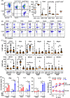
Figure 1
Figure 1
Cord blood-derived iNK T Cells are exclusively CD4+ subtype with Th2/-10 polarized cytokine production profile, yet maintain potent cytolytic activity. The iNKT cells were expanded from cord and adult donors, followed by phenotypic and functional assessment. (A) The representative phenotype analysis (left) and % expression of indicated markers on iNK T cells derived from cord blood (CB) and adult donor (AD)-iNKT cells. Each symbol represents a value from a single donor. (B) A representative expression of activating and inhibitory NK receptors on CB- and AB-iNKT cells. (C) The expression of indicated NK cell markers on CD4+ or CD4- AB-iNKT cells, and CB-iNKT cells. Each symbol represents a value from a single donor. (D) The production of soluble IL-4, IL-10, and IFNγ by CB- and AB-iNKT cells after stimulation with dendritic cells (DC) +/- αGC for 72 hours. The experiment was performed in triplicates, and one of three independent experiments with unique iNKT donor. Mean +/- SD was represented. (E) Cytolysis of Target cells by CB or AD-iNK T cells. CD1d+ C1R cells were pulsed with αGC and incubated with indicated iNKT cells at indicated ratios of Effector to Target for 16hrs. Specific killing was calculated by counting the number of live target cells in experimental conditions against the number of live target cells without effector cells. Experiment was performed in triplicate, and one representative experiment of two independent experiments with different iNK T donors. Values were represented by Mean +/- SD. Unpaired student t-test was used to compare values between AD- and CB-iNK T cells or between different conditions. Paired student t-test was used to compare values between CD4+ vs CD4- AD-iNK T cells derived from same donors. *, P < 0.05.
Figure 2
Figure 2
Highly pure CAR-iNK T cells are reliably produced from the cord blood to a clinically meaningful number. (A) Schematic of 8F4CAR-iNKT cell expansion protocol. The iNKT cells from cord blood units and adult peripheral blood were first isolated via magnetic cell sorting (MACS) with iNK-TCR-microbeads and stimulated by allogeneic dendritic cells pulsed with αGC. Activated iNK T cells were transduced with retrovirus harboring 8F4CAR and cultured for additional 14 days. Lastly, 8F4CAR-iNK T cells were re-expanded via single antigenic stimulation as described above. (B) Representative flow cytometry analysis of iNK T cells from cord and adult donor, in pre- and post-iNK T cell enrichment, and the final 8F4CAR expression (blue and red histogram indicate 8F4CAR-iNK T cells and grey is donor-matched UT-iNK T cells). (C) Absolute number of iNK T cells throughout the expansion protocol. (D) Total fold change of iNK T cells following the 28-day cell expansion protocol. Bolded numbers indicate the mean number of iNK T cells generated per 500M PBMC. Cells were counted by trypan blue exclusion. Mean +/- SD. ***, P < 0.001; ns, not significant; paired student t-test for intra-donor type comparison; unpaired student t-test for inter-donor type comparison. (E) iNK T cell purity of all live cells in culture determined by both CD3 and 6B11 positivity. Each point is a donor. (F) 8F4CAR expression at days 14 and 28 of the cell generation protocol by gating on 8F4CAR+ and 6B11+ iNKT cells. A symbol represents a value from a single donor. Unpaired student t-test was used to compare differences of values between CB vs AD-iNK T cells. Paired student t-test was used to compare differences of values between UT- or 8F4CAR-iNK T cells derived from the same donor. *, P < 0.05.
Figure 3
Figure 3
CB-8F4CAR-iNKT cells maintain a consistent CD4+ phenotype and are significantly enriched in CD62L+ and ICOS+ cells compared to AD-8F4CAR-iNKT cells. (A) A representative flow cytometric analysis of one cord and one adult donor UT- and 8F4CAR-iNK T cells. (B) The percentage of CD4+, CD8α+, and CD4-CD8α- UT- and 8F4CAR-iNK T cells at the expansion protocol conclusion. *, P < 0.05; ns, not significant; paired student t-test for intra-donor type comparison. Each point is a donor. (C) Representative flow cytometric analysis of the memory phenotype, defined by CD62L and CD45RA, of UT- and 8F4CAR-iNK T cells from one cord and adult donor. (D) Percentage of CD62L+ iNK T cells throughout the cell expansion protocol. A symbol represents a value from a single donor. Mean +/- SD. *, P < 0.05, **, P < 0.01, ****, P < 0.0001; ns, not significant; paired student t-test for intra-donor type comparison; unpaired student t-test for inter-donor type comparison. (E) The percentage of activation and exhaustion markers on UT- and 8F4CAR-CB- and AD-iNK T cells determined by flow cytometry. Each point is a donor, *, P < 0.05; paired student t-test for intra-donor type comparison; unpaired student t-test for inter-donor type comparison. (F) Cluster analysis of 8F4CAR+ CB and AD-iNK T cells based on markers listed in panel E was done using open access software CRUSTY (39), showing segregation of CB-8F4CAR-iNK T cells from AD-8F4CAR-iNK T cells (G) The percentage of 8F4CAR+ CB and AD-iNK T cells in each cluster, and a heatmap cluster defining markers as determined by CRUSTY software, demonstrating that CB-8F4CAR-iNK T cells are enriched with ICOS+ or CD27+ clusters compared to AD-8F4CAR-iNK T cells.
Figure 4
Figure 4
CB-8F4CAR-iNK T cells display effective lysis of PR1/HLA-A2+ leukemia in vitro. The indicated leukemia cell lines and primary AML patient samples were co-cultured with effector iNK T cells for 16 hours at indicated E:T ratios. Specific killing was calculated by counting the number of live target cells in experimental conditions against the number of live target cells without effector cells. (A) PR1/HLA-A2 expression on WT or HLA-A2+ U937 target cells used in cytotoxicity experiments (left panel), and specific killing of indicated target cells (right panel): WT U937, HLA-A2+ U937 cells, and αGC pulsed CD1d+ K562 cells by effector iNK T cells: CB-UT-iNK T cells, CB-8F4CAR-iNK T cells, AD-UT-iNK T cells and AD-8F4CAR-iNK T cells. One experiment from 3 independent experiments (each with a unique iNK T donor) was shown in triplicate, and represented with Mean +/- SD. (B) PR1/HLA-A2 expression on HLA-A2- or HLA-A2+ AML primary cells obtained from three patients (left panel) and specific killing of indicated target cells by effector iNK T cells (right panel): CB-UT-iNK T cells, CB-8F4CAR-iNK T cells, AD-UT-iNK T cells and AD-8F4CAR-iNK T cells. One of two independent experiments were shown in triplicates and represented by Mean +/- SD. (C) A schematic of the repeat challenge cytotoxicity experiment (top panel); U937 HLA/A2+ GFP+ cells were co-cultured with effector cells at a 1:1 E:T ratio with 50 IU/mL IL-2. Every 3–4 days iNKT cells and target cells were counted and analyzed by flow cytometry and fresh U937 cells were added to maintain an E:T ratio of 1:1. A representative flow cytometric analysis of iNK T cells and remaining GFP+ HLA-A2+ U937 target cells throughout the experiment is shown (bottom panel). (D) Absolute numbers and fold of changes of effector iNK T cells after each round of leukemia cell challenge, and cumulated fold of change of iNK T cells at the conclusion of experiment. Absolute numbers of GFP+ HLA-A2+ U937 target cells after each round of leukemia cell challenge, and absolute number of GFP+ HLA-A2+ U937 target cells at the conclusion of experiment. One of two independent experiments with different iNK T donors was shown in Mean +/- SD, and experiment was performed in triplicates. Student t-test was used to compare values between groups. *; p<0.05.
Figure 5
Figure 5
CB-8F4CAR-iNK T cells significantly decrease leukemia burden in vivo and extend survival of xenogeneic AML mice. NSG mice were intravenously injected with HLA/A2+luciferase+ U937 cells via tail vein followed by UT- or 8F4CAR-iNK T cells three days later. Leukemia progression was monitored by weekly bioluminescence imaging (BLI). (A) BLI images beginning at day 14 post leukemia cell injection for CB-8F4CAR-iNK T cells or CB-UT-iNK T cells in different doses. One mouse in the 2.5M CB-UT-iNK T cell group did not show successful leukemia engraftment. (B) BLI signal at the last time point where all mice were alive for mice depicted in panel (A). (C) Survival curves of the xenografted mice treated with UT- or 8F4CAR-CB-iNK T cells. (D-F) NSG mice were intravenously injected with HLA/A2+ luciferase+ U937 cells followed by UT- or CB-8F4CAR-iNK T cells or AD-8F4CAR-iNK T cells at a dose of 10e7 three days later. Leukemia progression was monitored by weekly BLI. (D) BLI images (D), BLI intensity (E), and percent survival (F) of xenografted mice treated with CB-UT-iNK T cells, CB-8F4CAR-iNK T cells or AD-8F4CAR-iNK T cells. Student t-test was used to compare differences between group, and Log-rank Mantel-Cox statistical assessment was used to calculate the differences of survival between groups. A symbol represents a value from a mouse. *, P < 0.05.
Figure 6
Figure 6
CB-8F4CAR-iNK T Cells maintain Th2/10-biased cytokine production profile upon iNK-TCR and 8F4CAR mediated activation. CB or AD-8F4CAR-iNK T cells were co-cultured with various target cells (WT-U937, HLA-A2+ U937, αGC/DC) for 8 hours at a ratio of 1 effector to target and subsequently stained for intracellular cytokines for flow cytometry analysis. (A) Representative flow cytometric analysis of intracellular IL-4, IL-10, and IFNγ production by effector iNK T cells after antigenic stimulation. (B) Percent IL-4+, IL-10+, and IFNγ+ CB- and AD-8F4CAR-iNK T cells after antigenic stimulation. (C) CB- or AD-8F4CAR iNK T cells were co-cultured with target cells (WT-U937, HLA-A2+ U937, DC, αGC/DC) for 24 hours, and culture supernatants were analyzed for soluble cytokine production. Experiments were performed in triplicates, and one of three independent experiments with unique iNK T donor was shown in mean +/- SD. Student t-test was used to compare values between groups. *; p < 0.05.

References

    1. Im A, Pavletic SZ. Immunotherapy in hematologic Malignancies: past, present, and future. J Hematol Oncol. (2017) 10:94. doi: 10.1186/s13045-017-0453-8 - DOI - PMC - PubMed
    1. Majzner RG, Mackall CL. Clinical lessons learned from the first leg of the CAR T cell journey. Nat Med. (2019) 25:1341–55. doi: 10.1038/s41591-019-0564-6 - DOI - PubMed
    1. Chen YJ, Abila B, Mostafa Kamel Y. CAR-T: what is next? Cancers (Basel). (2023) 15. doi: 10.3390/cancers15030663 - DOI - PMC - PubMed
    1. Kochenderfer JN, Dudley ME, Feldman SA, Wilson WH, Spaner DE, Maric I, et al. B-cell depletion and remissions of Malignancy along with cytokine-associated toxicity in a clinical trial of anti-CD19 chimeric-antigen-receptor-transduced T cells. Blood. (2012) 119:2709–20. doi: 10.1182/blood-2011-10-384388 - DOI - PMC - PubMed
    1. Morris EC, Neelapu SS, Giavridis T, Sadelain M. Cytokine release syndrome and associated neurotoxicity in cancer immunotherapy. Nat Rev Immunol. (2022) 22:85–96. doi: 10.1038/s41577-021-00547-6 - DOI - PMC - PubMed

MeSH terms

Substances