

Engineering Poly(lactic Acid)-Based Scaffolds for Abundant, Sustained, and Prolonged Lactate Release
- PMID: 40519951
- PMCID: PMC12163944
- DOI: 10.1021/acspolymersau.4c00097
Engineering Poly(lactic Acid)-Based Scaffolds for Abundant, Sustained, and Prolonged Lactate Release
Abstract
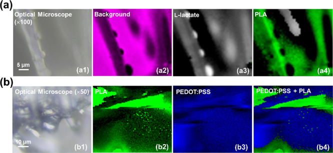
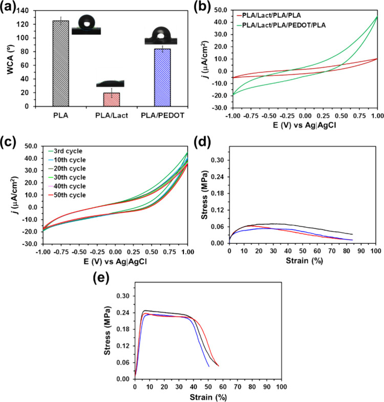
Recent studies have revealed that cardiac tissue regeneration is promoted by administering an initial dose of exogenous lactate and locally maintaining an abundant concentration of this compound for a prolonged period (i.e., around 10-14 days) through sustained release. The aim of this study is to develop a scaffold based on poly-(lactic acid) (PLA) for achieving a sustained daily release of lactate from the first day to the end of the recommended period. First, a five-layered electroresponsive scaffold has been engineered using three PLA layers (first, third, and fifth), each composed of electrospun microfibers (MFs), separated by spin coated lactate (second) and poly-(3,4-ethylenedioxythiophene):poly-(styrenesulfonate) (PEDOT:PSS) (fourth) intermediate layers. The hydrophobicity of the outer PLA layers (first and fifth) has been used to maintain the release of lactate from the intermediate second layer over 3 days, while the conducting fourth PEDOT:PSS layer has ensured a complete lactate release by electrostimulation. After that, in a second step, the same scaffold has been re-engineered to maintain the sustained release not only for a short period (3 days) but also for a prolonged period (>10 days). For this purpose, the PLA MFs of the intermediate third layer have been substituted by plasma-treated proteinase K-containing PLA MFs, obtained by electrospinning a PLA:enzyme mixture. The activity of the enzyme, which decomposes the ester bonds of PLA, combined with the effect of the plasma on the PLA structure, results in a prolonged sustained release that, in addition, can be modulated.
Keywords: cardiac tissue regeneration; conducting polymer; electroresponsive scaffolds; electrospinning; enzymatic degradation; poly(lactic acid).
© 2025 The Authors. Published by American Chemical Society.
Figures


Similar articles
-
Tuning multilayered polymeric self-standing films for controlled release of L-lactate by electrical stimulation.J Control Release. 2021 Feb 10;330:669-683. doi: 10.1016/j.jconrel.2020.12.049. Epub 2020 Dec 31. J Control Release. 2021. PMID: 33388340
-
Enzymatic Degradation of Polylactic Acid Fibers Supported on a Hydrogel for Sustained Release of Lactate.ACS Appl Bio Mater. 2023 Sep 18;6(9):3889-3901. doi: 10.1021/acsabm.3c00546. Epub 2023 Aug 22. ACS Appl Bio Mater. 2023. PMID: 37608579
-
Immediate-sustained lactate release using alginate hydrogel assembled to proteinase K/polymer electrospun fibers.Int J Biol Macromol. 2023 May 31;238:124117. doi: 10.1016/j.ijbiomac.2023.124117. Epub 2023 Mar 21. Int J Biol Macromol. 2023. PMID: 36948340
-
Conductive PEDOT:PSS coated polylactide (PLA) and poly(3-hydroxybutyrate-co-3-hydroxyvalerate) (PHBV) electrospun membranes: Fabrication and characterization.Mater Sci Eng C Mater Biol Appl. 2016 Apr 1;61:396-410. doi: 10.1016/j.msec.2015.12.074. Epub 2015 Dec 31. Mater Sci Eng C Mater Biol Appl. 2016. PMID: 26838866
-
Poly(lactic acid)-poly(ethylene oxide) block copolymers: new directions in self-assembly and biomedical applications.Curr Med Chem. 2011;18(36):5676-86. doi: 10.2174/092986711798347324. Curr Med Chem. 2011. PMID: 22172072 Review.
References
-
- CeCe R., Jining L., Islam M., Korvink J. G., Sharma B.. An Overview of the Electrospinning of Polymeric Nanofibers for Biomedical Applications Related to Drug Delivery. Adv. Eng. Mater. 2024;26:2301297. doi: 10.1002/adem.202301297. - DOI
-
- Mannai F., Elhleli H., Feriani A., Otsuka I., Belgacem M. N., Moussaoui Y.. Electrospun Cactus Mucilage/Poly(vinyl alcohol) Nanofibers as a Novel Wall Material for Dill Seed Essential Oil (Anethum graveolens L.) Encapsulation: Release and Antibacterial Activities. ACS Appl. Mater. Interfaces. 2023;15:58815–58827. doi: 10.1021/acsami.3c13289. - DOI - PubMed
LinkOut - more resources
Full Text Sources