. 2025 Feb;2(1):100030.
doi: 10.1016/j.bvth.2024.100030. Epub 2024 Sep 25.

Unfolded von Willebrand factor binds protein S and reduces anticoagulant activity

Affiliations

Unfolded von Willebrand factor binds protein S and reduces anticoagulant activity

Martha M S Sim et al. Blood Vessel Thromb Hemost. 2025 Feb.

Abstract

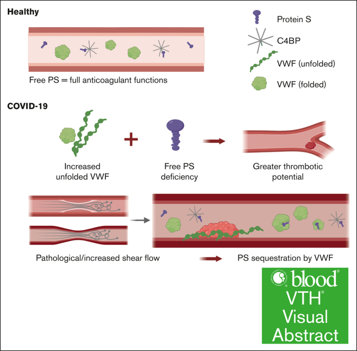
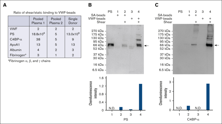
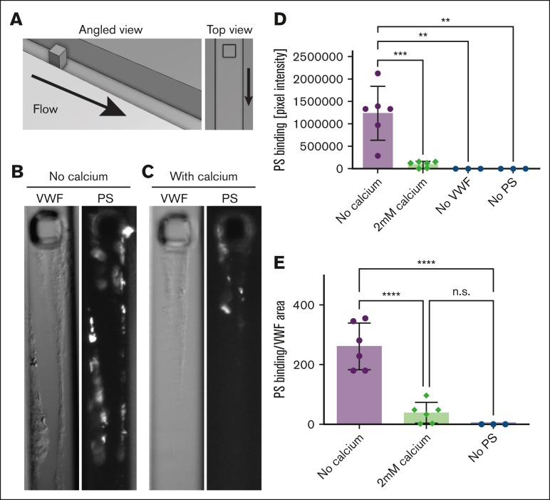
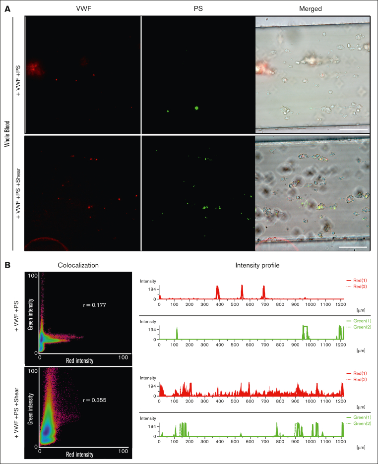
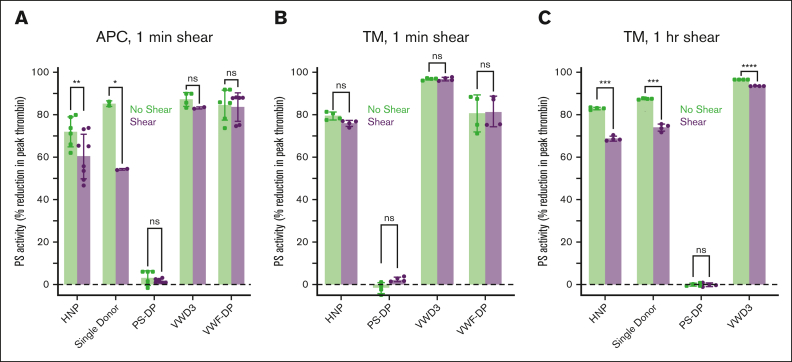
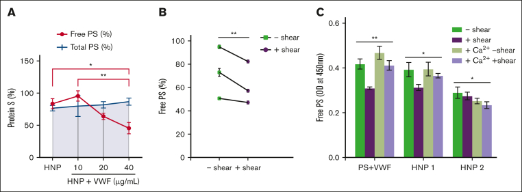
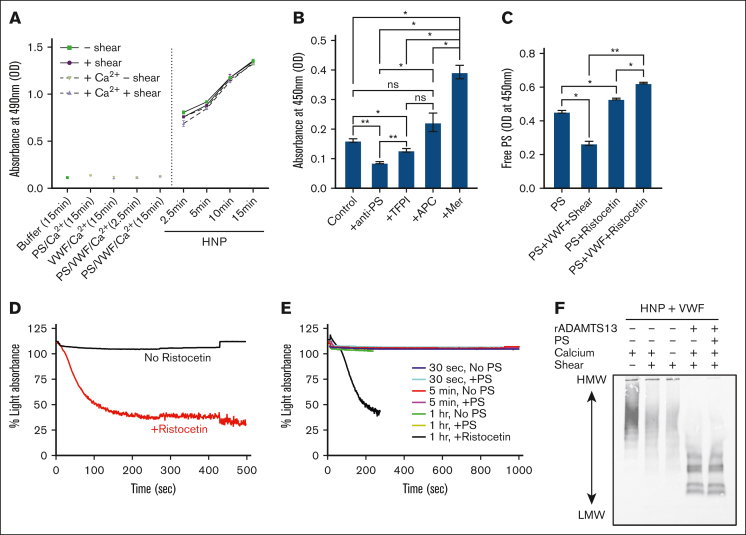
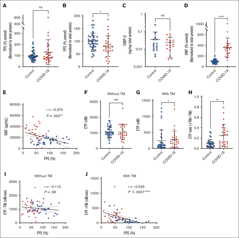
The critical plasma anticoagulant protein S (PS) circulates in 2 functionally distinct pools: free (anticoagulant) or bound to complement component 4b-binding protein (C4BP; anti-inflammatory). Acquired free PS deficiency is detected in several viral infections, but its cause is unclear. Here, we used biochemical approaches and human patient plasma samples to identify an interaction between PS and von Willebrand factor (VWF), which causes free PS deficiency and reduced PS anticoagulant activity. We first identified a shear-dependent interaction between PS and VWF by mass spectrometry. Consistently, PS and VWF could be crosslinked together in plasma, and plasma PS and VWF comigrated in gel electrophoresis. The PS/VWF interaction was blocked by and tissue factor pathway inhibitor but not activated protein C, suggesting an interaction with the sex hormone binding globulin region of PS. Microfluidic systems demonstrated that PS stably binds VWF as VWF unfolds under turbulent flow. PS/VWF complexes also localized to platelet thrombi under laminar arterial flow. In thrombin generation-based assays, shearing plasma decreased PS activity, an effect not seen in the absence of VWF. Finally, free PS deficiency in patients with COVID-19 correlated with changes in VWF, but not C4BP, and with thrombin generation. Our data indicate that PS binds to a shear-exposed site on VWF, thus sequestering free PS and decreasing its anticoagulant activity, which would account for the increased thrombin generation potential. Because many viral infections present with free PS deficiency, elevated circulating VWF, and increased vascular shear, we propose that the PS/VWF interaction reported here is a likely contributor to virus-associated thrombotic risk.


Conflict of interest statement

Conflict-of-interest disclosure: J.P.W. received an investigator-initiated grant through Pfizer Inc, unrelated to this project. The remaining authors declare no competing financial interests.

Figures

None
Graphical abstract
Figure 1.
Figure 1.
Sheared VWF interacts with PS in plasma and interferes with free PS measurement. Streptavidin beads–immobilized biotinylated VWF was exposed to pooled human plasma or single donor plasma, under static conditions or under shear (vortexing), and bound proteins were analyzed with nanoLC-MS/MS, and probed for PS and C4BP-α by immunoblot, with SA used as a control. (A) Mass spectrometry analysis of VWF pull down in pooled normal plasmas (pooled plasma 1 from Innovative Research; pooled plasma 2 from Precision Biologics) and single donor. (B-C) Western blot of VWF pull down probed for PS (B) and reprobed for C4BP-α (C). Arrows indicate the bands used for densitometric analysis. LC-MS, liquid chromatography-mass spectrometry, SA, serum albumin.
Figure 2.
Figure 2.
PS binds to VWF that unfolds during disrupted flow conditions in a calcium-modulated manner. (A) VWF and PS were perfused through a PDMS microfluidic device. (B-C) In the absence (B) or presence of 2 mM calcium (C), VWF self-association and Alexa Fluor 488-PS binding were visualized by DIC and epifluorescence microscopy, respectively. (D) Raw pixel intensity above noise was summed for these conditions and controls without VWF (with PS) and without PS (with VWF). Each data point indicates an independent run. (E) When accounting for the area of VWF self-association, PS binding was significantly higher than all other conditions. Error bars are standard deviation (SD). Statistical significance was determined with an analysis of variance (ANOVA) and Tukey post hoc test; n.s., nonsignificant; ∗∗P < .01; ∗∗∗P < .001; ∗∗∗∗P < .0001. Scale bar, 25 μm. DIC, differential interference contrast, PDMS, polydimethylsiloxane.
Figure 3.
Figure 3.
The PS/VWF complex is stable under flow. Whole blood was supplemented with 10 μg/mL AlexaFluor555-conjugated VWF and 200 nM AlexaFluor488-conjugated PS and perfused through collagen-coated channels at 35 dyn/cm2. Experiments were performed either without or with PS/VWF vortexing. (A) Images taken after flushing with buffer; scale bar, 100 μm. (B) Colocalization (shown is Pearson correlation r) and intensity profile analyses using the Nikon Instruments Software Element software.
Figure 4.
Figure 4.
Unfolded VWF reduces PS plasma anticoagulant activity. Plasma thrombin generation was measured in pooled healthy normal plasma (HNP), single donor plasma, PS-DP, plasma from a patient with VWD3, or VWF-DP in the presence or absence of exogenous APC (5 nM; A) or thrombomodulin (20 nM; B-C) and the presence or absence of short-term shear (1 minute; A-B) or long-term shear (1 hour; C). Paired t tests were performed with each individual data set, comparing plasma with or without shearing; ∗P < .05; ∗∗P < .01; ∗∗∗P < .001; ∗∗∗∗P < .001. PS-DP, PS-depleted plasma, VWD3, type 3 von Willebrand disease, VWF-DP, VWF-depleted plasma.
Figure 5.
Figure 5.
VWF reduces the measurement of free PS. (A) Free PS and total PS enzyme-linked immunosorbent assay (ELISA) measurements of HNP (from Siemens), with additions of purified VWF, under shear (∼2500 rpm for 30 seconds). Free and total PS are presented as percent of free and total PS in reference plasma, respectively. (B) Free PS ELISA measurements of healthy control plasmas from single donors with or without shear. P values are according to Wilcoxon matched-pairs signed rank test; ∗∗P < .01. (C) Free PS ELISA results of purified proteins (200 nM PS and 10 μg/mL VWF in HEPES-buffered saline, with albumin), HNP1 (Corgenix), and HNP2 (Siemens), with or without shear or 1 μM hirudin, 5 mM Gly-Pro-Arg-Pro peptide, and 5 mM CaCl2 supplementation; nondetectible. P values for panel A are according to 2-way ANOVA with Sidak multiple comparisons test. P values for panels B-C are according to Kruskal-Wallis with Dunn multiple comparison test; ∗P < .05; ∗∗P < .01. OD, optical density; rpm, revolutions per minute.
Figure 6.
Figure 6.
Direct measurement of the PS/VWF complex in plasma. (A) PS/VWF complex was measured by ELISA, using polyclonal antibodies to capture VWF and detect bound PS. Experiments were performed with purified proteins (200 nM PS, 10 μg/mL VWF, and 5 mM CaCl2, as indicated, in HEPES-buffered saline, with albumin) or HNP, with or without shear or 1 μM hirudin, 5 mM Gly-Pro-Arg-Pro peptide, and 5 mM CaCl2 supplementation. Samples were incubated on the plate for the indicated time. (B) Biotinylated PS (150 nM) was added to PS-immunodepleted plasma, and PS/VWF complex was detected as in panel A, except using streptavidin-HRP to detect. Experiments were performed in the presence or absence of saturating concentrations of an anti-PS polyclonal antibody, TFPIα, APC, or MerTK. (C) Free PS was measured, as in Figure 1, using purified PS (200 nM), with or without purified VWF (10 μg/mL) and ristocetin (2 mg/mL). (D-E) Washed platelets (2.5 × 108/mL) were aggregated in the presence or absence of VWF (10 μg/mL), PS (150 nM), and ristocetin (2 mg/mL). Experiments were performed in the absence (D) or presence (E) of vortexing to unfold VWF. In panel E, VWF was vortexed for 30 seconds, 5 minutes, or 1 hour. (F) VWF multimer blot of 0.25 μL HNP with additional 150 nM purified VWF, with or without 50 nM recombinant ADAMTS13, 200 nM PS, or 1 μM hirudin, 5 mM Gly-Pro-Arg-Pro peptide, and 5 mM CaCl2, supplementation with or without shear (∼2500 rpm for 60 minutes). Every data point is the average of 3 replicates (mean ± SD). P values for panels B-C are according to Kruskal-Wallis with Dunn multiple comparison test; ∗P < .05; ∗∗P < .01. For panels D-E, experiments were performed using platelets from 3 different donors. Shown are the average aggregation curves. HRP, horseradish peroxidase; rpm, revolutions per minute.
Figure 7.
Figure 7.
Free PS deficiency in patients with COVID-19 can be explained by changes in VWF but not C4BP. Citrated plasma samples were collected from healthy controls (n = 38) and patients with COVID-19 (n = 30). Due to sample limitations, some measurements could not be performed with all participants. (A-D) Total PS (A), free PS (B), C4BP-β (C), and VWF (D) were measured by ELISA. (E) Free PS plotted against VWF:Ag. (F-J) Plasma thrombin generation was measured using calibrated automated thrombography. Several patients were receiving heparin prophylactic dose at the time of blood collection. Shown are the ETPs in assays initiated with 4 μM phospholipids and 1 pM tissue factor (TF) (F) or with 20 nM thrombomodulin (TM) supplementation to evaluate the contribution of APC/PS activity (G), and the ETP ratio (value with TM/value without TM) (H). (I-J) Free PS plotted against ETP in the absence (I) or presence (J) of thrombomodulin. Every data point is the average of 3 replicates (mean ± SD). For panels A-H, P values are according to Mann-Whitney test; ∗P < .05; ∗∗P < .01. For panels I-J, P values and r correlation coefficients are according to Spearman correlation analysis; ∗∗∗P < .001; ∗∗∗∗P < .0001. Blue squares represent controls, and red circles represent patients with COVID-19. FPS, free protein S; TPS, total protein S.

Update of

Similar articles

Cited by

References

    1. Mann KG, Nesheim ME, Church WR, Haley P, Krishnaswamy S. Surface-dependent reactions of the vitamin K-dependent enzyme complexes. Blood. 1990;76(1):1–16. - PubMed
    1. Calzavarini S, Prince-Eladnani R, Saller F, et al. Platelet protein S limits venous but not arterial thrombosis propensity by controlling coagulation in the thrombus. Blood. 2020;135(22):1969–1982. - PMC - PubMed
    1. Mahasandana C, Suvatte V, Marlar RA, Manco-Johnson MJ, Jacobson LJ, Hathaway WE. Neonatal purpura fulminans associated with homozygous protein S deficiency. Lancet. 1990;335(8680):61–62. - PubMed
    1. Dahlbäck B, Stenflo J. High molecular weight complex in human plasma between vitamin K-dependent protein S and complement component C4b-binding protein. Proc Natl Acad Sci U S A. 1981;78(4):2512–2516. - PMC - PubMed
    1. Walker FJ. Regulation of activated protein C by protein S. The role of phospholipid in factor Va inactivation. J Biol Chem. 1981;256(21):11128–11131. - PubMed

LinkOut - more resources