Review
. 2025 Jun 12;11(2):e002475.
doi: 10.1136/bmjsem-2025-002475. eCollection 2025.

Tendon and ligament injuries of the finger and thumb in athletes: a narrative review

Affiliations
Review

Tendon and ligament injuries of the finger and thumb in athletes: a narrative review

Anaas Mergoum et al. BMJ Open Sport Exerc Med. .

Abstract

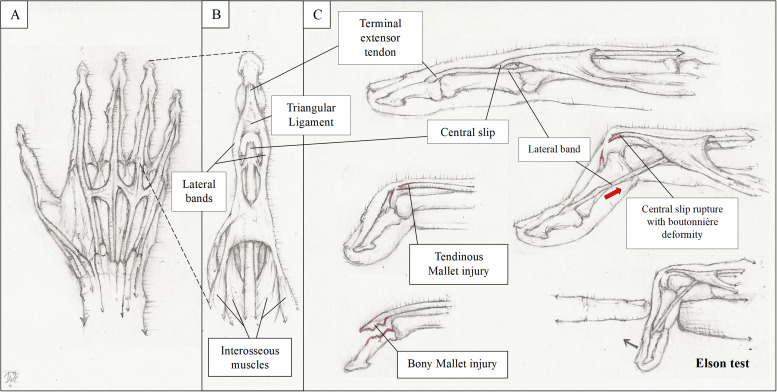
Acute finger and thumb injuries of the ligament and tendons are common. Indeed, a cross-sectional study in 2012 showed that they accounted for 38.4% of all upper extremity injury visits to the emergency room in the USA. Understanding the anatomy and mechanical functions of tendons and ligaments in the digits is crucial for recognising various types of injuries and their treatment. Treating an athlete with such conditions comes with the added pressure of ensuring a timely return to play. This review will cover the anatomy, diagnoses and management of select tendinous and ligamentous injuries of the fingers and thumb seen in athletes.

Keywords: Finger; Sporting injuries; Tendon.


Conflict of interest statement

None declared.

Figures

Figure 1
Figure 1. Anatomy and pathology of the extensors. (A) The extensor anatomy of the digits is depicted. (B) The dorsal side of the fourth finger is magnified to show the anatomy in detail. (C) Mallet injuries occur either through the rupture of the extensor tendon (tendinous mallet injury) or through an avulsion fracture with the tendon remaining intact (bony mallet injury). If a central slip injury is left untreated, a boutonnière deformity might develop; this results in volar subluxation of the lateral bands leading to flexion and extension of the PIP and DIP joints (red arrow), respectively. The Elson test may be used to assess for this injury. To perform the Elson test, the injured digit is flexed at a 90° over the edge of a table, the patient then tries to extend the PIP joint against resistance. The test is positive when there is an absence of extension force at the PIP joint and fixed extension at the DIP joint (black arrow). DIP, distal interphalangeal; PIP, proximal interphalangeal.
Figure 2
Figure 2. Anatomy and pathology of the flexors and collateral ligaments. The function of the FDP and FDS tendons is to flex the DIP and PIP joints, respectively. The vinculum brevis and longus compromise the vincular system that provides the blood supply to both these tendons. (B) The flexor anatomy of the digits is shown (the fifth finger dissected deeper with tendons retracted); FDP injury typically affects the fourth finger (highlighted in red). The zone classification (roman numerals) of flexor tendon injury is also depicted. (C) The lateral view of the fifth finger portrays the two collateral ligaments and volar plate of the PIP joint. Dorsal dislocation of the middle phalanx (arrow) often results in damage to both the collateral ligaments and the volar plate. An example of progressive extension using a dorsal blocking splint to treat a volar plate injury is also shown. (D) The anatomy of the pulley system of the finger is shown along with Schöffl’s injury grades. The ‘crimp-grip position’ (MCP and DIP joints in extension while PIP joints in flexion) puts a tremendous force across the pulley system, especially across the A2, A3 and A4 pulleys, which may lead to significant damage complicated by bowstringing. DIP, distal interphalangeal; FDP, flexor digitorum profundus; FDS, flexor digitorum superficialis; MCP, metacarpophalangeal; PIP, proximal interphalangeal.
Figure 3
Figure 3. Distal forearm squeeze test. This test is performed by squeezing the distal forearm while isolating the DIP joint of the suspected injured finger; a lack of flexion at the DIP joint (curved black arrow) is considered a positive test. The flexors are favoured in the squeeze test because there is a greater gap (straight black arrow) between the flexors and the arm bones—ie, radius and ulna—than the extensors and the arm bones. DIP, distal interphalangeal.
Figure 4
Figure 4. Ulnar collateral ligament injury of the thumb. (A) Ulnar collateral ligament (UCL) may be damaged from forced abduction (red arrow). (B) Stener lesions occur when the UCL is displaced outside the adductor aponeurosis. This leads to UCL entrapment and reduced likelihood of healing with conservative management. (C) To determine the degree of laxity, the metacarpophalangeal (MCP) joint is stabilised while applying a valgus stress.

Similar articles

References

    1. Ootes D, Lambers KT, Ring DC. The epidemiology of upper extremity injuries presenting to the emergency department in the United States. Hand (N Y) 2012;7:18–22. doi: 10.1007/s11552-011-9383-z. - DOI - PMC - PubMed
    1. Cheung JPY, Fung B, Ip WY. Review on mallet finger treatment. Hand Surg. 2012;17:439–47. doi: 10.1142/S0218810412300033. - DOI - PubMed
    1. Krastman P, de Schepper E, Bindels P, et al. Incidence and management of mallet finger in Dutch primary care: a cohort study. BJGP Open. 2024;8 doi: 10.3399/BJGPO.2023.0040. - DOI - PMC - PubMed
    1. Lamaris GA, Matthew MK. The Diagnosis and Management of Mallet Finger Injuries. Hand (N Y) 2017;12:223–8. doi: 10.1177/1558944716642763. - DOI - PMC - PubMed
    1. Netscher DT, Pham DT, Staines KG. Finger Injuries in Ball Sports. Hand Clin. 2017;33:119–39. doi: 10.1016/j.hcl.2016.08.018. - DOI - PubMed

LinkOut - more resources