Review
. 2025 May 31;11(2):97-106.
doi: 10.4103/bc.bc_4_23. eCollection 2025 Apr-Jun.

Cerebral arteritis in bacterial meningitis: Structural adaptations of the vascular wall in response to an infectious nidus - A narrative review

Affiliations
Review

Cerebral arteritis in bacterial meningitis: Structural adaptations of the vascular wall in response to an infectious nidus - A narrative review

Vivig Shantha Kumar et al. Brain Circ. .

Abstract

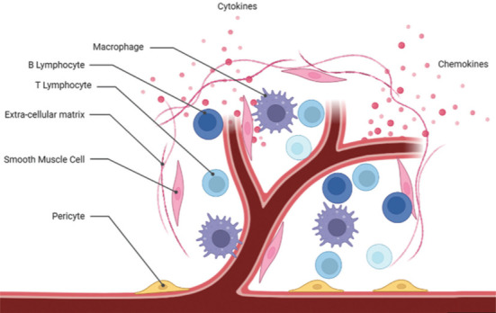
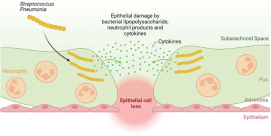
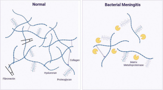
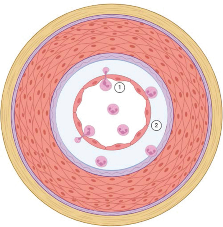
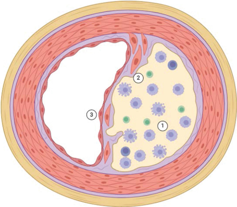
Despite dramatic improvements in diagnosis and antimicrobial treatment of bacterial meningitis over the last few decades, adverse postinfective sequelae and mortality remain exceedingly high in adults. Of note, the unfavorable clinical outcome is usually attributable to the presence of intracranial complications during the acute phase of infection, such as cerebral edema and increases in intracranial pressure and cerebral blood flow disturbances. Involvement of the cerebral vasculature during bacterial meningitis is overwhelmingly clear from clinical and laboratory evidence highlighting cerebral blood flow alterations with the use of Doppler blood flow analysis, angiographic studies of cerebral vessel wall structural irregularities and computed tomography/magnetic resonance imaging recording of cerebral infarctions. With the widespread agreement of cerebrovascular involvement in bacterial meningitis, very few studies have documented histopathological observations of cerebral vessel irregularities affecting the various layers of the vascular wall. In an attempt to understand the arterial wall changes that take place before the occurrence of cerebral ischemic consequences in bacterial meningitis, we have investigated the sequential changes affecting the arterial vasculature, beginning with early reflexive modifications of the adventitia and culminating in late proliferative lesions of the intima.

Keywords: Bacterial meningitis; adventitia; cerebral arteritis; internal elastic lamina; intima; media.


Conflict of interest statement

There are no conflicts of interest.

Figures

Figure 1
Figure 1
Cerebrovascular involvement in bacterial meningitis by immune cells. From several pieces of histological evidence in the 19th century, it is clear that the mechanisms responsible for cerebral vasculitis during bacterial meningitis involve infiltration of arterial walls by immunological cells primarily involving macrophages and lymphocytes (T & B) and the vasculopathic actions of proinflammatory cytokines.
Figure 2
Figure 2
Early reflexive adventital involvement on contact with the surrounding purulent exudate. The adventitia represents the initial contact between the purulent exudate accumulation in the cerebrospinal fluid and the vessel wall during bacterial meningitis, with early adventitial epithelial injury setting the foundation for early vasculitic changes seen involving the inner medial and intimal layers.
Figure 3
Figure 3
Adventitial microenvironment with inflammatory cell infiltration resulting in fibrosis. Following adventitial involvement by the surrounding purulent exudate, a local inflammatory response, consisting of neutrophils, macrophages, T-lymphocytes, is initiated leading to adventitial fibrosis and proliferation that progressively invades into the media.
Figure 4
Figure 4
Hypocellular tunica media with upregulation of matrix degradative proteases and resultant loss of extracellular matrix structural components. In contrast to a normal media, which is rich in fibronectin, proteoglycan, collagen and hyaluronan, the media during bacterial meningitis is considerably devoid of these extracellular matrix components as a result of increased matrix metalloproteinase mediated degeneration resulting in a markedly hypocellular media subsequently accompanied by internal elastic lamina degradation.
Figure 5
Figure 5
Reflexive intimal edema and neutrophilix infiltration promote intimal narrowing and early luminal stenosis. During the early stages of vasculitis, endothelial degeneration accompanied by upregulation of leukocyte adhesion molecules on the endothelial surface leads to subendothelial trafficking of circulating neutrophils (1) and intimal edema (2) contributing to a circumferential reflexive luminal narrowing.
Figure 6
Figure 6
Organized smooth muscle cell and lymphocyte infiltration and proliferation accelerate intimal fibrosis resulting in organic stenosis. With vasculitis progression, subendothelial neutrophils are replaced by macrophages and T-lymphocytes (1), which accelerate intimal proliferation through their mitotic actions on remaining smooth muscle cells (2) resulting in an eccentric and progressive luminal narrowing (3).

References

    1. Loewenstein Ueber die veränderungen des gehirns und rückenmarks bei meningitis cerebrospinalis. Beitr Path Anat Allg Path. 1910;47:282.
    1. Ernst P. Das nervensystem. In: Aschoff L, editor. Pathologische Anatomie. Jena: Gustav Fischer; 1928.
    1. MacCallum WG. Philadelphia: W. B. Saunders Company; 1926. Textbook of Pathology.
    1. Wertham F. The cerebral lesions in purulent meningitis. Arch Neurol Psychiatry. 1931;26:549.
    1. Buzzard EF, Greenfield JG. London: Constable and Company, Ltd; 1921. Pathology of the Nervous System.

LinkOut - more resources