. 2025 May 30;6(7):1174-1183.
doi: 10.1039/d5cb00086f. eCollection 2025 Jul 2.

Biochemical characterization of Bifidobacterium bifidum peptidoglycan d,l-endopeptidase BbMep that generates NOD2 ligands

Affiliations

Biochemical characterization of Bifidobacterium bifidum peptidoglycan d,l-endopeptidase BbMep that generates NOD2 ligands

Jeric Mun Chung Kwan et al. RSC Chem Biol. .

Abstract

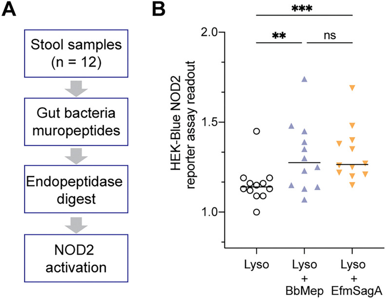
Soluble peptidoglycan fragments produced by the gut bacteria are key effectors in microbiota-host crosstalk. Here, we biochemically characterized BbMep, an NlpC/p60 domain-containing peptidoglycan d,l-endopeptidase from Bifidobacterium bifidum, which efficiently digests Lys- or Orn-type sacculi. Digestion of human stool-derived muropeptides by BbMep enhances NOD2 activation.


Conflict of interest statement

There are no conflicts to declare.

Figures

Fig. 1
Fig. 1. (A) Peptidoglycan d,l-endopeptidases cleave bacterial sacculi or larger muropeptides into dipeptide (GMDPs) and terminal peptide products (TerPs). GMDPs are liberated only if the sacculus is further treated with a muramidase (e.g. lysozyme and mutanolysin). (B) GMDPs have been previously found to be prominent in the peptidoglycan composition of B. adolescentis and B. bifidum through LC-MS/MS analysis. The two GMDP products shown are (NAG)(NAM)-Ae (C27H44N4O17, [M + H]+ = 697.277) and (NAG)(NAM)-Aq (C27H45N5O16, [M + H]+ = 696.293). (C) Both Bifidobacterium species encode a d,l-endopeptidase that contains the NlpC/p60, which are referred to as BaMep and BbMep. The specific domains in BaMep and BbMep are determined by SignalP, MobiDB, and InterPro. SP: signal peptide. (D) Histograms showing the distributions of 364 putative NlpC/p60 endopeptidases in the Bifidobacterium genus in terms of sequence similarity (to EfmSagA) (left) and protein lengths (right).
Fig. 2
Fig. 2. (A) Schematic showing biochemical assay of endopeptidases. (B) and (C) LC-MS/MS analysis of quantification of GMDP and TerP products released by endopeptidase digestion of various bacterial sacculi. Bb_SD contains the NlpC/p60 domain only (residues 156–268). The muropeptide (NAG)(NAM)-A was used as an internal standard for peak area normalization. Error bars represent the standard deviations of the mean of three independent replicates. p-Values were calculated with a two-tailed student's t-test (α = 0.05, n = 3), comparing each enzyme with the negative control (Nil). p-Values were adjusted for multiple comparisons with the Bonferroni method. *p < 0.05; **p < 0.01; ***p < 0.001; ****p < 0.0001; ns: not significant.
Fig. 3
Fig. 3. (A) and (B) Multiple sequence alignment and phylogenetic tree of BaMep and BbMep with other peptidoglycan d,l-endopeptidases with known activity. Numbers on the phylogenetic tree indicate the computed BLOSUM62 distance. Enteroccocus: Efm = E. faecium, Emu = E. mundtii, and Edu = E. durans; Lactobacillus: Lpc = L. paracasei and Lsa = L. salivarius; Bifidobacterium: Ba = B. adolescentis and Bb = B. bifidum; Mycobacterium: Mtb = M. tuberculosis. (C) Percentage of BbMep residue conservation (full or partial) across 364 putative Bifidobacterium endopeptidases. The black arrows indicate residues aligned to W429 (Trp1) and W458 (Trp2) from EfmSagA. The catalytic residues C186 and H233 in BbMep are bolded in red. The predicted glycan- and peptide-binding residues in BbMep (from the docking analysis in next section) are indicated in magenta and green, respectively. The grey area indicates the running average for five residues.
Fig. 4
Fig. 4. (A) Sacculi binding assay revealed that BbMep and its NlpC/p60 domain-only construct, BbMep_SD (10 μM) bind to E. faecium sacculi (20 mg mL−1), whereas EfmSagA (10 μM) does not. (B) and (C) Prediction of potential PGN-interacting residues in BbMep based on in silico molecular docking. The AlphaFold predicted BbMep structure was docked with (NAG)(NAM)-AqKAA using AutoDock Vina. The best docking poses from 3 runs × 10 poses were selected for further analysis (see Fig. S10, ESI†). We selected the docked poses with a binding affinity < −6 kcal mol−1 and a catalytic distance < 7.5 Å between C186 and the peptide bond undergoing cleavage. The BbMep residues involved in glycan- (magenta) or peptide-binding (green) meet two criteria: (1) they form one or more hydrogen bonds, and (2) they differ from Efm residues. (D) Construction of BbMep_Mut1, Mut2, and Mut3 to experimentally validate the proposed PGN-interacting residues. (E) and (F) Both Mut1 and Mut2 exhibit slightly reduced binding to bacterial sacculi (E) and significantly reduced endopeptidase activity in LC-MS analysis (E). The muropeptide (NAG)(NAM)-A was used as an internal standard for peak area normalization in LC-MS. Error bars represent the standard deviations of the mean of three independent replicates. p-Values were calculated with a two-tailed student's t-test (α = 0.05, n = 3), comparing each group with the wild-type BbMep. p-Values were adjusted for multiple comparisons with the Bonferroni method. *p < 0.05; ****p < 0.0001; ns: not significant.
Fig. 5
Fig. 5. BbMep, similar to EfmSagA, generates active NOD2 ligands in human stool samples. (A) Protocol of workflow: soluble muropeptides were first extracted from human stool by lysozyme and then treated with BbMep or EfmSagA, followed by analysis using HEK-Blue NOD2 reporter assay. (B) A significant increase in NOD2 induction was observed in both BbMep- and Efm-treated samples compared to non-endopeptidase control. p-Values were calculated with a paired t-test (α = 0.05, n = 12). **p < 0.01; ****p < 0.0001; ns: not significant.

Similar articles

References

    1. Li C. Liang Y. Qiao Y. Front. Microbiol. 2022;13:863407. doi: 10.3389/fmicb.2022.863407. - DOI - PMC - PubMed
    1. Sommer F. Bäckhed F. Nat. Rev. Microbiol. 2013;11:227–238. doi: 10.1038/nrmicro2974. - DOI - PubMed
    1. Agus A. Clément K. Sokol H. Gut. 2021;70:1174–1182. doi: 10.1136/gutjnl-2020-323071. - DOI - PMC - PubMed
    1. Gabanyi I. Lepousez G. Wheeler R. Vieites-Prado A. Nissant A. Wagner S. Moigneu C. Dulauroy S. Hicham S. Polomack B. Verny F. Rosenstiel P. Renier N. Boneca I. G. Eberl G. Lledo P.-M. Science. 2022;376:eabj3986. doi: 10.1126/science.abj3986. - DOI - PubMed
    1. Schwarzer M. Gautam U. K. Makki K. Lambert A. Brabec T. Joly A. Šrůtková D. Poinsot P. Novotná T. Geoffroy S. Courtin P. Hermanová P. P. Matos R. C. Landry J. J. M. Gérard C. Bulteau A.-L. Hudcovic T. Kozáková H. Filipp D. Chapot-Chartier M.-P. Šinkora M. Peretti N. Boneca I. G. Chamaillard M. Vidal H. De Vadder F. Leulier F. Science. 2023;379:826–833. doi: 10.1126/science.ade9767. - DOI - PubMed

LinkOut - more resources