. 2025 May 30:12:1537164.
doi: 10.3389/fmolb.2025.1537164. eCollection 2025.

Flexible iron: disorder in the ironome brings order to protein structure and function

Affiliations

Flexible iron: disorder in the ironome brings order to protein structure and function

Vladimir N Uversky et al. Front Mol Biosci. .

Abstract

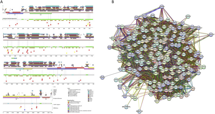
Iron is one of the most abundant elements on earth. The most recognized role of iron in living organisms is its incorporation in the heme-containing protein hemoglobin, which is abundantly found in the red blood cells that facilitate the oxygen transportation throughout the body. In fact, about 70% of organism's iron is found in hemoglobin. However, besides being essential for oxygen transport and serving as a crucial component of the molecular oxygen-carrying proteins hemoglobin and myoglobin, iron has a wide range of other biological functions. It is involved in numerous metabolic and regulatory processes and therefore is indispensable for almost all living organisms. Since iron enzymes are responsible for most of the redox metallo-catalysts, it is not surprising that 6.5% of all human enzymes are expected to be iron-dependent. Furthermore, iron-binding proteins account for about 2% of the entire proteome. The ironome encompasses heme-binding proteins, proteins binding individual iron ions, and iron-sulfur cluster-binding proteins. Although the structure-function relations of ordered iron-binding proteins are rather well understood, the prevalence and functionality of intrinsic disorder in iron-binding proteins remain to be evaluated. To fill this knowledge gap, in this study, we evaluate the intrinsic disorder of the human ironome. Our analysis revealed that the human ironome contains a noticeable level of functional intrinsic disorder, with most noticeable applications in protein-protein interactions, posttranslational modifications, and liquid-liquid phase separation.

Keywords: intrinsically disordered proteins; iron; iron-binding proteins; iron-sulfur center; liquid-liquid phase transition; proteinprotein interactions.


Conflict of interest statement

The authors declare that the research was conducted in the absence of any commercial or financial relationships that could be construed as a potential conflict of interest.

Figures

FIGURE 1
FIGURE 1
Distribution of the human heme-binding proteins (A and B), iron ion-binding proteins (C and D), iron-sulfur cluster-binding proteins (E and F), and the human calcium-binding (G and H) proteins based on their PPIDR (A, C, E, and G) and MDS (B, D, F, and H) values evaluated by various per-residue disorder predictors utilized in this study.
FIGURE 2
FIGURE 2
Nested box-plots comparing the overall disorder predispositions of human heme- (yellow bars), iron ion- (green bars), and iron-sulfur cluster-binding proteins (violet bars) with human calceome (gray bars) and entire human proteome (red bars) evaluated by their PPIDR (A) and MDS (B) values derived based on the outputs of six commonly used per-residue disorder predictors, PONDR® VLXT, PONDR® VSL2, PONDR® VL3, PONDR® FIT, IUPred_Short, and IUPred-Long, as well as mean disorder prediction (MDP) calculated as an average of outputs of these six predictors.
FIGURE 3
FIGURE 3
Evaluation of the global disorder status of the human ironome (data for heme-, iron ion-, and iron-sulfur cluster-binding proteins are shown by yellow, green and purple colors, respectively) in comparison with the calceome (gray circles). (A) PONDR® VSL2 score vs PONDR® VSL2 (%) plot. Here, each point corresponds to a query protein, coordinates of which are evaluated from the corresponding PONDR® VSL2 data as its mean disorder score (MDS) and percent of the predicted intrinsically disordered residues (PPIDR). Color blocks are used to visualize proteins based on the accepted classification, with red, pink/light pink, and blue/light blue regions containing highly disordered, moderately disordered, and ordered proteins, respectively (see the text). Dark blue or pink regions correspond to the regions, where PPIDR agrees with MDS, whereas areas in which only one of these criteria applies are shown by light blue or light pink. (B) CH-CDF plot, where coordinates for a protein are calculated as the average distance of its CDF curve from the CDF boundary (X-axis) and its distance from the CH boundary. Protein classification is based on the quadrant, where the proteins are located: Q1, proteins predicted to be ordered by both predictors; Q2, proteins predicted to be ordered to by CH-plot and disordered by CDF; Q3, proteins predicted to be disordered by both predictors; and Q4, proteins predicted to be disordered by CH-plot and ordered by CDF. [Color code is the same as in panel A].
FIGURE 4
FIGURE 4
STRING-generated protein-protein interaction networks of entire human ironome (A) and sets of human heme- (B), iron ion- (C) and iron-sulfur cluster-binding proteins (D).
FIGURE 5
FIGURE 5
(A) Dependence of the number of interactors within the ironome PPI network on the PPIDR level of the human heme-, iron ion-, and iron-sulfur cluster-binding proteins. Vertical lines show 10% and 30% thresholds. (B) Comparison of the LLPS predisposition of the members of human ironome with their intrinsic disorder propensity. Vertical lines show 10% and 30% thresholds, whereas the horizontal line corresponds to the 0.6 pLLPS threshold.
FIGURE 6
FIGURE 6
Functional disorder analysis of human PER1 (UniProt IDs: O15534). (A) Functional disorder profile generated by the D2P2 platform. Here, the IDR localization predicted by IUPred, PONDR® VLXT, PONDR® VSL2, PrDOS, PV2, and ESpritz is shown with nine differently colored bars at the top of the plot, whereas the agreement between the outputs of these disorder predictors is indicated by the middle green-white bar, with the consensus disordered regions shown in blue and green. The two lines with colored and numbered bars above the disorder consensus bar show the positions of functional SCOP domains (Murzin et al., 1995; Andreeva et al., 2004) predicted using the SUPERFAMILY predictor (de Lima Morais et al., 2011). Positions of the predicted disorder-based binding sites (MoRF regions) identified by the ANCHOR algorithm are shown by yellow zigzagged bars (Meszaros et al., 2009). Locations of the sites of different posttranslational modifications (PTMs) identified by the PhosphoSitePlus platform (Hornbeck et al., 2012) are shown at the bottom of the plot with the differently colored circles. (B) Protein-protein interaction (PPI) network of human PER1 generated by STRING using seven types of evidence shown by differently colored lines: a black line represents co-expression evidence; a blue line–co-occurrence evidence; a green line - neighborhood evidence; a light blue line–database evidence; a purple line–experimental evidence; a red line–the presence of fusion evidence; and a yellow line–text mining evidence (Szklarczyk et al., 2011). (C) 3D structure for PER1 as modeled by AlphaFold. Structure is colored based on the AlphaFold-generated per-residue confidence score (pLDDT) that ranges between 0 and 100, where orange, yellow, cyan, and blue colors correspond to the segments predicted by AlphaFold with very high very low (pLDDT < 50), low (70 > pLDDT > 50), high (90 > pLDDT > 70), and (pLDDT > 90) confidence.
FIGURE 7
FIGURE 7
Functional disorder analysis of human BACH1 (UniProt IDs: O14867). (A) Functional disorder profile produced by the D2P2 platform. (B) STRING-generated PPI network centered at BACH1. (C) 3D structure for BACH1 as modeled by AlphaFold. Structure is colored based on the pLDDT values.
FIGURE 8
FIGURE 8
Functional disorder analysis of human TET3 (UniProt IDs: O43151). (A) Per-residue disorder profile generated by RIDAO. The outputs of PONDR® VLXT, PONDR® VSL2, PONDR® VL3, PONDR® FIT, IUPred long, and IUPred short are shown by black, red, green, pink, blue, and yellow lines, respectively. Mean disorder profile (or mean disorder prediction, MDP) calculated as an average of outputs of these six predictors is shown by dashed dark pink line, whereas error distribution are shown as light pink shadow. In this per-residue disorder analysis, a disorder score was assigned to each residue. A residue with disorder score equal to or above 0.5 is considered as disordered and a residue with disorder score below 0.5 is predicted as ordered. Residues/regions with disorder scores between 0.15 and 0.5 were considered as ordered but flexible. The corresponding thresholds are shown by solid (0.5) and long-dashed lines (0.15). (B) Protein-protein interaction (PPI) network of human TET3 generated by STRING using seven types of evidence shown by differently colored lines: a black line represents co-expression evidence; a blue line–co-occurrence evidence; a green line - neighborhood evidence; a light blue line–database evidence; a purple line–experimental evidence; a red line–the presence of fusion evidence; and a yellow line–text mining evidence (Szklarczyk et al., 2011). (C) 3D structure for TET3 as modeled by AlphaFold. Structure is colored based on the pLDDT values.
FIGURE 9
FIGURE 9
Functional disorder analysis of human KDM6B (UniProt IDs: O15054). (A) Functional disorder profile produced by the D2P2 platform. (B) STRING-generated PPI network centered at KDM6B. (C) 3D structure modeled by AlphaFold.
FIGURE 10
FIGURE 10
Functional disorder analysis of human anamorsin (UniProt IDs: Q6FI81). (A) Functional disorder profile generated by the D2P2 platform. (B) STRING-generated PPI network centered at anamorsin. (C) 3D structure modeled by AlphaFold.
FIGURE 11
FIGURE 11
Functional disorder analysis of human REV3L (UniProt IDs: O60673). (A) Functional disorder profile generated by the D2P2 platform. (B) STRING-generated PPI network centered at REV3L.

Similar articles

References

    1. Akahori H., Guindon S., Yoshizaki S., Muto Y. (2015). Molecular evolution of the TET gene family in mammals. Int. J. Mol. Sci. 16, 28472–28485. 10.3390/ijms161226110 - DOI - PMC - PubMed
    1. Alavattam K. G., Maezawa S., Andreassen P. R., Namekawa S. H. (2021). Meiotic sex chromosome inactivation and the XY body: a phase separation hypothesis. Cell Mol. Life Sci. 79, 18. 10.1007/s00018-021-04075-3 - DOI - PMC - PubMed
    1. Albagli O., Dhordain P., Deweindt C., Lecocq G., Leprince D. (1995). The BTB/POZ domain: a new protein-protein interaction motif common to DNA- and actin-binding proteins. Cell Growth Differ. 6, 1193–1198. - PubMed
    1. Alberti S. (2017). The wisdom of crowds: regulating cell function through condensed states of living matter. J. Cell. Sci. 130, 2789–2796. 10.1242/jcs.200295 - DOI - PubMed
    1. Alberti S., Dormann D. (2019). Liquid-liquid phase separation in disease. Annu. Rev. Genet. 53, 171–194. 10.1146/annurev-genet-112618-043527 - DOI - PubMed

LinkOut - more resources