. 2025 May 23:32:101880.
doi: 10.1016/j.mtbio.2025.101880. eCollection 2025 Jun.

Transplantation of human embryonic stem cell-derived retinal pigment epithelial cells via injectable microfluidic-templated microgels for retinal regeneration

Affiliations

Transplantation of human embryonic stem cell-derived retinal pigment epithelial cells via injectable microfluidic-templated microgels for retinal regeneration

Ying Wei et al. Mater Today Bio. .

Abstract

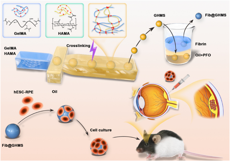
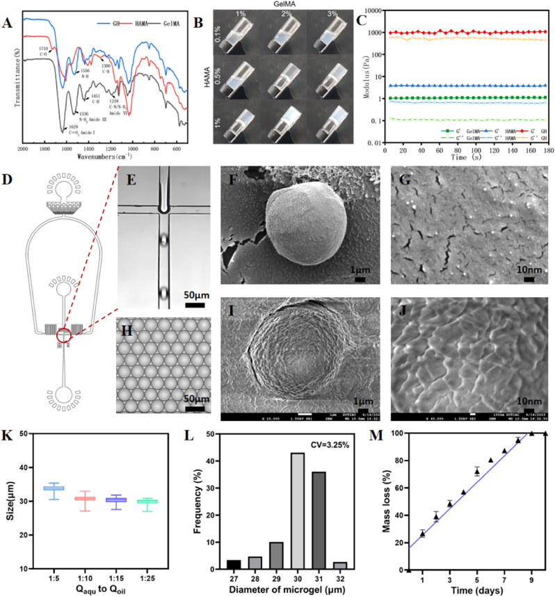
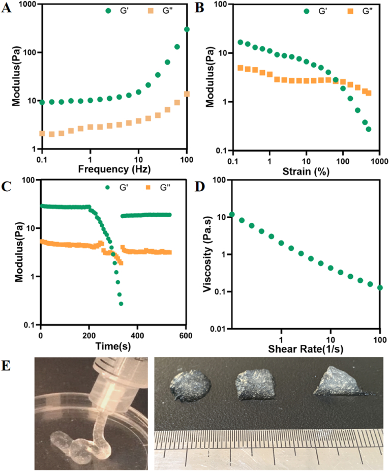
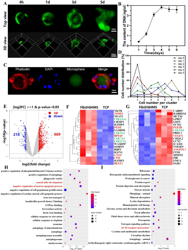
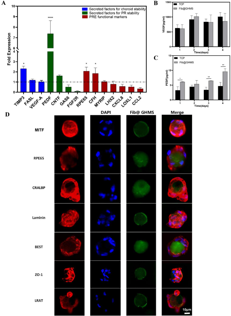
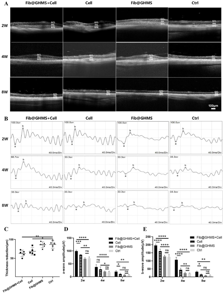
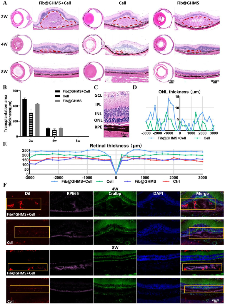
Retinal pigment epithelial (RPE) cells are specialized neural cells crucial for vision, while human embryonic stem cell-derived retinal pigment epithelial (hESC-RPE) cells hold great potential as a viable cell source for treating retinal degenerative diseases like retinitis pigmentosa (RP). However, the transplantation efficiency and viability of hESC-RPE cell suspensions are relatively low due to detrimental shear-force during operations and host immune-clearance. We herein develop an alternative transplantation strategy with the aid of a microfluidic-templating microgel cell carrier to achieve substantially enhanced loading and delivery efficiency of hESC-RPE cells, thereby promoting visual function recovery after subretinal injection in the RP model Royal College of Surgeons (RCS) rats. Specifically, injectable monodispersed microgels consisting of gelatin-methacryloyl/Hyaluronic acid-methacryloyl core coated with fibrin shell (denoted as Fib@GHMS) were fabricated in a high-throughput and controllable manner, facilitating the adhesion and proliferation of hESC-RPE cells. RCS rats treated with microcarriers showed significantly improved visual function, evidenced by increased B-wave amplitudes and the preservation of the inner nuclear layer at 8 weeks post-surgery. In conclusion, our innovative delivery system Fib@GHMS for hESC-RPE cell transplantation presents a potential therapeutic graft for retinal tissue engineering. It may open a new avenue for clinical transplantation of minimally invasive cell-based treatments in retinal degenerative diseases.

Keywords: Cell transplantation; Cell-laden microgel; Injectable granular gel; Microfluidics; hESC-RPE.


Conflict of interest statement

The authors declare that they have no known competing financial interests or personal relationships that could have appeared to influence the work reported in this paper.

Figures

Image 1
Graphical abstract
Fig. 1
Fig. 1
Scheme showing the transplantation strategy using minimally invasive injection of hESC-RPE cell-loaded Fib@GHMS for the treatment of retinal degeneration diseases. Monodisperse GelMA/HAMA microgels (GHMS) were manufactured via microfluidic droplet-templating method followed by photopolymerization to solidify hydrogel precursor. Thereafter, the microgels were further coated with a thin layer of fibrin via immerging the microgels in aqueous solution containing fibronectin, which were subsequently washed with thrombin to generate fibrin-coated GHMS (Fib@GHMS). hESC-RPE cells were cultured on the microgel surface in vitro to proliferate to a sufficient cell density, they were then injected into the subretinal space of RCS rats for transplantation.
Fig. 2
Fig. 2
Microfluidic-templating fabrication of Fib@GHMS microgels. A: FTIR spectra of HAMA, GelMA and composite GH hydrogels, showing the reduced peak at 1556 cm−1 corresponding to the GH double network hydrogel. B: Screening the minimum concentrations required of the GelMA and HAMA precursors to form a stable gel. C: Time sweeps of 2 % GelMA, 0.5 % HAMA, and G2H0.5 hydrogels to evaluate the mechanical performance. D: AutoCAD design of the microfluidic device. E: Microscopic images of the microchannel showing the preparation of uniformly sized aqueous droplets containing GelMA and HAMA precursors. H: Light microscopic images showing the generated monodisperse droplets. SEM images showing the morphology of GHMS (F, G) and Fib@GHMS microgels (I, J). K: The droplet size control via varying the flow ratio of Qaqu/Qoil. L: Statistical analysis of the microgel diameter range to analyze particle size uniformity. M: In vitro degradation of microgels.
Fig. 3
Fig. 3
Formulating shear-thinning, self-healing granular gels based on Fib@GHMS. A: Oscillatory frequency sweep (1 % strain) tests of granular gels of Fib@GHMS. B: Strain sweep (1 Hz) tests of Fib@GHMS. C: Evolution of Fib@GHMS storage and loss modulus during one destructive shearing cycle (strain from 0.5 % to 500 %, 1 Hz), showing granular gel self-healing. D: Granular gel viscosity as a function of shear rate revealed shear-thinning behaviour. E: Images of Fib@GHMS injectability and moldability.
Fig. 4
Fig. 4
The potential of hESC-RPE cell-Loaded Fib@GHMS microgels to restore retinal function. A: Cellular spreading and viability at different timepoints (4 h, 1 d, 3 d, 5 d) on the microcarriers. B: dsDNA cell proliferation assay of hESC-RPE cells cultured on the microcarriers. C: Cell cytoskeleton staining of hESC-RPE cells cultured on the microcarriers on day 5. D: Quantitative analysis of cell numbers on the microcarriers at different timepoints. E: Volcano plot depicting DEGs between the Fib@GHMS and TCP groups. F: Heatmap of upregulated genes and G: downregulated genes in the Fib@GHMS group compared with the TCP group in the retinal function recovery microenvironment (p < 0.05). KEGG pathway enrichment analysis of DEGs. H: Twenty significantly enriched GO terms for the differential genes. I: Twenty significantly enriched KEGG pathways for the differential genes.
Fig. 5
Fig. 5
Specific functional expression of hESC-RPE cells cultured on Fib@GHMS microgels. A: qRT–PCR comparing the expression profiles of key genes between hESC-RPE cells on Fib@GHMS and in TCP cultures (n = 3). The results are presented as 'fold expression' values to highlight the enhanced expression levels of certain genes in Fib@GHMS cultures compared with adherent cultures (dashed line). Gene expression was normalized to the endogenous reference gene (GAPDH). Wilcoxon paired signed-rank tests were used to compare ΔCt values for each gene in Fib@GHMS and adherent cultures. B, C: The levels of secreted PEDF and VEGF in conditioned culture media. The error bars represent standard deviations. D: Immunostaining of hESC-RPE cells on the surface of Fib@GHMS using antibodies against MITF, RPE65, CRALBP, Laminin, Best1, ZO-1, and LRAT. ∗p < 0.05, ∗∗p < 0.01, ∗∗∗∗p < 0.0001.
Fig. 6
Fig. 6
Subretinal injection of hESC-RPE cell-loaded Fib@GHMS restores visual function. A: OCT evaluation of the biosafety and degradation status of hESC-RPE cell-loaded Fib@GHMS microcarrier in the subretinal space of RCS rats at 2, 4, and 8 weeks. Scale bar, 100 μm. GCL: ganglion cell layer; INL: inner nuclear layer; RPE: retinal pigment epithelium layer, including the transplanted RPE region. B: ERG assessment of the visual function recovery status of hESC-RPE cell-loaded Fib@GHMS in the subretinal space of RCS rats at 2, 4, and 8 weeks. C: Comparison of the relative decrease in retinal thickness (μm) in different groups at 8 weeks. Comparison of retinal a-wave(D) and b-wave(E) amplitudes (μV) for different groups at 2, 4, and 8 weeks. ∗p < 0.05, ∗∗p < 0.01, ∗∗∗∗p < 0.0001.
Fig. 7
Fig. 7
Retinal morphological analysis and tissue immunofluorescence staining after hESC-RPE cell-loaded Fib@GHMS transplantation in RCS rats. A: Representative hematoxylin and eosin-stained images of the whole eye and the transplanted area of rats from each group at 2, 4, and 8 weeks. Observation of degradation in the transplanted area and assessment of graft biocompatibility. The red dashed area indicates the subretinal transplantation bulge region. B: Comparison of transplantation area thickness (μm) for different groups at 2, 4, and 8 weeks. C: Close up image of HE stained histological sections where the injection was administered, revealing the healthy retina. D: The horizontal thickness distributions of the ONL near the optic disc in RCS rats of each group at 8 weeks, summarized from the results of serial sections of eyes. E: The retina thickness at 8 weeks. F: Representative immunofluorescence images at 4 and 8 weeks. Evaluation of the short-term survival of transplanted cells and visual function recovery in RCS rats after the subretinal transplantation of Fib@GHMS and cells. Selected areas (orange box) were marked to further observe the grafted hESC-RPE. Red: Transplanted cells labeled with Dil; Pink: RPE65 staining to identify RPE cells; Green: Cralbp staining to identify RPE cells overlying photoreceptor cells; DAPI: Nuclear staining. (For interpretation of the references to colour in this figure legend, the reader is referred to the Web version of this article.)

Similar articles

References

    1. Yang S., Zhou J., Li D. Functions and diseases of the retinal pigment epithelium. Front. Pharmacol. 2021;12 - PMC - PubMed
    1. Lakkaraju A., Umapathy A., Tan L.X., et al. The cell biology of the retinal pigment epithelium. Prog. Retin. Eye Res. 2020 - PMC - PubMed
    1. George S.M., Lu F., Rao M., et al. The retinal pigment epithelium: development, injury responses, and regenerative potential in mammalian and non-mammalian systems. Prog. Retin. Eye Res. 2021;85 - PMC - PubMed
    1. Sparrow J.R., Hicks D., Hamel C.P. The retinal pigment epithelium in health and disease. Curr. Mol. Med. 2010;10(9):802–823. - PMC - PubMed
    1. Intartaglia D., Giamundo G., Conte I. Autophagy in the retinal pigment epithelium: a new vision and future challenges. FEBS J. 2021;289(22):7199–7212. - PMC - PubMed

LinkOut - more resources