. 2025 Feb 20;33(2):200954.
doi: 10.1016/j.omton.2025.200954. eCollection 2025 Jun 18.

Preclinical development and clinical safety assessment of a synthetic peptide conjugate enabling endogenous antibody binding to promote innate receptor engagement

Affiliations

Preclinical development and clinical safety assessment of a synthetic peptide conjugate enabling endogenous antibody binding to promote innate receptor engagement

Erika A K Fletcher et al. Mol Ther Oncol. .

Abstract

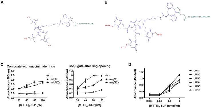
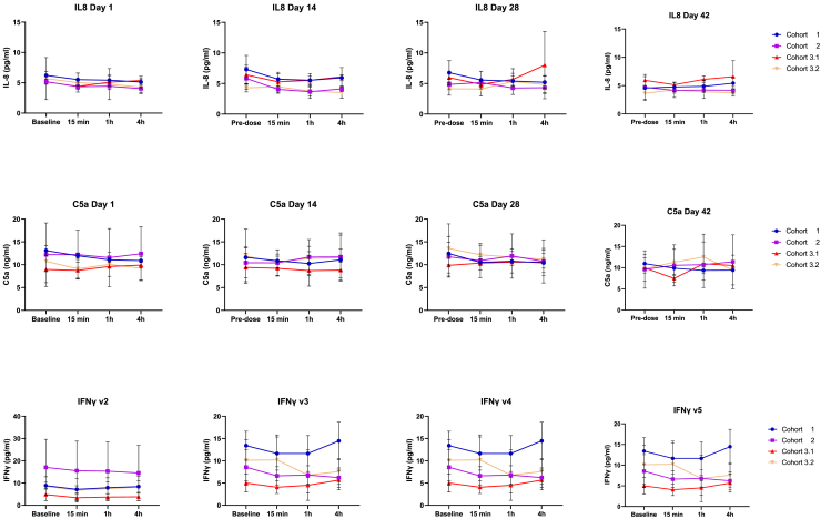
Peptide-based vaccines can be used to deliver tumor-specific antigens to dendritic cells (DCs), leading to tumor-directed T cell responses. We previously developed a peptide-peptide conjugate technology enabling in vivo cross-linking of pre-existing tetanus toxin-directed antibodies, facilitating antigen delivery to, and activation of DCs. To achieve this, multiple identical tetanus toxin-derived B cell epitopes (MTTEs) are conjugated to synthetically produce target antigens of choice. Herein, we describe the generation of a prostate cancer vaccine candidate (TENDU) based on this technology. It includes long synthetic peptides harboring epitopes (CD4 and CD8) from prostate-specific antigen (PAP) and prostate-specific membrane antigen (PSMA). The preclinical efficacy of TENDU was assessed in experimental systems, and safety was evaluated in a rabbit toxicity study and a human whole blood loop assay. We also report the first clinical safety assessment of TENDU. Experimental studies showed that prostate cancer patients mounted anti-MTTE antibodies in response to tetanus vaccination with recall T cell responses detected in two patients. Transgenic humanized HLA-DR4 mice displayed T cell responses and increased anti-MTTE IgG levels after vaccination with a peptide construct including an HLA-DR4 epitope. The vaccine candidate was found safe, and a positive correlation between T cell responses and anti-MTTE antibodies was noted in the first-in-human study.

Keywords: MT: Regular Issue; cancer vaccine; dendritic cells; immunotherapy; peptide-conjugate vaccine; prostate cancer; tetanus toxoid.


Conflict of interest statement

The individual affiliations stated describe the potential conflicts of interest to the industrial parties involved. I.D. holds a current position with AstraZeneca but the work was performed under a separate affiliation and AstraZeneca has no connection to the work presented herein. W.L. has a consultant agreement with Ultimovacs ASA. We have registered patents and patent applications related to this work.

Figures

None
Graphical abstract
Figure 1
Figure 1
Maintained function of MTTE-peptide conjugates after succinimide ring opening MTTE-peptide conjugates ([MTTE]3-SLP) were synthesized and are illustrated either with (A) succinimide rings or (B) after ring opening, both variants with an OVA-derived CD8+ epitope (LEQLESIINFEKLAAAAAK) in the SLP peptide. Red: MTTE, blue: core structure, green: SLP. (C) The biological effect of [MTTE]3- SIINFEKL conjugates, comparing intact and opened succinimide rings, was measured as β-galactosidase activity of SIINFEKL-specific T cells (B3Z cells) co-cultured with dendritic cells and activity was assessed by the level of substrate cleavage at 595nm. mIgG1 = mouse IgG1; mIgG2a = mouse IgG2a; MTTE = minimal tetanus toxin epitope; SLP = synthetic long peptide. Results are shown as mean ± SD. (D) Maintained binding of GMP LUG-1-6 constructs to recombinant anti-MTTE IgG1 antibody as assessed by ELISA.
Figure 2
Figure 2
Anti-MTTE antibody titers in plasma from prostate cancer patients and healthy donors pre- and post-DTP vaccination Blood samples were collected from prostate cancer patients from the urology department and the oncology department and from healthy donors before and approximately 2 weeks after a DTP vaccination. Anti-MTTE antibody levels in plasma were determined using an in-house ELISA. The antibody titers are expressed by the dilution whereby the absorbance of MTTE is divided to the respective ETTM background signal. Levels of IgG (A), IgG1 (B), IgG4 (C), and IgM (D) in prostate cancer patients (n = 17) before and after DTP vaccination presented as absorbance (450–570 nm) at dilution 1:200 for IgG and 1:10 of plasma for IgG1, IgG4, and IgM. Anti-MTTE antibody titers post vaccination were analyzed in age-matched male healthy volunteers (n = 5) and prostate cancer patients (n = 5): (E) IgG titers and (F) IgG1 titers shown as median with 95% confidence interval. Antibody titers in (E) and (F) were determined based on how much the samples could be diluted before the antibodies could no longer be detected. A cutoff value for detection was set to optical absorbance = 0.100. Significance was tested with Wilcoxon matched-pairs test (A)–(D) or unpaired t test with Welch’s correction, (E) and (F); ∗∗p < 0.01; ∗∗∗p < 0.001.
Figure 3
Figure 3
IFNγ and TNFα-release by memory T cells in response to LUR1-6 conjugates Peptides and LUR1-6 conjugates were incubated in human whole blood from prostate cancer patients and healthy donors, pre and post a DTP vaccination, (A) and (B), or with and without a mouse anti-MTTE IgG2a antibody, (C) and (D), in a circulating blood loop assay. T cell surface markers and intracellular IFN-γ and TNF-α were analyzed with flow cytometry. The conjugates tested were LUR1-6, LUG1-6 (see Table 2), and SLP1-6, which are the free synthetic long peptides of respective conjugates. The [MTTE]3-CMV conjugate contains the HLA-A∗0201-restricted epitope pp65(NLV) from CMV and CMV lysates were used as positive controls. MTTE3-irrelevant (MTTE-irrel.) contains a scrambled SLP sequence (DGLQGLLLGLRQRIETLEGK) without any know human T cell epitopes. Representative flow cytometry dot plots of the three responding donors from (A) and (C) are displayed in (B) and (D). The cells were gated as CD45RO + CD3+CD4-CD8+ and the % of IFN-γ+ and TNF-α+ cells are displayed. (E) SLP-specific IFN-γ producing T cells upon LUG2 vaccination in seropositive HLA-DR4 humanized transgenic mice were measured in a recall-ELISpot assay. Result shown as mean ± SD. Significance was tested with Mann Whitney-test ∗p < 0.05.
Figure 4
Figure 4
No systemic risk of cytokine release or complement activation by TENDU vaccination Plasma levels of IL-8, C5a, and IFN-γ before and after four doses of TENDU vaccination administered with a 2-week interval between each dose. At each visit, blood was collected at four time points (baseline [time 0], 15 min, 1 h, and 4 h). Cytokines were assessed using a Mesoscale V-plex kit, result expressed as pg/mL (mean values +SD). Lower and upper limit of quantifications (LLOQ and ULOQ) are verified by MSD and calculated from the standard curve and percentage recovery of diluent standards with precision of 20% and accuracy of 80%–120%. Complement activation (C5a) analyzed using ELISA kits from Hycult Biotech, results expressed as ng/mL (mean values +SD). Blue symbols (circle) represent cohort 1 (TENDU dose 40 μg); purple symbols (square) represent cohort 2 (TENDU dose 400 μg); red symbols (triangle) represent cohort 3.1 (TENDU dose 960 μg); yellow symbols (upside down triangle) represent cohort 3.2 (TENDU dose 960 μg; co-administered with tetanus toxoid in the same anatomical location).
Figure 5
Figure 5
Anti-tetanus- and MTTE antibody titers, T cell responses, and preliminary anti-tumor activity after TENDU vaccination (A) Schematic diagram of the trial design. (B) Anti-tetanus (TTd) and (C) anti-MTTE antibody levels in plasma (mean ± SD) before and after tetanus toxoid-containing vaccination and after four doses of TENDU vaccination administered with a 2-week interval between each dose (n = 3 in each cohort). The antibody titers are expressed by the dilution whereby the absorbance of MTTE is divided to the respective ETTM background signal. (D) IFN-γ production in response to stimulation with mixes of peptides corresponding to CD4 and CD8 epitopes derived from PSA and PSMA. Each pair of lines represents one patient; solid lines show CD4-responses, dotted lines show CD8-responses. (E) T cell responses over time for the best Ab-responder patient in cohort 3.1. (F) Number of circulating tumor cells expressing PDL1, PSMA, and PSAP, respectively, at baseline and end of trial (mean ± SD, (n = 3 in each cohort). V1 = visit 1; EOT = end of trial; FU = follow-up. Blue symbols represent cohort 1 (TENDU dose 40 μg); purple symbols represent cohort 2 (TENDU dose 400 μg); red symbols represent cohort 3.1 (TENDU dose 960 μg); yellow symbols represent cohort 3.2 (TENDU dose 960 μg; co-administered with tetanus toxoid in the same anatomical location).

References

    1. Bray F., Laversanne M., Sung H., Ferlay J., Siegel R.L., Soerjomataram I., Jemal A. Global cancer statistics 2022: GLOBOCAN estimates of incidence and mortality worldwide for 36 cancers in 185 countries. CA. Cancer. J. Clin. 2024;74:229–263. doi: 10.3322/caac.21834. - DOI - PubMed
    1. Sandhu S., Moore C.M., Chiong E., Beltran H., Bristow R.G., Williams S.G. Prostate cancer. Lancet. 2021;398:1075–1090. doi: 10.1016/S0140-6736(21)00950-8. - DOI - PubMed
    1. Wadosky K.M., Koochekpour S. Molecular mechanisms underlying resistance to androgen deprivation therapy in prostate cancer. Oncotarget. 2016;7:64447–64470. doi: 10.18632/oncotarget.10901. - DOI - PMC - PubMed
    1. Davies A., Conteduca V., Zoubeidi A., Beltran H. Biological Evolution of Castration-resistant Prostate Cancer. Eur. Urol. Focus. 2019;5:147–154. doi: 10.1016/j.euf.2019.01.016. - DOI - PubMed
    1. Essand M. Gene therapy and immunotherapy of prostate cancer: adenoviral-based strategies. Acta. Oncol. 2005;44:610–627. doi: 10.1080/02841860510007477. - DOI - PubMed

LinkOut - more resources