. 2025 Jun 12;9(2):026125.
doi: 10.1063/5.0250953. eCollection 2025 Jun.

Modeling nitric oxide diffusion and plasticity modulation in cerebellar learning

Affiliations

Modeling nitric oxide diffusion and plasticity modulation in cerebellar learning

Alessandra Maria Trapani et al. APL Bioeng. .

Abstract

Nitric oxide (NO) is a versatile signaling molecule with significant roles in various physiological processes, including synaptic plasticity and memory formation. In the cerebellum, NO is produced by neural NO synthase and diffuses to influence synaptic changes, particularly at parallel fiber-Purkinje cell synapses. This study aims to investigate NO's role in cerebellar learning mechanisms using a biologically realistic simulation-based approach. We developed the NO Diffusion Simulator (NODS), a Python module designed to model NO production and diffusion within a cerebellar spiking neural network framework. Our simulations focus on the eye-blink classical conditioning protocol to assess the impact of NO modulation on long-term potentiation and depression at parallel fiber-Purkinje cell synapses. The results demonstrate that NO diffusion significantly affects synaptic plasticity, dynamically adjusting learning rates based on synaptic activity patterns. This metaplasticity mechanism enhances the cerebellum's capacity to prioritize relevant inputs and mitigate learning interference, selectively modulating synaptic efficacy. Our findings align with theoretical models, suggesting that NO serves as a contextual indicator, optimizing learning rates for effective motor control and adaptation to new tasks. The NODS implementation provides an efficient tool for large-scale simulations, facilitating future studies on NO dynamics in various brain regions and neurovascular coupling scenarios. By bridging the gap between molecular processes and network-level learning, this work underscores the critical role of NO in cerebellar function and offers a robust framework for exploring NO-dependent plasticity in computational neuroscience.


Conflict of interest statement

The authors have no conflicts to disclose.

Figures
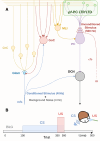
FIG. 1.
FIG. 1.
Cerebellar microcircuit with plasticity domains and EBCC protocol: (a) the network is composed of mossy fibers (mfs), granule cells (GrCs), Golgi cells (GoCs), stellate and basket cells (SCs and BCs as MLIs), Purkinje cells (PCs), inferior olivary nuclei (IO), climbing fibers (cfs), and the deep cerebellar nucleus (DCN). The cerebellar circuit expresses different forms of plasticity, we focus on pfs-PC LTP and LTD (green area and dots in the PC dendritic tree). (b) EBCC protocol where CS and US are identified, respectively, as tone and air puff stimulus. During the time course of one trial (500 ms), mfs receive a background noise (e.g., at 4 Hz), whereas CS is modeled as a burst of 40 Hz spanning 280 ms, while US is conveyed as a rapid (30 ms) burst of 500 Hz to IO nuclei.
FIG. 2.
FIG. 2.
Comparison of NODS and NEURON Reaction&Diffusion module: (a) [NO] reaction cascade characterized by instantaneous Ca2+ events, following spikes, causing a smooth buildup of NO. Time (b) and space (c) profile of the NO signal, produced by a single source stimulated with different frequencies (color-coded by darkening the shade of green): single spike, 10, 20, 50, 100, 300, and 500 Hz. All stimuli have been delivered at ti=0 ms and last for 200 ms. (d) Reaction cascade leading to NO production. (1) Spike train arriving at the pf presynaptic site leads to depolarization, which causes Ca2+ entrance (2) through NMDA receptors. Ca2+ reacts with calmodulin to form the Calm2C complex (3) that activates nNOS and thus NO production (4). (e) Scheme of NO diffusive cloud following production, where its radius ( 15μm) is highlighted.
FIG. 3.
FIG. 3.
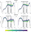
Comparison of PC-CS SDF and DCN-CS SDF over trials with standard STDP and NO-dependent STDP: both panels report the SDF in PC population (averaging across cell SDFs) for each trial. The progress of the trials is color-mapped by the horizontal color bar below. The vertical lines represent the CS-onset (light blue), US-onset (light red), and CS-US co-termination (lilac). In gray are shown the two windows, baseline and CR window, for evaluation of functional plasticity. Regarding the DCN-CS SDF, the threshold for calculating the CR% is represented as a black dashed line. On panels (a) and (c), the results of EBCC are simulated using the standard STDP rule. On panels (b) and (d), the results of EBCC are simulated using the NO-dependent STDP rule. PC-CS activity shapes the DCN-CS one by inhibiting it. Thus, the depression along trials for PC-CS is reflected in a higher spiking activity for DCN-CS. Regarding network learning, the main difference between the results for the standard and the NO-dependent STDP is given by the flattening of the curve in the baseline window for both PC-CS and DCN-CS with NO.
FIG. 4.
FIG. 4.
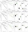
SDF-change for different levels of background noise: standard STDP is color-coded in black, and NO-dependent STDP is color-coded in green. (a), (c), and (e) Dashed lines connect the SDF-change computed in the baseline window in each trial, while continuous lines connect the SDF-change computed in the CR response window. The overimposed gray lines represent the linear fitting of the SDF-change curves, which help visualize the average change during the trials. (b), (d), and (f) SDF-change in PC from the last five trials, for EBCC protocols with a background noise of 0, 4, and 8 Hz. Standard STDP is color-coded in black, and NO-dependent STDP is color-coded in green. The red * corresponds to p < 0.01 in the Wilcoxon test.
FIG. 5.
FIG. 5.
CR% for different levels of background noise: standard STDP is color-coded in black, and NO-dependent STDP is color-coded in green. The curves are obtained by calculating for each trial the amount of relevant learning (as 0 or 1) and then applying a moving average (left zero-padding). Then, from the distribution of the values of each trial for the five simulations, we extracted the median value, plotted together with an error bar of the first and third quartiles.
FIG. 6.
FIG. 6.
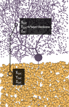
nNOS placement in PC dendritic tree: nNOS placement is based on the relative connected GrC (yellow) and PC (purple) location in the cerebellar microcircuit volume. Thus, the nNOS coordinates depend on Grc and PC ones as xnNOS=xGrC,ynNOS=yGrC+k*layerthickness,znNOS=zPC.

Similar articles

References

    1. Lundberg J. O. and Weitzberg E., “Nitric oxide signaling in health and disease,” Cell 185, 2853–2878 (2022). 10.1016/j.cell.2022.06.010 - DOI - PubMed
    1. Picón-Pagès P., Garcia-Buendia J., and Muñoz F. J., “Functions and dysfunctions of nitric oxide in brain,” Biochim. Biophys. Acta 1865, 1949–1967 (2019). 10.1016/j.bbadis.2018.11.007 - DOI - PubMed
    1. Koshland D. E., “The molecule of the year,” Science 258, 1861–1861 (1992). 10.1126/science.1470903 - DOI - PubMed
    1. Garthwaite J., “From synaptically localized to volume transmission by nitric oxide,” J. Physiol. 594, 9–18 (2016). 10.1113/JP270297 - DOI - PMC - PubMed
    1. Hardingham N., Dachtler J., and Fox K., “The role of nitric oxide in pre-synaptic plasticity and homeostasis,” Front. Cell. Neurosci. 7, 1–19 (2013). 10.3389/fncel.2013.00190 - DOI - PMC - PubMed

LinkOut - more resources