. 2025;3(1):15.
doi: 10.1038/s44182-025-00028-1. Epub 2025 Jun 13.

A soft robotic "Add-on" for colonoscopy: increasing safety and comfort through force monitoring

Affiliations

A soft robotic "Add-on" for colonoscopy: increasing safety and comfort through force monitoring

Viola Del Bono et al. Npj Robot. 2025.

Abstract

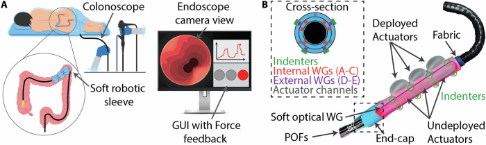
Colonoscopy is vital for diagnosing colorectal cancer, but limitations in instrument dexterity and sensor feedback can affect safety and patient comfort. We propose a disposable soft robotic "add-on" that attaches to existing endoscopic tools, enhancing safety without requiring custom instruments or workflow changes. The robot features soft optical sensors for 3D shape detection and force monitoring. If excessive force is detected, soft actuators redistribute pressure. A graphical interface provides real-time force data alongside the endoscope camera view. Validation experiments show accurate 3D shape reconstruction (8.51% curvature error, 9.67% orientation error) and force estimation up to 6 N with 3.38% accuracy. In-vitro tests confirm effective force redistribution, while ex-vivo tests on a bovine colon demonstrate smooth integration with minimal impact on the user learning curve. In-vivo swine studies validate safety and feasibility, confirming compatibility with existing tools and minimal disruption to clinical workflows, ensuring an efficient colonoscopy experience.

Keywords: Biomedical engineering; Materials for devices; Mechanical engineering; Soft materials.


Conflict of interest statement

Competing interestsThe authors declare no competing interests.

Figures

Fig. 1
Fig. 1. Enhanced colonoscopy with the soft robotic sleeve.
A The robot, mounted on a commercial endoscope, inflates when a high contact force is detected, reducing patient discomfort and risk of tissue perforation. The force feedback is displayed on a GUI in real time. B Soft robotic sleeve overview showing both inflated and deflated actuator lines. An external U-shaped optical waveguide is highlighted in purple along with its set of indenters, circled in green. The stiffer end-cap encapsulates the connection between the soft waveguides and the plastic optical fibers (POFs). The internal fabric layer ensures a low friction when inserted onto the endoscope. The cross-sectional view depicts the layout of the five waveguides (three internal and two external), indenters, and actuation channels.
Fig. 2
Fig. 2. 3D shape validation.
A Reconstructed shape by using constant curvature modeling. B The sleeve tip position is recorded with an EM tracker and subsequently transformed into model parameters k and ϕ. C, D Comparison between actual and estimated curvature and orientation over time, showing an accurate shape reconstruction throughout the workspace.
Fig. 3
Fig. 3. Force estimation validation.
A Test setup. The soft robot is placed on the curvature testing platform. An ATI-nano 17 force sensor is mounted on the end-effector of a UR-5 robot arm, along with a 3D printed pressing fixture. For each of the calibrated functional directions (FDs), the sleeve is bent up to 10 m−1 curvature, and the robot arm gently presses on the soft robot. B Estimated versus actual force applied to the soft sleeve in the four FDs. In all cases, the error between the true and estimated force is less than 1 N.
Fig. 4
Fig. 4. Shape estimation under external forces.
A Sleeve bent at 7 m−1 and oriented at 60° (FD 1). B Sleeve bent at 5 m−1 and oriented at 120° (FD 2). C Sleeve bent at 7 m−1 and oriented at 240° (FD 3). D Sleeve bent at 10 m−1 and oriented at 300° (FD 4).
Fig. 5
Fig. 5. In-vitro validations.
A Setup with a TPE colon inserted in a Kyoto Kagaku simulator. B Low force sensed and green indicator displayed (F < 3 N). C An excessive force is reached: the indicator turns red and the actuators starts inflating. D A warning zone with medium-high force (3 N ≤F < 5 N) is reached and the indicator turns yellow. E Three FSR sensors are bonded to the internal lumen for the force redistribution experiment. F Validation of redistribution: starting by pressing on one specific sensor (FSR 1), inflation is triggered, and the other two sensors start experiencing force (FSR 2 and 3).
Fig. 6
Fig. 6. Ex-vivo validation.
A Test setup including the ColoEASIE-2 Simulator with the bovine tissue placed inside it, the endoscope with the robot mounted on, the internal and external camera views, and the GUI view. Examples of tests performed by B a novice user (novice 1 trial 2), and C an expert user (expert 1 trial 4), showing the force estimated from the soft sleeve over time.
Fig. 7
Fig. 7. Ex-vivo metric.
AC Learning curves for novices, experts, and all users combined (mean ± standard deviation for each trial). DF Average navigation times for novices, experts, and all users combined (mean ± standard deviation for all trials). GI Average NASA TLX score for novices, experts, and all users combined (mean ± standard deviation for all trials). JL TLX sub-scores for for novices, experts, and all users combined.
Fig. 8
Fig. 8. In-vivo testing.
A Endoscopy unit setup. The animal is under anesthesia and its vital signs are continuously monitored. The sleeve control box is placed on a movable cart next to the operating table, as well as the colonoscopy tower. A laptop is connected to the control box to record the robot data during the navigation. B Soft robotic sleeve on the colonoscope, in its deflated and inflated state. C Average learning curves for the users with and without the sleeve attached to the colonoscope (mean ± standard deviation for each trial). D Average navigation times with and without soft sleeve (mean ± standard deviation for all trials) only differ by 3.6 s (Wilcoxon Sign-Ranked, p < 0.05). E Average TLX subscores for the users combined. F, G, H TLX total scores for each user separately.
Fig. 9
Fig. 9. Soft sleeve fabrication.
A Three aluminum molds are machined for the bending layer (1), contact layer(2), and mask for indenters (3). Silicone is poured into (1) and (2). (3) is placed on top of (2), and all the molds are degassed and cured. B The base layer is spin-coated. The section view of the three layers is shown. C The layers are stacked together by spin-coating steps. D The sleeve body is bonded to a fabric and NOA 65 is injected into the 5 channels to create the WG cores, and UV cured. E A portion of the cores is exposed. F The sleeve is wrapped into a cylinder and silicone is injected into the seam to reinforce it. G Anti-buckling layer injection. H The soft cores and the POFs are encapsulated in a PTFE tube. I The end-cap is molded to secure the soft-rigid connection. J Indenters placement.

References

    1. Siegel, R. L., Wagle, N. S., Cercek, A., Smith, R. A. & Jemal, A. Colorectal cancer statistics, 2023. CA: Cancer J. Clin.73, 233–254 (2023). - PubMed
    1. Siegel, R. L., Giaquinto, A. N. & Jemal, A. Cancer statistics. CA: Cancer J. Clin74, 12–49 (2024). - PubMed
    1. Santucci, C. et al. European cancer mortality predictions for the year 2024 with focus on colorectal cancer. Ann. Oncol.35, 308–316 (2024). - PubMed
    1. Siegel, R. L., Miller, K. D., Fuchs, H. E. & Jemal, A. Cancer statistics, 2023. CA: Cancer J. Clin.73, 17–48 (2023). - PubMed
    1. Maes-Carballo, M. et al. A comprehensive systematic review of colorectal cancer screening clinical practices guidelines and consensus statements. Br. J. Cancer128, 946–957 (2023). - PMC - PubMed

LinkOut - more resources