. 2025 May 25;10(22):22735-22746.
doi: 10.1021/acsomega.4c11296. eCollection 2025 Jun 10.

Fabrication of Aligned Polyhydroxybutyrate Fibrous Scaffolds via a Touchspinning Apparatus

Affiliations

Fabrication of Aligned Polyhydroxybutyrate Fibrous Scaffolds via a Touchspinning Apparatus

Md Mazbah Uddin et al. ACS Omega. .

Abstract

Poly-(3-hydroxybutyrate) (PHB) fibers ranging from nano- to microscale were successfully fabricated using a touchspinning apparatus. The optimization of key spinning parametersincluding solution concentration (5-11% w/v), rotational speed (1300-2100 rpm), and feed rate (5-20 μL/min)enabled the production of aligned fibrous scaffolds. Morphological analysis via field emission scanning electron microscopy (FE-SEM) revealed fiber diameters in the range of 0.831-1.273 μm, which were influenced by spinning conditions. Thermal stability was confirmed using thermogravimetric analysis (TGA), with an onset degradation temperature of ∼290 °C. Differential scanning calorimetry (DSC) showed a melting peak of ∼172 °C and a crystallinity increase from 37.9% in the pellet to 42.5% in fibers of PHB. The scaffolds were functionalized with collagen to enhance bioactivity, and fibroblast (NIH3T3) viability was assessed through alamarBlue and Live/Dead assays. Metabolic activity increased significantly over 5 days (p < 0.05), particularly in collagen-modified scaffolds, confirming excellent cell adhesion and proliferation. Immunofluorescent microscopy demonstrated cell elongation along the fiber axis, indicating scaffold-guided cellular orientation. The results establish the feasibility of touchspun PHB scaffolds for tissue engineering applications, offering a scalable alternative to the conventional electrospinning process.


Figures

1
1
Touchspinning apparatus for the PHB scaffold fabrication.
2
2
Fabrication of TS PHB fibrous scaffolds. (a) Collection of TS fibers, (b, c) transparent ABS printed bottom and top scaffold frames, (d) TS unidirectional fiber mat sandwiched between the ABS frames using ABS glue (2 g/10 mL ABS in acetone), and (e) final scaffold for cell culture studies.
3
3
SEM images of the TS PHB fibers. TS1–TS4 fibers were spun using 5, 7, 9, and 11% w/v PHB solutions at 1700 rpm and a feed rate of 5 μL/min. TS5–TS7 fibers were spun with a 7% w/v PHB solution at 1700 rpm, with feed rates of 10, 15, and 20 μL/min, respectively. TS8 and TS9 fibers were spun with a 7% w/v PHB solution at 10 μL/min with 1300 and 2100 rpm, respectively (Scale bar: 10 μm).
4
4
Summary of SEM analysis of the TS PHB fibers. Effect of (a) solution percentage on fiber diameter at 1700 rpm and 5 μL/min, (b) feed rate at 7% w/v and 1700 rpm, and (c) speed at 7% w/v and 10 μL/min.
5
5
Thermal and crystallinity measurements of TS PHB fibers. (a) % Weight loss and (b) derivative curves from TGA. (c) DSC and (d) XRD profiles of the TS PHB fibers.
6
6
FTIR spectra of PHB pellet, TS5, and collagen-treated TS5 fibers of PHB. PHB fibers. (a) PHB pellet and (a, b­(iv)) TS5 fibers. (b­(i)) Neat collagen, (b­(ii)) TS5 treated with 2.0 mg/mL, and (b­(iii)) 0.05 mg/mL collagen solutions.
7
7
Viability of fibroblast cells on the control and PHB scaffolds. In merged images, live cells (green, calcein AM) and dead cells (red, ethidium homodimer-I) are shown over days 1, 3, and 5 using the inverted fluorescence microscope (EVOS M5000) at 20× magnification (Scale bar: 50 μm).
8
8
Metabolic activity of the fibroblast cells on control and PHB scaffolds at days 1, 3, and 5 using a Varioskan LUX multimode microplate reader (Thermo Fischer Scientific) at an excitation wavelength of 540 nm and an emission wavelength of 590 nm. The error bars represent the standard deviation.
9
9
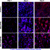
Morphology of fibroblast cells on the control and PHB scaffolds. F-actin (purple, Rhodamine phalloidin) and nuclei (blue, Hoechst) visualize cell morphology on control, neat, and collagen-modified scaffolds at days 1, 3, and 5 using the inverted fluorescence microscope (EVOS M5000) at 20× magnification (Scale bar: 50 μm).

References

    1. Han S., Nie K., Li J., Sun Q., Wang X., Li X., Li Q.. 3D Electrospun Nanofiber-Based Scaffolds: From Preparations and Properties to Tissue Regeneration Applications. Stem Cells Int. 2021;2021(1):8790143. doi: 10.1155/2021/8790143. - DOI - PMC - PubMed
    1. Ghimire U., Kandel R., Ko S. W., Adhikari J. R., Kim C. S., Park C. H.. Electrochemical technique to develop surface-controlled polyaniline nano-tulips (PANINTs) on PCL-reinforced chitosan functionalized (CS-f-Fe2O3) scaffolds for stimulating osteoporotic bone regeneration. Int. J. Biol. Macromol. 2024;264:130608. doi: 10.1016/j.ijbiomac.2024.130608. - DOI - PubMed
    1. Huang L., Nagapudi K., Apkarian R. P., Chaikof E. L.. Engineered collagen–PEO nanofibers and fabrics. J. Biomater. Sci., Polym. Ed. 2001;12(9):979–993. doi: 10.1163/156856201753252516. - DOI - PubMed
    1. Jin H.-J., Fridrikh S. V., Rutledge G. C., Kaplan D. L.. Electrospinning Bombyx mori Silk with Poly­(ethylene oxide) Biomacromolecules. 2002;3(6):1233–1239. doi: 10.1021/bm025581u. - DOI - PubMed
    1. Kenawy E.-R., Bowlin G. L., Mansfield K., Layman J., Simpson D. G., Sanders E. H., Wnek G. E.. Release of tetracycline hydrochloride from electrospun poly­(ethylene-co-vinylacetate), poly­(lactic acid), and a blend. J. Controlled Release. 2002;81(1):57–64. doi: 10.1016/S0168-3659(02)00041-X. - DOI - PubMed

LinkOut - more resources