. 2025 Jul;34(7):e70181.
doi: 10.1002/pro.70181.

Uncovering protein conformational dynamics within two-component viral biomolecular condensates

Affiliations

Uncovering protein conformational dynamics within two-component viral biomolecular condensates

Alice Colyer et al. Protein Sci. 2025 Jul.

Abstract

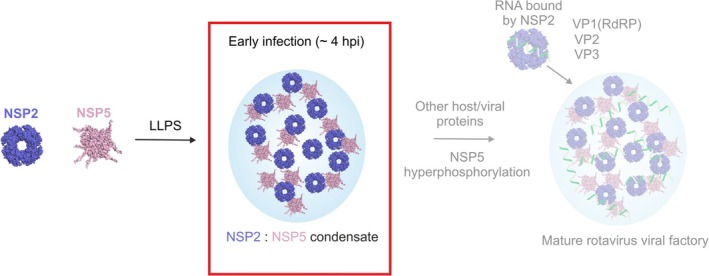
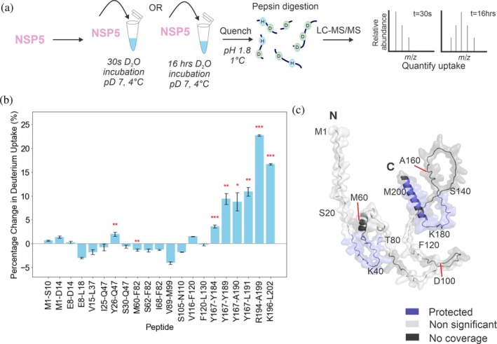
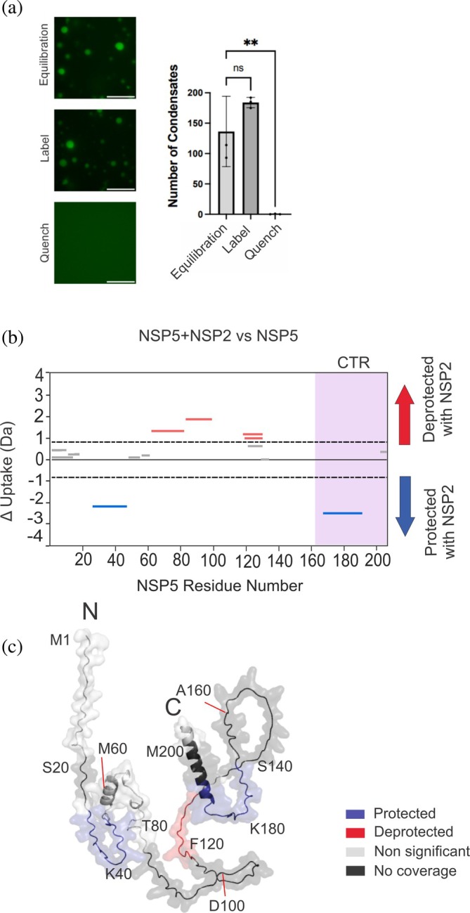
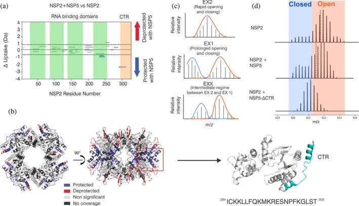
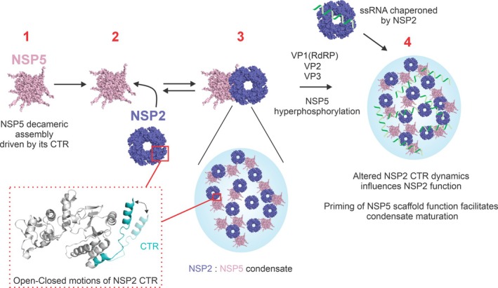
Biomolecular condensates selectively compartmentalize and organize biomolecules within the crowded cellular milieu and are instrumental in some disease mechanisms. Upon infection, many RNA viruses form biomolecular condensates that are often referred to as viral factories. The assembly mechanism of these viral factories remains poorly defined but involves transient, non-stoichiometric protein/RNA interactions, making their structural characterization challenging. Here, we sought to investigate the structural dynamics and intermolecular interactions of the key proteins responsible for condensate formation upon rotavirus infection, namely NSP2 (an RNA chaperone) and NSP5 (an intrinsically disordered protein [IDP]), using a combination of hydrogen-deuterium exchange mass spectrometry (HDX-MS), native MS, and biophysical tools. Our data reveal key structural features of intrinsically disordered NSP5 that are vital for condensate assembly and highlight inter/intra-protein interactions involved in condensate assembly. Moreover, we demonstrate that within a condensate there are altered conformational dynamics within the C-terminal region of NSP2, which has previously been shown to play a role in regulating its RNA chaperoning activity, and in the disordered regions of NSP5. We propose that altered conformational dynamics in NSP2 and NSP5 are critical for regulation of RNA annealing within a biomolecular condensate and for condensate assembly/client recruitment, respectively. Combined, our data demonstrate that the unique environment within a biomolecular condensate can tune functionally important protein conformational dynamics, which may play a crucial role in the replication of rotaviruses.

Keywords: biomolecular condensates; hydrogen–deuterium exchange mass spectrometry; native mass spectrometry; protein dynamics; rotavirus.


Conflict of interest statement

The authors declare no competing financial interest.

Figures

FIGURE 1
FIGURE 1
Biomolecular condensate formation driven by NSP2 and NSP5. NSP2 and NSP5 interact at low micromolar concentrations in vitro and form biomolecular condensates in RV‐infected cells at approximately 2–4 h post‐infection (hpi) (red box). Progression of viral infection is associated with RNA enrichment in VFs, likely bound by the RNA chaperone NSP2, and phosphorylation of NSP5. These VFs accumulate the viral pre‐genomic ssRNA, along with other components of the viral replicative machinery, including the RNA Polymerase (VP1), the inner capsid protein VP2, the capping enzyme (VP3), and additional capsid‐forming proteins (for simplicity, not shown). Here, we have focused on investigating the interactions driving early biomolecular condensate formation by NSP2 and NSP5 (red box).
FIGURE 2
FIGURE 2
Identifying structured regions in the IDP NSP5 using HDX‐MS. (a) Experimental schematic. NSP5 was incubated with deuterium for either 30 s or 16 h before quenching of the exchange reaction by lowering the pH and temperature (see Methods). Digestion with pepsin followed by LC–MS/MS enabled the mass increase from deuterium incorporation to be quantified at the peptide level. (b) The percentage change in deuterium uptake for each peptide was calculated by dividing the difference in deuterium uptake (between 30 s and 16 h for each peptide) by the maximum deuterium uptake for each peptide. The standard error was propagated to account for uncertainty with uptake measurements. A t‐test was performed to identify statistically significant differences in uptake for each peptide (*p < 0.05, **p < 0.02, and ***p < 0.01). (c) AlphaFold2 model of monomeric NSP5 with protected regions (where there is a significant increase in deuterium uptake at 16 h vs. 30 s labelling) in blue. Regions where there was no significant increase in deuterium uptake at 16 h are shown in light gray and regions of no coverage are shown in dark gray. For clarity, the position of every twentieth residue is labeled in the chain.
FIGURE 3
FIGURE 3
The C‐terminal region of NSP5 drives NSP5 oligomerization and is essential for biomolecular condensate formation by NSP2 and NSP5. (a) Native mass spectrum of NSP5. Red triangles represent the decameric assembly, while purple circles represent the charge‐stripped nonamer formed in the gas‐phase via collision‐induced dissociation (Belov et al., 2013). (b) Native mass spectrum of NSP5‐ΔCTR. Native MS experiments were performed using a ThermoFisher Q‐Exactive UHMR (see Methods). (c) Experimental schematic, demonstrating the method used to uncover the role of the NSP5‐CTR in biomolecular condensate formation driven by NSP2 and NSP5. NSP2 labeled with Atto‐488 was mixed with NSP5 or NSP5‐ΔCTR at 15 and 30 μM, respectively, and immediately imaged using an ONI Nanoimager S. (d) Wide‐field fluorescence microscopy images of spherical biomolecular condensates formed by NSP2 and NSP5 (scale bar = 10 μm, left panel). Removal of the CTR results in NSP5 that fails to form biomolecular condensates when mixed with NSP2 (right panel).
FIGURE 4
FIGURE 4
HDX‐MS analysis of NSP5 within a biomolecular condensate. (a) Fluorescence microscopy of NSP5 + NSP2‐Atto‐488 in equilibration (H2O‐containing), label (D2O‐containing), and quench HDX‐MS buffers. Biomolecular condensates form under deuterium labeling conditions and dissociate upon quenching of the HDX reaction (scale bar = 10 μm) (left). Quantification of the number of condensates per field of view (right). (b) Cumulative Woods' plot showing the summed differences in deuterium incorporation over all timepoints. Peptides from NSP5 that were significantly protected and deprotected when incubated with deuterium in the presence of NSP2 are shown in blue and red, respectively (confidence interval of 98%, see Methods). Wood's plots were produced using Deuteros (Lau et al., 2019). (c) AlphaFold2 model of monomeric NSP5 with regions of protection (blue), deprotection (red), non‐significant peptides (light gray), and no coverage (dark gray), mapped on the proposed structure. For clarity, the position of every twentieth residue is labeled in the chain.
FIGURE 5
FIGURE 5
HDX‐MS analysis of NSP2 within a biomolecular condensate. (a) Cumulative Woods' plot showing the differences in deuterium incorporation over 0.5–10 min timepoints. Peptides from NSP2 that were significantly protected and deprotected when incubated with deuterium in the presence of NSP5 are shown in blue and red, respectively (confidence interval of 98%, see Methods). Produced using Deuteros (Lau et al., 2019). Green regions represent proposed RNA‐binding regions (Bravo et al., 2020), and the C‐terminal region (CTR) is represented in orange. (b) NSP2 octameric structure (PDB: IL9V) with regions of protection (blue), deprotection (red), non‐significant peptides (light gray), and no coverage (dark gray), mapped on the structure. A monomer subunit is indicated (red box). The structure of a monomeric subunit from NSP2 octamer is shown and CTR is highlighted in blue. The corresponding peptide sequence (residues 284–305) is shown below the monomeric structure. (c) Schematic of theoretical isotopic envelopes for EX2, EX1, and EXX kinetics detected by HDX‐MS. (d) Representative isotopic envelopes for a peptide spanning residues 284–305 of NSP2, for NSP2 alone and NSP2 + NSP5, and NSP2 + NSP5‐ΔCTR after incubation with D2O. Closed and open populations are represented by blue and orange boxes, respectively.
FIGURE 6
FIGURE 6
Proposed mechanism of biomolecular condensate formation by NSP2 and NSP5. (1) NSP5 assembles into a decameric oligomer driven by its CTR; (2) NSP5 recruits NSP2; (3) the proteins form biomolecular condensates. Binding of NSP5 within a biomolecular condensate induces a change in conformational dynamics of the NSP2 CTR, which may contribute to RNA dissociation from NSP2 or influence inter‐octamer interactions. (4) ssRNA transcripts are bound by VP1 and NSP2, and they partition into condensates along with VP2 and other capsid‐forming components. Opening (measured by deprotection in HDX‐MS) of the disordered regions of NSP5 facilitates client recruitment. During the late stages of infection, VFs undergo maturation that correlates with NSP5 hyperphosphorylation.

Similar articles

References

    1. Alenquer M, Vale‐Costa S, Etibor TA, Ferreira F, Sousa AL, Amorim MJ. Influenza A virus ribonucleoproteins form liquid organelles at endoplasmic reticulum exit sites. Nat Commun. 2019;10(1):1629. 10.1038/s41467-019-09549-4 - DOI - PMC - PubMed
    1. Baggett DW, Medyukhina A, Tripathi S, Shirnekhi HK, Wu H, Pounds SB, et al. An image analysis pipeline for quantifying the features of fluorescently‐labeled biomolecular condensates in cells. Front Bioinforma. 2022;2:897238. 10.3389/fbinf.2022.897238 - DOI - PMC - PubMed
    1. Banani SF, Lee HO, Hyman AA, Rosen MK. Biomolecular condensates: organizers of cellular biochemistry. Nat Rev Mol Cell Biol. 2017;18(5):285–298. 10.1038/nrm.2017.7 - DOI - PMC - PubMed
    1. Banani SF, Rice AM, Peeples WB, Lin Y, Jain S, Parker R, et al. Compositional control of phase‐separated cellular bodies. Cell. 2016;166(3):651–663. 10.1016/j.cell.2016.06.010 - DOI - PMC - PubMed
    1. Belov ME, Damoc E, Denisov E, Compton PD, Horning S, Makarov AA, et al. From protein complexes to subunit backbone fragments: a multi‐stage approach to native mass spectrometry. Anal Chem. 2013;85(23):11163–11173. 10.1021/ac4029328 - DOI - PubMed

MeSH terms

Substances

LinkOut - more resources