. 2025;17(1):2513881.
doi: 10.1080/17590914.2025.2513881. Epub 2025 Jun 16.

Cortical Stimulation-Based Transcriptome Shifts on Parkinson's Disease Animal Model

Affiliations

Cortical Stimulation-Based Transcriptome Shifts on Parkinson's Disease Animal Model

Johyeon Nam et al. ASN Neuro. 2025.

Abstract

Parkinson's disease is the second most prevalent neurodegenerative disorder and is characterized by the degeneration of dopaminergic neurons. Significant improvements in gait balance, particularly in step length and velocity, were observed with less invasive wireless cortical stimulation. Transcriptome sequencing was performed to demonstrate the cellular mechanism, specifically targeting the primary motor cortex, where stimulation was applied. Our findings indicated that 38 differentially expressed genes (DEGs), initially downregulated following Parkinson's disease induction, were subsequently restored to normal levels after cortical stimulation. These 38 DEGs are potential targets for the treatment of motor disorders in Parkinson's disease. These genes are implicated in crucial processes, such as astrocyte-mediated blood vessel development and microglia-mediated phagocytosis of damaged motor neurons, suggesting their significant roles in improving behavioral disorders. Moreover, these biomarkers not only facilitate the rapid and accurate diagnosis of Parkinson's disease but also assist in precision medicine approaches.

Keywords: Electrotherapy; Parkinson’s disease; genetic treatment; neurodegenerative disorders; transcriptome sequencing; wireless cortical stimulation.


Conflict of interest statement

No potential conflict of interest was reported by the author(s).

Figures

Figure 1.
Figure 1.
Overview of experimental design. (A) Graphene-based electrode array neural implant configuration and the wireless device. (B) Hemi-parkinsonian rat implanted with wireless devices and electrodes. (C) Process involved vertical sectioning of the rat’s brain, followed by the sampling of the primary motor cortex for mRNA sequencing.
Figure 2.
Figure 2.
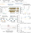
Alleviation of gait balance using 130 Hz cortical stimulation on hemi-parkinsonian rats. (A) Overview of the timeline of the experiment. Following a recovery period after 6-OHDA-induced PD, a 30-minute apomorphine-induced rotation test (0.5 mg/kg) was performed. Rats rotating at least three revolutions per minute were classified as well-lesioned, and a graphene-based electrode array was implanted into the motor cortex of control or unilateral PD groups. (B) Apomorphine administration resulted in the measurement of unilateral rotations. (C) Schematic images of TH-positive staining in substantia nigra (SN) and striatum (STR) slices at Ctrl group and unilateral PD group. (D) Significant differences were observed between Ctrl and PD groups’ area of TH-positive cells. (E, F) Gait balance of three groups from the first to second weeks of stimulation. The stimulation effect led to the restoration of the step length and velocity ratio in the Stim on PD group to levels comparable to the Ctrl group. All data are presented as the mean ± standard deviation (SD). *p < 0.05, **p < 0.01, ***p < 0.005, ****p < 0.001.
Figure 3.
Figure 3.
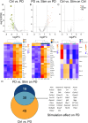
DEGs in Ctrl vs. PD groups, PD vs. Stim on PD groups and Ctrl vs. Stim on ctrl groups identified by edgeR (FDR < 0.05, |log2FC| > 2). (A) Volcano plot (top) representing the gene expression difference between Ctrl and PD groups. The green dots stand for downregulated DEGs, and the red dots stand for upregulated DEGs. Heatmap (bottom) constructed with top 20 DEGs between Ctrl and PD groups. The color of each block indicates the degree of differential expression in gene in sample. The green color indicates downregulation, while red color indicates upregulation. (B) Volcano plot (top) and Heatmap (bottom) constructed with top 20 DEGs between PD and Stim on PD groups. (C) Volcano plot (top) and Heatmap (bottom) constructed with 7 DEGs between Ctrl and stim on ctrl groups. (D) Venn diagram of 38 DEGs showing decreased expression following PD treatment and increased expression upon cortical stimulation.
Figure 4.
Figure 4.
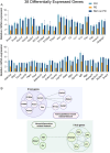
The mRNA expression levels, and protein-protein interaction (PPI) network of the overlapped 38 DEGs between the Ctrl vs. PD groups and PD vs. Stim on PD groups. (A) The mRNA expression level of 38 genes recovered in the Stim on PD group after cortical stimulation. X-axis represents the 38 DEGs, and Y-axis shows the expression values, which are normalized to the control group. (B) The two networks identified with hierarchical clustering implemented in STRING. First network was associated with astrocyte and blood vessel development, and the other was associated with neuroinflammation.
Figure 5.
Figure 5.
Schematic illustration presenting a conceptual model of how cortical surface stimulation might modulate astrocytic, microglial, and vascular interactions to improve motor function in PD. This model, derived from our transcriptomic and network analyses, is intended to generate hypotheses for future studies rather than serve as definitive evidence of the underlying mechanisms.

References

    1. Aleksander, S. A., Balhoff, J., Carbon, S., Cherry, J. M., Drabkin, H. J., Ebert, D., Feuermann, M., Gaudet, P., Harris, N. L., Hill, D. P., Lee, R., Mi, H., Moxon, S., Mungall, C. J., Muruganugan, A., Mushayahama, T., Sternberg, P. W., Thomas, P. D., Van Auken, K., … Westerfield, M, Gene Ontology Consortium . (2023). The gene ontology knowledgebase in 2023. Genetics, 224(1). 10.1093/genetics/iyad031 - DOI - PMC - PubMed
    1. Allen, N. J. (2014). Astrocyte regulation of synaptic behavior. Annual Review of Cell and Developmental Biology, 30(1), 439–463. 10.1146/annurev-cellbio-100913-013053 - DOI - PubMed
    1. Antonini, A., Moro, E., Godeiro, C., & Reichmann, H. (2018). Medical and surgical management of advanced Parkinson’s disease. Movement Disorders: official Journal of the Movement Disorder Society, 33(6), 900–908. 10.1002/mds.27340 - DOI - PubMed
    1. Araque, A., Carmignoto, G., Haydon, P. G., Oliet, S. H., Robitaille, R., & Volterra, A. (2014). Gliotransmitters travel in time and space. Neuron, 81(4), 728–739. 10.1016/j.neuron.2014.02.007 - DOI - PMC - PubMed
    1. Ashburner, M., Ball, C. A., Blake, J. A., Botstein, D., Butler, H., Cherry, J. M., Davis, A. P., Dolinski, K., Dwight, S. S., Eppig, J. T., Harris, M. A., Hill, D. P., Issel-Tarver, L., Kasarskis, A., Lewis, S., Matese, J. C., Richardson, J. E., Ringwald, M., Rubin, G. M., & Sherlock, G. (2000). Gene ontology: tool for the unification of biology. The gene ontology consortium. Nature Genetics, 25(1), 25–29. 10.1038/75556 - DOI - PMC - PubMed

Publication types

LinkOut - more resources