. 2025 Jun 16;21(6):e1011687.
doi: 10.1371/journal.pgen.1011687. eCollection 2025 Jun.

The Aspergillus nidulans velvet domain containing transcription factor VeA is shuttled from cytoplasm into nucleus during vegetative growth and stays there for sexual development, but has to return into cytoplasm for asexual development

Affiliations

The Aspergillus nidulans velvet domain containing transcription factor VeA is shuttled from cytoplasm into nucleus during vegetative growth and stays there for sexual development, but has to return into cytoplasm for asexual development

Anja Strohdiek et al. PLoS Genet. .

Abstract

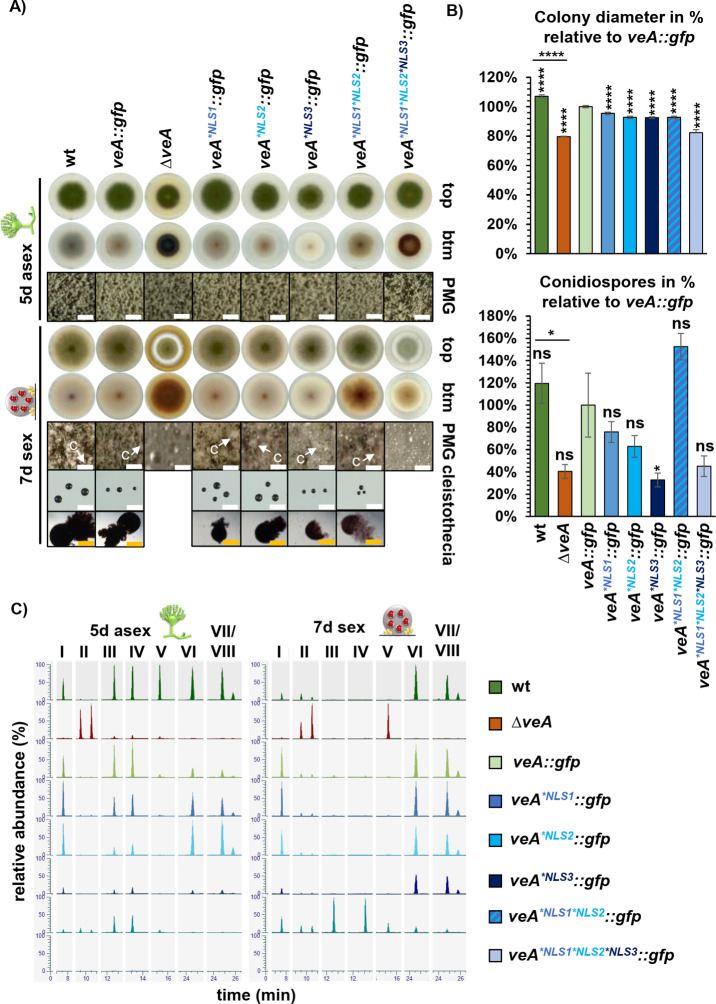
Survival of multicellular organisms requires the coordinated interplay between networks regulating gene expression and controlled intracellular transport of respective regulators. Velvet domain proteins are fungal transcription factors, which form various heterodimers and play key roles in controlling early developmental decisions towards more either asexual or sexual differentiation. VeA is the central subunit of the trimeric velvet complex VelB-VeA-LaeA, which links transcriptional to epigenetic control for the coordination of fungal developmental programs to specific secondary metabolite synthesis. Nuclear localization of the VeA bridging factor is carefully controlled in fungi. In this work we demonstrate that VeA carries three nuclear localization signals NLS1, NLS2 and NLS3, which all contribute to nuclear import. We show that VeA has an additional nuclear export sequence (NES) which provides a shuttle function to allow the cell to relocate VeA to the cytoplasm. VeA is nuclear during vegetative growth, but has to be exported from the nucleus to allow and promote asexual development. In contrast, progression of the sexual pathway requires continuous nuclear VeA localization. Our work shows that an accurate nuclear import and export control of velvet proteins is further connected to specific stability control mechanism as prerequisites for fungal development and secondary metabolism. These results illustrate the various complex mutual dependencies of velvet regulatory proteins for coordinating fungal development and secondary metabolism.


Conflict of interest statement

The authors have declared that no competing interests exist.

Figures

Fig 1
Fig 1. A prediction revealed NLS1 and NES in VeAs velvet domain and C-terminally exposed NLS2/ NLS3.
(A) NLS prediction with cNLS mapper [18] revealed the bipartite NLS1 (blue) in the velvet domain (light green) as well as the monopartite NLS2 (turquoise) and bipartite NLS3 (dark blue) in the C-terminal part of the VeA protein. In addition, a potential nuclear export signal (red) is located at the end of the velvet domain [17]. The full wild type amino acid sequences of each motifs are indicated in the box. Functional studies of all NLS and the NES sequences were conducted by codon exchanges for the indicated amino acids (wt: wild type, mt: mutant). (B) Crystal structure of VeA velvet domain (green) [19] combined with the predicted structure of the C-terminus of VeA using AlphaFold1 [20] and PyMOL (PyMOL Molecular Graphics System, Version 2.0 Schrödinger, LLC.). The NLS and NES are highlighted as in A.
Fig 2
Fig 2. VeA NLS are additionally required to exclude VeA from the nucleus to promote asexual development.
(A) Scheme of VeA-GFP with its domains and predicted NES and NLS motifs as well as different truncated versions of VeA-GFP. VeA∆velvet-GFP is missing the velvet domain. VeA46-573-GFP is missing the first 45 amino acids together with the NLS1. VeA1-225-GFP is missing the C-terminus of VeA with the NLS2 and NLS3 motifs. VeA46-225-GFP lacks all three NLS motifs. (B) Fluorescence images of strains expressing VeA-GFP and VeA-GFP NLS versions with amino acid substitutions grown on agar slant surfaces after 18 h illumination for promoting asexual (condiophore = light) or darkness (cleistothecia = darkness) for sexual development. Nuclei are visualized with RFP-H2A (size bars: 10 µm). C) Fluorescence microscopy was performed with VeA-GFP, VeA missing the velvet domain and VeA truncation strains (shown in A) after 18 h vegetative growth (hyphae) from an illuminated liquid culture as well as from cultures grown on tilted agar slides with illumination (conidiophore) for promoting asexual or darkness (cleistothecia) for sexual development. The nucleus is visualized with RFP-H2A (size bars: 10 µm).
Fig 3
Fig 3. Interplay between NES and NLS1 for combined action on VeA cellular localization.
Fluorescence images of strains with VeA-GFP and VeA-GFP with amino acid substitutions in NES or in NES/NLS motif combinations were compared after 18 h of vegetative growth and on agar slants after either 18 h in light (conidiophore) to promote asexual or in darkness (cleistothecia) to promote sexual development. Nuclei are visualized with RFP-H2A. A defective NES motif leads to an accumulated VeA-GFP signal in the nucleus. Combinations of defective NES and NLS reduce nuclear VeA accumulation, especially if NLS1 is dysfunctional. NES, NLS1 and NLS2 amino acid substitution strain shows less cytoplasmic signals (size bars: 10 µm).
Fig 4
Fig 4. All three NLS of VeA are required for wild type like development in A. nidulans.
(A) Asexual development of veA deletion (ΔveA) and complementation (veA::gfp) strains as well as strains with amino acid substitutions in the NLS motifs (veA*NLS1::gfp, veA*NLS2::gfp, veA*NLS3::gfp, veA*NLS1*NLS2::gfp, veA*NLS1*NLS2*NLS3::gfp) and the wild type were analyzed after 5 days of incubation at 37°C in light. Plates were incubated in the dark and with limited oxygen for 7 days at 37°C for sexual development. (PMG: photomicrographs, c: cleistothecia, size bar: 200 µm (white), cleistothecia 50 µm (orange)). (B) Measurement of colony diameters and quantification of spores from wild type, veA deletion and mutant strains. The diagram summarizes quantification results from three biological replicates (3 technical replicates each). Error bars represent standard error of the mean, and the significances were compared to veA::gfp complementation strain or between strains with connecting lines (p > 0.05: ns, p ≤ 0.05: *, p ≤ 0.0001: ****). (C) Ethyl acetate extracts were obtained from the indicated strains that were cultivated as described in A. Samples were measured by LC-MS. Extracted ion chromatograms (EIC, [M-H]-) of the most prominent identified masses show differences in peak intensities between the analyzed strains. Identified metabolites are chichorine (I), F-9775A/B (II), Austinol (III), Dehydroaustinol (IV), Sterigmatocystin (V), Emericellin (VI), Shamixanthone (VII), and Epishamixanthone (VIII). Detailed information about identified metabolites is given in S1 Table. A tolerated mass deviation of 0.0010 was used to visualize the metabolites.
Fig 5
Fig 5. Nuclear export of VeA is required for A. nidulans development and coordinated secondary metabolism.
(A) Wild type, the veA deletion strain as well as strains with amino acid substitutions in NES and NLS motifs were analyzed five days after point inoculation on solid MM and incubation at 37°C in light to induce asexual development. For sexual development, plates were incubated in the dark and with limited oxygen for 7 days at 37°C. (PMG: photomicrographs, c: cleistothecia, size bar: 200 µm (white), cleistothecia 50 µm (orange)). (B) Measurement of colony diameters and quantification of spore amounts from wild type, veA deletion and mutant strains. Quantification results from three biological replicates (3 technical replicates each). Error bars represent standard error of the mean, and the significances were compared to veA::gfp complementation strain (p > 0.05: ns, p ≤ 0.0001: ****). (C) Ethyl acetate extracts were obtained from the indicated strains that were cultivated as described in A. Samples were measured by LC-MS. Extracted ion chromatograms (EIC, [M-H]-) of the most prominent identified masses show differences in peak intensities between the analyzed strains. Identified metabolites are chichorine (I), F-9775A/B (II), Austinol (III), Dehydroaustinol (IV), Sterigmatocystin (V), Shamixanthone (VII), and Epishamixanthone (VIII). Detailed information about identified metabolites is given in S1 Table. A tolerated mass deviation of 0.0010 was used to visualize the metabolites.
Fig 6
Fig 6. Transcription and VeA protein levels depend on VeAs NES/NLS during vegetative growth or sexual development.
(A) qRT-PCR was performed to analyze gene expression of veA::gfp and mutant strains. Fungal strains were grown for 20 h in liquid cultures for vegetative growth. RNA was isolated from these samples and transcribed to cDNA. qRT-PCRs were performed with h2A and gpdA as reference genes. Expression levels of the mutants were quantified relative to veA::gfp expression. Quantification results from three biological replicates (3 technical replicates each). (B) Western experiments of fungal protein extracts from VeA-GFP and VeA NES and NES/NLS mutant strains were performed. All strains were grown for 20 h at 37°C in liquid minimal medium for vegetative growth. Mycelia were shifted onto 30 ml minimal medium agar plates for 6 to 48 h at 37°C in dark for sexual development. Samples were collected at identical time points and protein crude extracts were prepared for western experiments with α-GFP antibody. Signal quantification was performed using BioID software, signals of VeA-GFP (red arrow) were normalized to Ponceau S staining. Wild type (wt) VeA protein amount at the 20 h vegetative time point was set to 1 and used as reference for protein amounts of following time points of wild type VeA or VeA with amino acid substitutions in NES and NLS motifs. Quantification results from three biological replicates (2 technical replicates each). Error bars for western experiments and qRT-PCRs represent standard error of the mean (p > 0.05: ns, p ≤ 0.05: *, p ≤ 0.01: **, p ≤ 0.001: ***, p ≤ 0.0001: ****).
Fig 7
Fig 7. VeA shuttles between cytoplasm and nucleus to allow A. nidulans fungal development.
VeA is channeled into the nucleus during germination and vegetative growth. External signals as darkness or decreased oxygen levels lead to VeA-induced sexual development forming, cleistothecia as hibernating structures. Light or increased oxygen levels lead to nuclear export of VeA to the cytoplasm for accurate asexual development. The nuclear exchange of VeA is connected with a stability control mechanism of the velvet protein for both developmental programs.

References

    1. Pöggeler S, Nowrousian M, Teichert I, Beier A, Kück U. Fruiting-body development in ascomycetes. In: Physiology and genetics. 2nd ed. The Mycota XV; 2018. p. 1–56.
    1. de Vries R, Riley R, Wiebenga A, Aguilar-Osorio G, Amillis S, Uchima C, et al. Comparative genomics reveals high biological diversity and specific adaptations in the industrially and medically important fungal genus Aspergillus. Genome Biol. 2017;18:1–45. - PMC - PubMed
    1. Moon H, Han K, Yuk J. Upstream regulation of development and secondary metabolism in Aspergillus species. Cells. 2023;12(2):1–19. - PMC - PubMed
    1. Noble LM, Andrianopoulos A. Reproductive competence: a recurrent logic module in eukaryotic development. Proc Biol Sci. 2013;280(1766):20130819. doi: 10.1098/rspb.2013.0819 - DOI - PMC - PubMed
    1. Blumenstein A, Vienken K, Tasler R, Purschwitz J, Veith D, Frankenberg-Dinkel N, et al. The Aspergillus nidulans phytochrome FphA represses sexual development in red light. Curr Biol. 2005;15(20):1833–8. doi: 10.1016/j.cub.2005.08.061 - DOI - PubMed

MeSH terms

LinkOut - more resources