

Leptomeningeal hemangioblastoma: illustrative case
- PMID: 40523339
- PMCID: PMC12171096
- DOI: 10.3171/CASE25204
Leptomeningeal hemangioblastoma: illustrative case
Abstract
Background: Hemangioblastoma is a slow-growing vascular tumor commonly found in the posterior fossa. It is associated with von Hippel-Lindau disease, yet most cases arise sporadically. Resection and belzutifan are highly effective in the treatment of hemangioblastoma. Rarely, leptomeningeal dissemination may occur months to years after resection.
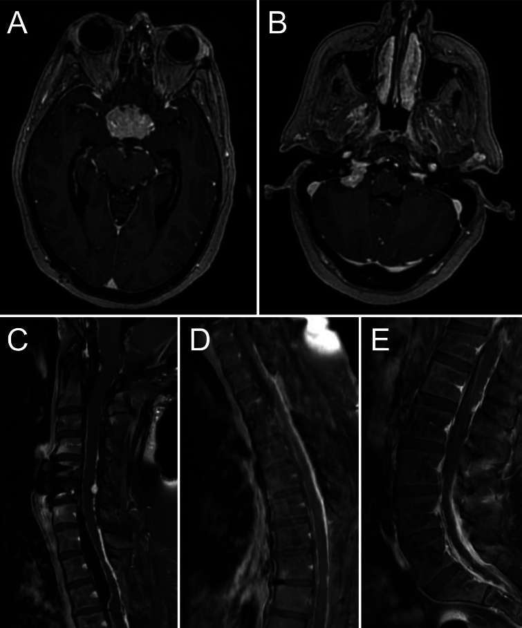
Observations: A 56-year-old female with cerebellar hemangioblastoma resected in 2016 presented 8 years later with left hemiparesis. Updated neuroimaging revealed new leptomeningeal disease, confirmed via tissue from a right cerebellopontine angle mass. The authors conducted a review of 33 patients diagnosed with leptomeningeal disease; two-thirds were male with a median age of 48 years. Thirty-two patients presented initially with a solitary mass, and 96% of these underwent resection. The mean time from initial lesion resection to dissemination was 94 months.
Lessons: After resection, hemangioblastoma recurrence with leptomeningeal spread is a rare but dangerous possible complication. Management is complex, yet a combination of pan-CNS radiation therapy, targeted resection, and belzutifan (a small-molecule inhibitor that selectively targets and blocks the function of HIF-2α) may represent an effective treatment combination. https://thejns.org/doi/10.3171/CASE25204.
Keywords: case report; hemangioblastoma; leptomeningeal.
Figures

References
-
- Koo HW, Park JE, Cha J.Hemangioblastomas with leptomeningeal dissemination: case series and review of the literature. Acta Neurochir. 2016;158(6):1169-1178. - PubMed
-
- Hottinger AF Khakoo Y.. Neurooncology of familial cancer syndromes. J Child Neurol. 2009;24(12):1526-1535. - PubMed
-
- Binderup MLM Jensen AM Budtz-Jørgensen E Bisgaard ML.. Survival and causes of death in patients with von Hippel–Lindau disease. J Med Genet. 2017;54(1):11-18. - PubMed
LinkOut - more resources
Full Text Sources
