

Intraoperative neurovascular considerations for efficient intraventricular meningioma surgery: illustrative case
- PMID: 40523341
- PMCID: PMC12171106
- DOI: 10.3171/CASE2569
Intraoperative neurovascular considerations for efficient intraventricular meningioma surgery: illustrative case
Abstract
Background: Intraventricular meningiomas (IVMs) are a rare subtype of brain tumors. Typically slow growing, these tumors can occasionally reach a substantial size, causing ventricular obstruction and hydrocephalus. Resection remains the treatment of choice; however, the deep location and proximity to critical neurovascular structures can pose significant challenges. Various surgical strategies and adjuncts have been described. Here, the authors highlight the benefits of early intraoperative tumor devascularization to minimize blood loss and enable safe, efficient removal through a minimally disruptive transsulcal approach.
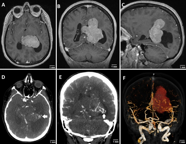
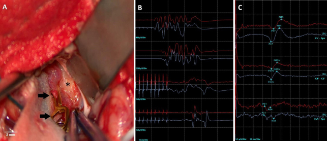
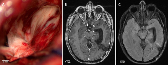
Observations: A 59-year-old woman presented with symptoms of increased intracranial pressure due to a large atrial IVM causing temporal horn entrapment. The tumor was hypervascular, with prominent arterial feeders. Early intraoperative microsurgical devascularization was favored over preoperative embolization, combined with temporary clipping of adjacent arterial feeders and intraoperative neurophysiological monitoring. This strategy facilitated piecemeal gross-total tumor resection within a relatively short surgical duration, minimizing brain retraction. The patient was neurologically intact after gross-total resection.
Lessons: Early and strategic tumor devascularization is an effective approach to achieve safe and efficient resection of large intraventricular tumors with minimal brain retraction. https://thejns.org/doi/10.3171/CASE2569.
Keywords: intraoperative neuromonitoring; intraventricular tumors; meningioma; microneurosurgery.
Figures
Similar articles
-
The Black Book of Psychotropic Dosing and Monitoring.Psychopharmacol Bull. 2024 Jul 8;54(3):8-59. Psychopharmacol Bull. 2024. PMID: 38993656 Free PMC article. Review.
-
Percutaneous Endoscopic Decompression for Lumbar Central and Lateral Recess Spinal Stenosis: A Combined Uni-Portal and Bi-Portal Approach.JBJS Essent Surg Tech. 2025 Jul 17;15(3):e24.00002. doi: 10.2106/JBJS.ST.24.00002. eCollection 2025 Jul-Sep. JBJS Essent Surg Tech. 2025. PMID: 40678179 Free PMC article.
-
Management of urinary stones by experts in stone disease (ESD 2025).Arch Ital Urol Androl. 2025 Jun 30;97(2):14085. doi: 10.4081/aiua.2025.14085. Epub 2025 Jun 30. Arch Ital Urol Androl. 2025. PMID: 40583613 Review.
-
Short-Term Memory Impairment.2024 Jun 8. In: StatPearls [Internet]. Treasure Island (FL): StatPearls Publishing; 2025 Jan–. 2024 Jun 8. In: StatPearls [Internet]. Treasure Island (FL): StatPearls Publishing; 2025 Jan–. PMID: 31424720 Free Books & Documents.
-
Pediatric intraparenchymal meningioma in the basal ganglia treated with gross total resection: a case report and review of the literature.Childs Nerv Syst. 2023 Dec;39(12):3595-3600. doi: 10.1007/s00381-023-06056-2. Epub 2023 Jun 27. Childs Nerv Syst. 2023. PMID: 37369950 Review.
References
-
- Pereira BJA de Almeida AN Paiva WS de Aguiar PHP Teixeira MJ Marie SKN.. Natural history of intraventricular meningiomas: systematic review. Neurosurg Rev. 2020;43(2):513-523. - PubMed
-
- Lyngdoh BT Giri PJ Behari S Banerji D Chhabra DK Jain VK.. Intraventricular meningiomas: a surgical challenge. J Clin Neurosci. 2007;14(5):442-448. - PubMed
-
- Grujicic D, Cavallo LM, Somma T.Intraventricular meningiomas: a series of 42 patients at a single institution and literature review. World Neurosurg. 2017;97:178-188. - PubMed
-
- Bertalanffy A, Roessler K, Koperek O.Intraventricular meningiomas: a report of 16 cases. Neurosurg Rev. 2006;29(1):30-35. - PubMed
LinkOut - more resources
Full Text Sources
