. 2025 Jul;301(7):110384.
doi: 10.1016/j.jbc.2025.110384. Epub 2025 Jun 14.

Targeted incorporation of collagen IV to the basement membrane: A step forward for developing extracellular protein therapies

Affiliations

Targeted incorporation of collagen IV to the basement membrane: A step forward for developing extracellular protein therapies

Elena N Pokidysheva et al. J Biol Chem. 2025 Jul.

Abstract

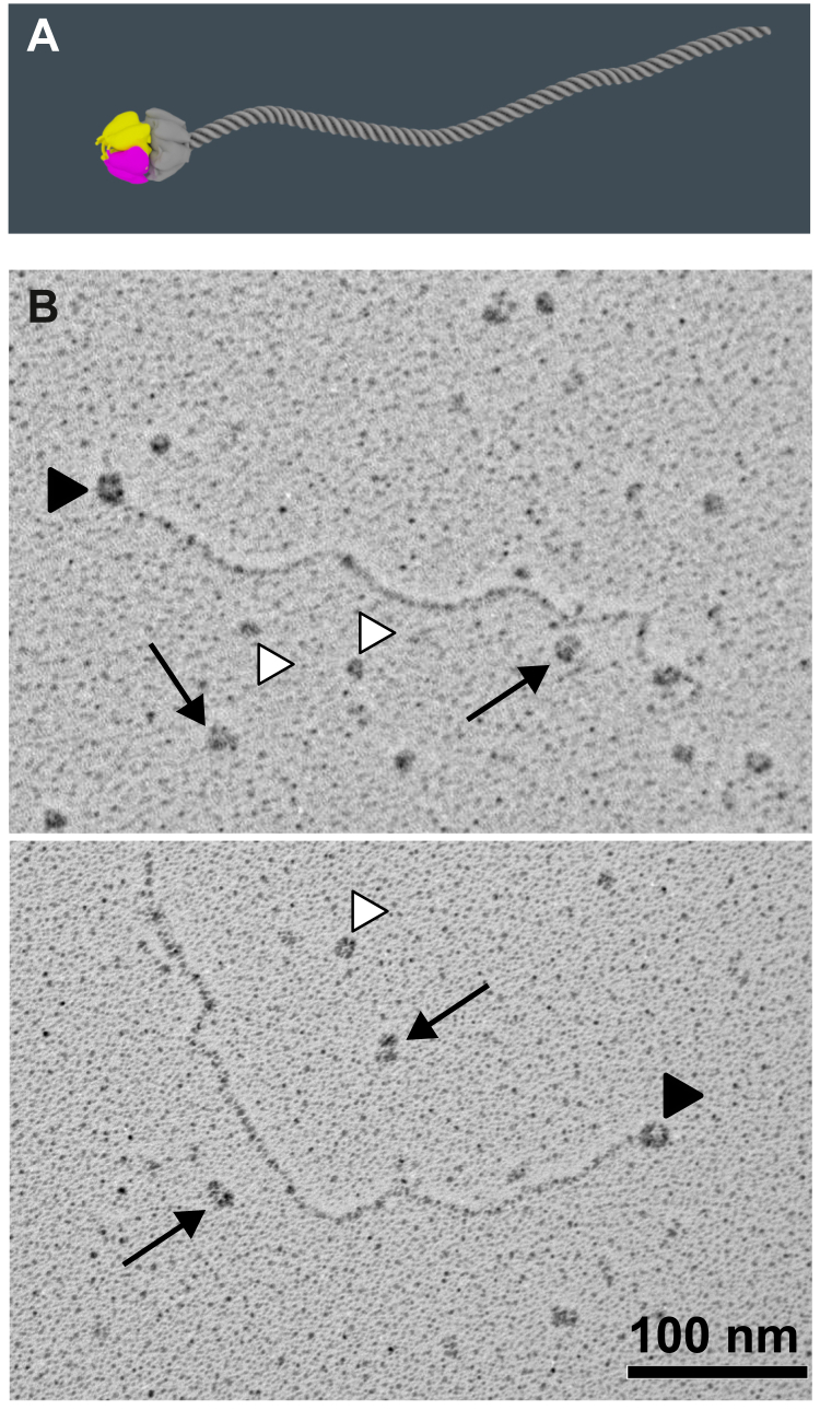
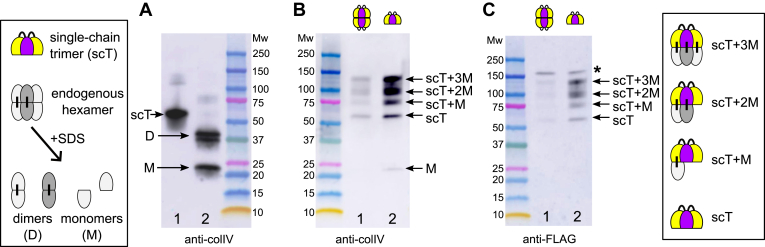
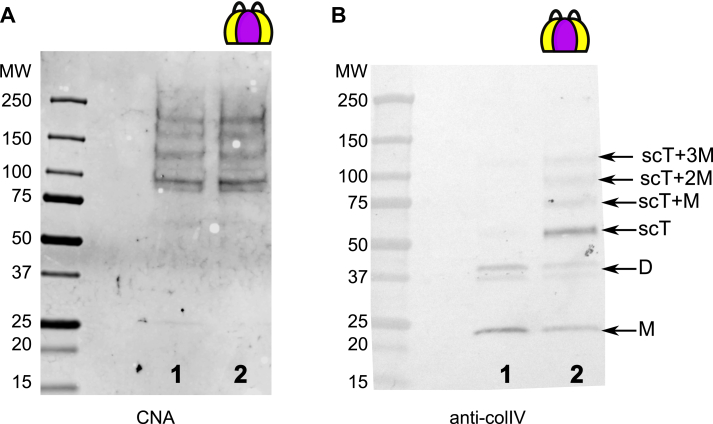
The collagen IV scaffold serves as a fundamental structural unit of the basement membrane (BM). Understanding its structure, assembly, and function is essential for tissue engineering, the design of organoid models, and developing therapies for diseases such as Alport syndrome, Gould syndrome, psoriasis, eye abnormalities, hearing loss, and others, where collagen IV is required for structural integrity and functionality of the BM. The collagen IV molecule is a 400 nm long heterotrimer, comprising non-collagenous 1 (NC1), collagenous, and 7S domains. The assembly of the collagen IV scaffold involves oligomerization of the C-terminal NC1 and the N-terminal 7S domains, along with lateral associations within the collagenous domain. However, the detailed architecture and assembly mechanisms of the collagen IV scaffold remain unclear. Here, we investigated the potency and mechanism of recombinant single-chain NC1 trimer incorporation into the collagen IV scaffold. We discovered that the NC1 trimer influences the overall assembly of the basement membrane by affecting the quality of the developing collagen IV scaffold in a dose-dependent manner, without impacting already established scaffolds. This interference occurs through the hexamerization of supplemented NC1 trimers with endogenous NC1 domains, as the NC1 trimer becomes sulfilimine crosslinked with the existing chains. Overall, the single-chain NC1 trimer of collagen IV is crucial for developing novel extracellular therapies in two main ways: (1) facilitating the delivery and incorporation of functional replacements like collagen IV fragments and (2) inhibiting the formation of new basement membranes in conditions such as tumor growth and detrimental vascularization.

Keywords: Alport syndrome; Gould syndrome; NC1 domain; basement membrane; collagen IV; molecular orthotics.


Conflict of interest statement

Conflict of interest The authors declare that they have no conflicts of interest with the contents of this article.

Figures

Figure 1
Figure 1
The NC1 trimer associates with the endogenous full-length collagen IV protomer.A, schematic drawing of the recombinant NC1 trimer (colored) assembled with the native full-length collagen IV protomer (gray). B, chloride-free PFHR-9 conditioned media was pulled down using anti-FLAG resin and analyzed by rotary shadowing EM. The NC1 trimer pulls full-length collagen IV protomers. Black arrows point to self-assembled recombinant NC1 trimers into hexamers. Black arrowheads point to NC1 hexamers assembled from recombinant and protomer trimers. White arrowheads point to recombinant NC1 trimers.
Figure 2
Figure 2
Experimental setup to study basement membrane in PFHR-9 cell culture. PFHR-9 cells were grown to confluency in the high glucose DMEM medium and then medium was switched to the low glucose DMEM containing 50 μg/ml of ascorbic acid. The cross-sections were analyzed by transmission electron microscopy (TEM). n another set of experiments cells were removed with lysis buffer and deposited matrix was washed with EDTA-containing buffer to remove most BM component but not collagen IV. The resulting surface of stripped collagen IV scaffold was imaged using scanning electron microscopy (SEM).
Figure 3
Figure 3
Collagen IV scaffold deposited by PFHR-9 cell culture.A, transmission EM analysis of the PFHR-9 model basement membrane. B, scanning EM of the collagen IV scaffold after removal of cells and other components than collagen IV BM. C, immuno-gold labeling of type IV collagen NC1 domain analyzed by scanning EM.
Figure 4
Figure 4
The NC1 trimer disrupts the organization of the model basement membrane.A, cross-section of the model basement membrane grown under normal conditions. B, matrix of the PFHR-9 cells stably transfected with a GFP-containing version of the single-chain NC1 trimer showing significant disruption of the matrix. C, matrix deposited by regular PFHR-9 cells grown in the presence of 80 μg/ml of the NC1 trimer. DF, concentration-dependent effect of the presence of the NC1 trimer in the medium on disruption of the basement membrane deposited by the PFHR-9 cells. Cells were initially grown under normal conditions for 3 days, and then the NC1 trimer was added to the medium for three more days at various concentrations. The degree of disruption correlates with increasing concentration of the added protein.
Figure 5
Figure 5
Trimer but not hexamer disturbs the basement membrane organization. The PFHR-9 cells were first grown under normal conditions for 3 days, and then either recombinant NC1 hexamer (A) or trimer (B) was added to the medium for three more days at 80 μg/ml concentration. In (B), the dashed line indicates the visual border between normal and affected BM structure. The hexamer has no obvious effect on the BM structure (A), while trimer (B) produces defects identical to those observed in Figure 7.
Figure 6
Figure 6
Incorporation of the recombinant NC1 trimer into the collagen IV scaffold. Scanning EM of collagen IV scaffolds immuno-labeled against the FLAG-tag found in the recombinant NC1 domain. A, cell culture was supplemented with the hexamer assembled from the recombinant NC1 trimer. B, cell culture was supplemented with the recombinant NC1 trimer.
Figure 7
Figure 7
Disruption of the collagen IV scaffold with the recombinant NC1 trimer. Immuno-gold scanning EM images of collagen IV scaffolds grown for 5 days in the presence of the NC1 trimer at 80 μg/ml concentration. A, sample was immuno-gold labeled with the anti-FLAG antibody, therefore identifying the recombinant single-chain NC1 trimer only. B, the sample was labeled using the antibody against the NC1 domain, which recognizes both endogenous and recombinant NC1 domains. A varying degree of scaffold disruption is observed. White arrows are pointing to the filament breaks that are capped with the NC1 domain. Binding of the recombinant NC1 trimer along the collagen IV filament is evident.
Figure 8
Figure 8
The NC1 trimer is cross-linked with the endogenous NC1 domain of collagen IV.A, schematic representation (left panel) and Western blot analysis of the recombinant single-chain NC1 trimer (scT, lane 1) and endogenous NC1 hexamer isolated from the PFHR-9 matrix (lane 2). The hexamer self-assembled in vitro from the single-chain NC1 trimer dissociates into trimers under the denaturing conditions of the SDS-PAGE and thus undistinguished from the trimer sample (not shown). The hexamer isolated from the matrix deposited by cells dissociates into monomers (M) and dimers (D) on SDS-PAGE. The primary antibody used was against the NC1 domain. B, basement membrane grown in the presence of either assembled hexamer (lane 1) or trimer (lane 2) of the recombinant single-chain NC1 trimer was collagenized to release the NC1 domain, then soluble material was pulled down using anti-FLAG resin and analyzed on Western blot with the same anti-NC1 antibody. C, the same as in (B) but probed with the anti-FLAG antibody to identify recombinant NC1. ∗ indicates anti-FLAG reactive impurity. This analysis demonstrates that the recombinant NC1 trimer added into the growth media of PFHR-9 cells incorporates into the matrix via hexamerization with the endogenous NC1 domain of collagen IV protomer. The crosslinks are formed between the recombinant and endogenous proteins as indicated. The right panel shows all possible crosslinking of the endogenous NC1 domains and the single-chain trimer.
Figure 9
Figure 9
The NC1 trimer does not affect the formation of the 7S domain in the collagen IV scaffold. Basement membrane grown under regular conditions (lane 1) or in the presence of 100 μg/ml of the recombinant single-chain NC1 trimer (lane 2) was collagenized to release the 7S and NC1 domains. Samples were run under reducing conditions. The blot was probed with CNA35tri (65) to detect triple helical peptides within the 7S domain (A) and anti-colIV antibody to visualize the NC1 domains (B).

Similar articles

References

    1. Fidler A.L., Boudko S.P., Rokas A., Hudson B.G. The triple helix of collagens - an ancient protein structure that enabled animal multicellularity and tissue evolution. J. Cell Sci. 2018;131 - PMC - PubMed
    1. Page-McCaw P.S., Pokidysheva E.N., Darris C.E., Chetyrkin S., Fidler A.L., Gallup J., et al. Collagen IV of basement membranes: I. Origin and diversification of COL4 genes enabling metazoan multicellularity, evolution, and adaptation. J. Biol. Chem. 2025;301 - PMC - PubMed
    1. Brown K.L., Cummings C.F., Vanacore R.M., Hudson B.G. Building collagen IV smart scaffolds on the outside of cells. Protein Sci. 2017;26:2151–2161. - PMC - PubMed
    1. Fidler A.L., Darris C.E., Chetyrkin S.V., Pedchenko V.K., Boudko S.P., Brown K.L., et al. Collagen IV and basement membrane at the evolutionary dawn of metazoan tissues. Elife. 2017;6 - PMC - PubMed
    1. Parkin J.D., San Antonio J.D., Pedchenko V., Hudson B., Jensen S.T., Savige J. Mapping structural landmarks, ligand binding sites, and missense mutations to the collagen IV heterotrimers predicts major functional domains, novel interactions, and variation in phenotypes in inherited diseases affecting basement membranes. Hum. Mutat. 2011;32:127–143. - PMC - PubMed

LinkOut - more resources