

[Clinical analysis of changes in the position of the condyle and temporomandibular joint after repair of mandibular defects]
- PMID: 40523823
- PMCID: PMC12211498
- DOI: 10.7518/hxkq.2025.2024337
[Clinical analysis of changes in the position of the condyle and temporomandibular joint after repair of mandibular defects]
Abstract
Objectives: This retrospective study aimed to investigate factors influencing positional changes of the condyle and temporomandibular joint (TMJ) following mandibular defect reconstruction with bone flaps, and to evaluate the biomechanical impacts of flap reconstruction on condylar positioning, thereby providing evidence for optimizing surgical protocols and TMJ functional rehabilitation.
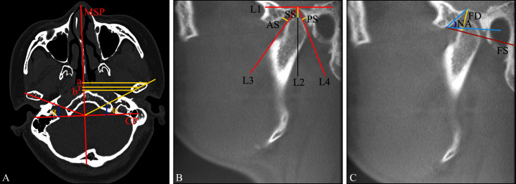
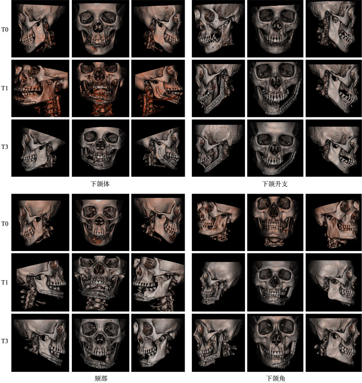
Methods: A retrospective study was conducted on 90 patients undergoing mandibular segmental resection with immediate bone flap reconstruction at Guizhou Medical University Affiliated Stomatological Hospital (June 2019 to May 2024). After strict screening, 50 cases with complete data were analyzed. Clinical parameters (defect size, location, reconstruction method) and craniofacial CT scans at four timepoints [preoperative (T0), 7-10 days (T1), 3 months (T2), and 6 months (T3) postoperatively] were collected. Mimics 20 software facilitated 3D reconstruction for measuring TMJ anterior/posterior/superior joint spaces (Kamelchuk method) and calculating condylar position via the Pullinger index [Ln (posterior/anterior space)]. Vitral and Krisjane methods quantified mandibular linear parameters (ramus length, condylar pole distances to the sagittal plane, angulation) and glenoid fossa morphology. Statistical analyses were performed using SPSS 21.0.
Results: Mandibular defect size and location were significant factors influencing postoperative condylar position changes (P<0.05). Compared to preoperative measurements, postoperative condylar anterior, posterior, and superior joint spaces were significantly increased (P<0.001). The most pronounced anterior condylar displacement occurred within 7-10 days postoperatively (P<0.05). In patients with condyle resection, postoperative joint space and angle changes were significant; in patients with condyle preservation, only superior and anterior joint space changes were statistically significant (P<0.05). Additionally, from T1 to T2, the changes in condylar medial-lateral distance, superior joint space, and anterior joint space were negatively correlated with the preoperative condylar position. Compared with preoperative,in the T0-T1 period, condylar medial-lateral distance, posterior joint space, and articular tubercle angle changes were significantly negatively correlated with time (P<0.05). Notably, the angle between the condylar long axis and the coronal axis showed a sustained negative trend from T1 to T3 (P<0.05).
Conclusions: Condylar position changes after mandibular defect repair with bone flap reconstruction are associated with the size and location of the defect. Additionally, adaptive remodeling of the temporomandibular joint (TMJ) joint space occurs postoperatively. The phenomenon of anterior displacement of the condyle in the early postoperative period (7-10 days) shows a trend of reduction with prolonged follow-up time, and further sample size research is needed.
目的: 通过回顾性分析探讨下颌骨缺损修复术后髁突及颞下颌关节位置改变的影响因素,并评估骨瓣重建后对髁突及颞下颌关节位置的影响,以期为确定下颌骨缺损修复方案和颞下颌关节重建提供临床参考。方法: 本研究选取贵州医科大学附属口腔医院2019年6月—2024年5月收治的90例下颌骨截断切除并同期行骨瓣修复术的患者作为研究对象。经严格筛选后,最终纳入50例完整病例进行回顾性分析。收集患者基本资料、骨缺损大小、位置及修复方式等临床信息,并获取术前(T0)、术后7~10 d(T1)、术后3月(T2)及术后6月(T3)4个时间节点的颌面部CT数据。运用Mimics 20软件进行三维重建后,通过Kamelchuk法测量颞下颌关节前间隙、后间隙及上间隙,根据Pullinger公式计算Ln值(后间隙/前间隙)大小以确定髁突在关节窝中的位置。同时Vitral法和Krisjane法测量下颌骨线距(下颌升支长度、髁突内外极点到矢状面的距离及角度)和关节窝形态指标,并使用SPSS 21.0进行统计分析。结果: 下颌骨缺损大小和位置是术后髁突位置变化的重要因素(P<0.05)。与术前相比,术后髁突前间隙、后间隙及上间隙均显著增加(P<0.001)。在术后7~10 d内,髁突位置前移现象最为显著,差异有统计学意义(P<0.05)。对于切除髁突的患者,术后关节间隙和角度显著增大;而保留髁突的患者,仅关节上间隙及前间隙变化差异有统计学意义(P<0.05)。此外,在T1~T2时间段内,髁突内极点到矢状面的距离、关节上间隙、前间隙的变化量与术前髁突位置呈负相关(P<0.05);在T0~T1时间段内,髁突内极点到矢状面的距离、关节后间隙、关节结节角的变化量呈显著负相关(P<0.05);值得注意的是,髁突长轴与冠状轴的夹角的变化量从T1至T3时间段内持续表现出负向变化趋势(P<0.05)。结论: 下颌骨缺损骨瓣修复术后,髁突位置的改变与骨缺损的大小和位置相关,同时术后颞下颌关节间隙发生适应性改建。术后早期(7~10 d)髁突前移现象随随访时间延长呈现复位趋势,需进一步扩大样本量研究。.
Keywords: bone flap repair; condylar position; mandibular defect; maxillofacial CT; temporomandibular joint position change.
Conflict of interest statement
作者声明本文无利益冲突。
Figures

Similar articles
-
Anterior mandibular distraction osteogenesis: a retrospective study of condylar stability in the treatment of mandibular retrognathia secondary to idiopathic condylar resorption.J Stomatol Oral Maxillofac Surg. 2025 Sep;126(4S):102391. doi: 10.1016/j.jormas.2025.102391. Epub 2025 Apr 25. J Stomatol Oral Maxillofac Surg. 2025. PMID: 40288534
-
Do Surgical Intervention Type and Baseline Condylar Position Affect Spatial Dimension Changes of the Temporomandibular Joint in the Surgical Correction of Skeletal Class II Deformities?J Oral Maxillofac Surg. 2024 Aug;82(8):931-943. doi: 10.1016/j.joms.2024.04.016. Epub 2024 Apr 26. J Oral Maxillofac Surg. 2024. PMID: 38750659
-
Age and Gender-related Morphometric Assessment and Degenerative Changes of Temporomandibular Joint in Symptomatic Subjects and Controls using Cone Beam Computed Tomography (CBCT): A Comparative Analysis.Curr Med Imaging. 2024;20:e15734056248617. doi: 10.2174/0115734056248617231002110417. Curr Med Imaging. 2024. PMID: 38389339
-
Three-Dimensional Mandibular Condyle Remodeling Post-Orthognathic Surgery: A Systematic Review.Medicina (Kaunas). 2024 Oct 14;60(10):1683. doi: 10.3390/medicina60101683. Medicina (Kaunas). 2024. PMID: 39459470 Free PMC article.
-
Changes in temporomandibular joint morphology in class II patients treated with fixed mandibular repositioning and evaluated through 3D imaging: a systematic review.Orthod Craniofac Res. 2015 Nov;18(4):185-201. doi: 10.1111/ocr.12099. Epub 2015 Aug 11. Orthod Craniofac Res. 2015. PMID: 26260422
References
-
- Kurlander DE, Garvey PB, Largo RD, et al. The cost utility of virtual surgical planning and computer-assisted design/computer-assisted manufacturing in mandible reconstruction using the free fibula osteocutaneous flap[J]. J Reconstr Microsurg, 2023, 39(3): 221-230. - PubMed
-
- van Baar GJC, Forouzanfar T, Liberton NPTJ, et al. Accuracy of computer-assisted surgery in mandibular reconstruction: a systematic review[J]. Oral Oncol, 2018, 84: 52-60. - PubMed
-
- Nham TT, Koudougou C, Piot B, et al. Prosthetic rehabilitation in patients with jaw reconstruction by fibula free flap: a systematic review[J]. J Stomatol Oral Maxillofac Surg, 2024, 125(3): 101735. - PubMed
-
- Tang QC, Li YX, Yu T, et al. Association between condylar position changes and functional outcomes after condylar reconstruction by free fibular flap[J]. Clin Oral Investig, 2021, 25(1): 95-103. - PubMed
Publication types
MeSH terms
LinkOut - more resources
Full Text Sources