. 2025 Jun 16;3(1):44.
doi: 10.1038/s44276-025-00150-5.

The influence of clinical risk factors on the classification of human cancer-associated fibroblasts in PDAC and pancreatitis patients

Affiliations

The influence of clinical risk factors on the classification of human cancer-associated fibroblasts in PDAC and pancreatitis patients

Viktoria Boeker et al. BJC Rep. .

Abstract

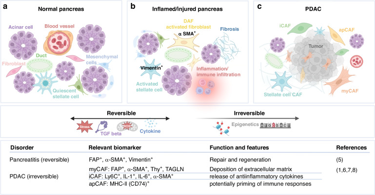
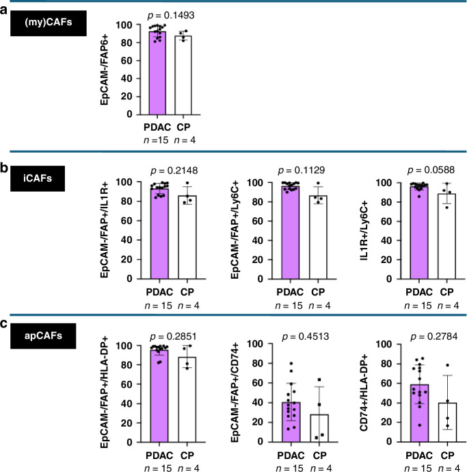
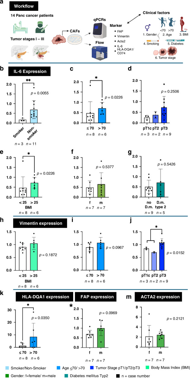
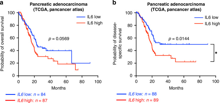
Cancer-associated fibroblasts (CAFs) constitute an important cell population in the microenvironment of pancreatic cancer. They can arise from disease-associated fibroblasts (DAFs) to support or restrain tumor growth. How many CAF subtypes exist and what signals drive their development is unclear. Currently, there are three commonly accepted subtypes, namely myofibroblast-like (myCAF), immunomodulatory (iCAF), and antigen-presenting (apCAF). Here, we analyzed the correlation between clinical risk factors with the proportion of each CAF subtype. In our patient cohort (n = 21), we investigated DAFs from patients with chronic pancreatitis (CP) and CAFs from pancreatic ductal adenocarcinoma (PDAC) patients after surgical resection via flow cytometry and RNA expression analysis. The expression of iCAF marker Interleukin-6 displayed significant differences depending on lifestyle factors, such as smoking status, age, and Body Mass Index (BMI). The apCAF marker HLA-DQA1 correlated with age. The largest difference showed the quantitative difference of apCAF markers in ~40% of PDAC- and ~20% of CP patients. In conclusion, clinical risk factors may influence the prevelance of specific CAF subsets. Unraveling the complex interplay between CAFs and tumor cells is crucial for novel therapies to improve long-term survival for pancreatic cancer patients.


Conflict of interest statement

Competing interests: The authors declare no competing interests. Ethics approval and consent to participate: This study was approved by the ethics committees of the University Hospital Carl Gustav Carus Dresden and the University Hospital Halle (Ethical approval EK 76032013 Dresden, Ethical approval 037 Halle). Written informed consent was obtained from all patients. The study was performed in accordance with the Declaration of Helsinki.

Figures

Fig. 1
Fig. 1. Fibroblasts in the normal pancreatic microenvironment, during activation and transition to cancer-associated fibroblasts.
In the healthy pancreatic microenvironment, fibroblasts and their subgroups like pancreatic stellate cells are, e.g., responsible for storing lipids and vitamin A (a). Due to factors like stress, ROS, and others, they get reversibly activated as disease-associated fibroblasts (DAFs) α-SMA+, FAP+, Vimentin+ (b). Functionally, they repair and regenerate injured tissue. After further epigenetic activation, they can irreversibly transition to cancer-associated fibroblasts. The most common subgroups are myCAFs: FAP+, α-SMA+, Thy+, TAGLN+, iCAFs: Ly6C+, IL-1+, IL-6+, α-SMA+ and apCAFs: MHC-II (CD74) + (c). Created with BioRender.com
Fig. 2
Fig. 2. Immunocytochemistry (ICC) of DAFs and CAFs confirm α - SMA, FAP and Vimentin expressions in cells from PDAC and chronic pancreatitis patients.
Representative staining displays cytoplasmatic α-SMA (a), FAP (b), and Vimentin (c). Expression levels of α-SMA (a), FAP (b), and Vimentin (c) are compared in CAFs of PDAC (n = 16) vs. DAFs of chronic pancreatitis/ pre-disease patients (n = 6). Magnification: x4.
Fig. 3
Fig. 3. Flow cytometry analysis for fibroblast subtype classification.
Percentages of EpCam-/ FAP+ fibroblasts in PDAC (n = 15) and CP (n = 4) samples (a). Percentages of EpCam-/FAP+/IL1R+ -, EpCam-/FAP+/Ly6C+-, and EpCam-/FAP+/IL1R+/Ly6C+ fibroblasts in PDAC (n = 15) and CP (n = 4) samples (b). EpCam-/FAP+/HLA-DP+-, EpCam-/FAP+/CD74+-, and EpCam-/FAP+/HLA-DP+/CD74+ fibroblasts in PDAC (n = 15) and CP (n = 4) samples (c).
Fig. 4
Fig. 4. Correlation of clinical risk factors with the expression of CAF subset markers.
Clinical risk factors for pancreatic diseases and tumor outcomes like Age, BMI, smoking, diabetes type2, and tumor stage were correlated with markers for the CAF subgroups myCAFs, iCAFs, and apCAFs (a). Expression levels of IL-6 (bg), Vimentin (hj). HLA-DQA1 (k), FAP (l), and ACTA2 (m) are shown. Unpaired t-tests were applied depending on experimental results. Otherwise, Kolmogorov–Smirnov tests were applied. Created with BioRender.com.
Fig. 5
Fig. 5. Survival analysis in patients expressing high- or low IL6 after RNA sequencing of tumor bulk tissues.
Kaplan–Meier plot showing Disease-specific Survival (n = 171) (a) and Overall Survival (n = 177) (b) of patients with PDAC having a low IL6 or high IL6 level in RNA of tumor bulk tissues. P values are as indicated in the respective graph.

Similar articles

References

    1. Halbrook CJ, Lyssiotis CA, Pasca Di Magliano M, Maitra A. Pancreatic cancer: advances and challenges. Cell. 2023;186:1729–54. - PMC - PubMed
    1. Rahib L, Wehner MR, Matrisian LM, Nead KT. Estimated projection of US cancer incidence and death to 2040. JAMA Netw Open. 2021;4:e214708. - PMC - PubMed
    1. Kleeff J, Korc M, Apte M, La Vecchia C, Johnson CD, Biankin AV, et al. Pancreatic cancer. Nat Rev Dis Primer. 2016;2:16022. - PubMed
    1. Sherman MH, Beatty GL. Tumor microenvironment in pancreatic cancer pathogenesis and therapeutic resistance. Annu Rev Pathol Mech Dis. 2023;18:123–48. - PMC - PubMed
    1. Huang H, Wang Z, Zhang Y, Pradhan RN, Ganguly D, Chandra R, et al. Mesothelial cell-derived antigen-presenting cancer-associated fibroblasts induce expansion of regulatory T cells in pancreatic cancer. Cancer Cell. 2022;40:656–673.e7. - PMC - PubMed

LinkOut - more resources