. 2025 Jun 16;9(1):186.
doi: 10.1038/s41698-025-01004-6.

Targeting SIX2 as a novel sensitization strategy of sorafenib treatment on advanced hepatocellular carcinoma through modulating METTL9-SLC7A11 axis

Affiliations

Targeting SIX2 as a novel sensitization strategy of sorafenib treatment on advanced hepatocellular carcinoma through modulating METTL9-SLC7A11 axis

Junren Lu et al. NPJ Precis Oncol. .

Abstract

Sorafenib is the main treatment for advanced hepatocellular carcinoma (HCC), but drug resistance limits its effectiveness. Evidence increasingly indicates that, in addition to targeting tyrosine kinases, sorafenib also induces ferroptosis. However, current studies have not fully clarified the relationship between ferroptosis and sorafenib treatment sensitivity. Our bioinformatics analysis identified that SIX Homeobox 2 (SIX2), known for maintaining cellular stemness via the Wnt signaling pathway, was significantly upregulated in sorafenib-resistant tissues. Overexpression and knockdown experiments revealed that altering SIX2 expression affected HCC cell sensitivity to sorafenib and involved the ferroptosis pathway, suggesting a regulatory role for SIX2 in ferroptosis. RNA sequencing and CUT&Tag analysis showed that SIX2 directly regulated methyltransferase 9 (METTL9) expression. Co-immunoprecipitation (Co-IP) assays confirmed that METTL9 bound to SLC7A11, enhancing its stability and reducing degradation, thus regulating ferroptosis. Importantly, the role of SIX2 in ferroptosis operated independently of the classical glutathione peroxidase 4 (GPX4) pathway. In vitro studies further supported these findings, demonstrating that SIX2 knockdown increased sorafenib-induced ferroptosis in HCC, while METTL9 overexpression largely counteracted the effects of SIX2 knockdown. In mouse models, overexpression of SIX2 increased tumor resistance to sorafenib. Our findings suggest that modulating the ferroptosis pathway through SIX2 could enhance sorafenib sensitivity. This study provides the first evidence that SIX2 influences ferroptosis via the METTL9-SLC7A11 axis, thereby sensitizing HCC cells to sorafenib. Reducing SIX2 expression could thus represent a promising strategy to improve the efficacy of sorafenib in advanced HCC.


Conflict of interest statement

Competing interests: The authors declare no competing interests.

Figures
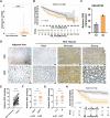
Fig. 1
Fig. 1. High SIX2 expression is associated with poor prognosis in hepatocellular carcinoma.
A SIX2 expression levels analyzed using the Cancer Genome Atlas (TCGA) database (374 HCC tissues and 50 non-tumorous tissues). B Kaplan-Meier analysis showing the relationship between SIX2 expression and overall patient survival in hepatocellular carcinoma (n = 346). C SIX2 expression levels in sorafenib-resistant and control cell lines from the GSE248769 dataset in the GEO database. D Representative immunohistochemical staining of SIX2 in HCC and adjacent non-tumorous tissues. Magnification: 100× (top), 400× (bottom). E Immunohistochemical staining scores of SIX2 in HCC and adjacent non-tumorous tissues. F-G SIX2 expression levels stratified by TNM stage and clinical stage, respectively. H Kaplan-Meier analysis of overall survival based on high and low SIX2 expression, determined using the optimal cutoff value.*p < 0.05, **p < 0.01, ***p < 0.001.
Fig. 2
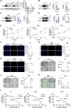
Fig. 2. SIX2 knockdown suppresses the malignant phenotype of HCC cells and increases sensitivity to sorafenib.
A Western blot analysis of SIX2 expression in normal liver and HCC cell lines. α-Tubulin served as a loading control. BD Western blot and qPCR analysis confirmed the knockdown efficiency of SIX2 in Li-7 and LM3 cell lines, as well as its overexpression in Huh7 cell lines. β-actin was used as a loading control. E Proliferation of HCC cells was measured by CCK-8 assay after SIX2 knockdown or overexpression. FH Proliferation of HCC cells was assessed using EdU assay following SIX2 knockdown or overexpression. IK Migration and invasion abilities of SIX2 knockdown or overexpression cell lines were evaluated using the transwell assay. L CCK-8 assay to determine the IC50 of sorafenib in SIX2 knockdown cell lines (Li-7 and LM3) and SIX2 overexpressing cell line (Huh7). All experimental results represent the mean values obtained from three independent experiments. *p < 0.05, **p < 0.01, ***p < 0.001. EV empty vector, OE overexpression of SIX2.
Fig. 3
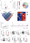
Fig. 3. SIX2 binding in the METTL9 promoter region and activation of transcription.
A Volcano plot of differentially expressed genes(DEGs) in SIX2 knockdown cell lines versus control cell lines. Red represents upregulated genes, and blue represents downregulated genes. And label the top 5 DEGs (up and down). B KEGG pathway enrichment analysis of differentially expressed genes. C Pie chart showing CUT&Tag sequencing of SIX2 binding regions. D Venn diagram displaying the overlap analysis of mRNA-Seq for differentially expressed genes, CUT&Tag-Seq for potential promoter-binding genes, and predicted data from the TCGA and GTRD public databases. E Heatmap of differentially expressed genes in SIX2 knockdown cell lines versus control cell lines. F Correlation analysis of the TCGA database indicating a positive correlation between SIX2 and METTL9 expression. G CUT&Tag sequencing results showing a binding peak of SIX2 in the METTL9 promoter region. The IgG group served as the control group. H CUT&Tag-qPCR showed enrichment of the METTL9 promoter region with SIX2 binding fragments in LM3 and Huh7 cell lines. IL Western blot and qPCR were performed to detect METTL9 expression in SIX2 knockdown or overexpressing cell lines. β-actin was used as a loading control. All experimental results represent the mean values obtained from three independent experiments. *p < 0.05, **p < 0.01, ***p < 0.001. EV empty vector, OE overexpression of SIX2.
Fig. 4
Fig. 4. SIX2 mediates ferroptosis in HCC cell lines.
AD Levels of lipid peroxides and lipid ROS in Li-7 and LM3 cells with SIX2 knockdown were detected by C11-BODIPY and DCFH-DA after 24 h of treatment with DMSO, IKE (20 μM), or sorafenib (10 μM). E, F Li-7 and LM3 cells with SIX2 knockdown were stained with Liperfluo and FerroOrange after 24 h of treatment with DMSO, IKE (20 μM), or sorafenib (10 μM). Intracellular lipid peroxides (green) and Fe²⁺(red) levels were visualized under a confocal microscope. G, H Knockdown of SIX2 in Li-7 and LM3 cells was followed by treatment with DMSO, IKE (20 μM), or sorafenib (10 μM) for 24 h. Intracellular MDA levels were detected by a colorimetric assay. I, J Intracellular lipid peroxides and lipid ROS levels in SIX2 overexpressing Huh7 cells were detected by C11-BODIPY and DCFH-DA after treatment with DMSO, IKE (10 μM), or sorafenib (5 μM) for 24 h. K Huh7 cells overexpressing SIX2 were stained with Liperfluo and FerroOrange after treatment with DMSO, IKE (10 μM), or sorafenib (5 μM) for 24 h. Intracellular lipid peroxides (green) and Fe²⁺(red) levels were visualized under a confocal microscope. L Intracellular MDA levels in Huh7 cells overexpressing SIX2 were detected by a colorimetric assay after treatment with DMSO, IKE (10 μM), or sorafenib (5 μM) for 24 h. All experimental results represent the mean values obtained from three independent experiments. *p < 0.05, **p < 0.01, ***p < 0.001. EV empty vector, OE overexpression of SIX2, IKE Imidazole ketone erastin, SOR sorafenib.
Fig. 5
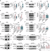
Fig. 5. METTL9 interacts with SLC7A11 and increases protein stability.
AC Efficiency of METTL9 knockdown or overexpression in HCC cell lines was detected by Western blot and qPCR. GAPDH was used as a loading control in the Western blot. DF SLC7A11, ACSL4, and GPX4 expression in HCC cell lines was detected by Western blot following METTL9 overexpression or knockdown. G Co-immunoprecipitation (co-IP) analysis of METTL9 and SLC7A11 was performed and detected by Western blot. H SLC7A11 protein expression in METTL9 knockdown and non-knockdown Huh7 cell lines was detected by Western blot after treatment with cycloheximide (CHX, 100 μg/mL). I Protein expression of SLC7A11 in METTL9 knockdown and non-knockdown Huh7 cell lines was detected by Western blot after treatment with MG-132 (10 μM) and bafilomycin A1 (Baf-A1) (10 μM) for 6 h. *p < 0.05, **p < 0.01, ***p < 0.001. MET METTL9, IKE Imidazole ketone erastin, SOR sorafenib, EV empty vector, OE overexpression of SIX2, NS not significant, co-IP co-immunoprecipitation, IB immunoblotting, CHX cycloheximide, Baf-A1 bafilomycin A1.
Fig. 6
Fig. 6. METTL9-SLC7A11 mediates ferroptosis in HCC cells caused by SIX2 knockdown.
AD Levels of lipid peroxides and lipid ROS in Li-7 and LM3 cells overexpressing METTL9 after SIX2 knockdown were detected by C11-BODIPY and DCFH-DA after 24 h of treatment with DMSO, IKE (20 μM), or sorafenib (10 μM). EF Li-7 and LM3 cells overexpressing METTL9 after SIX2 knockdown were stained with Liperfluo and FerroOrange after 24 h of treatment with DMSO, IKE (20 μM), and sorafenib (10 μM). The intracellular levels of lipid peroxides (green) and Fe²⁺(red) were visualized under a confocal microscope. GH Li-7 and LM3 cells with SIX2 knockdown followed by METTL9 overexpression were treated with DMSO, IKE (20 μM), or sorafenib (10 μM) for 24 h. Intracellular MDA levels were then detected by a colorimetric assay. IJ Intracellular lipid peroxides and lipid ROS levels in Huh7 cells with METTL9 knockdown after SIX2 overexpression were detected by C11-BODIPY and DCFH-DA after 24 h of treatment with DMSO, IKE (10 μM), or sorafenib (5 μM). K Intracellular lipid peroxides (green) and Fe²⁺ (red) levels were observed under a confocal microscope after treatment with DMSO, IKE (10 μM), or sorafenib (5 μM) for 24 h. Lipid peroxides were stained with Liperfluo, while Fe²⁺ was stained with FerroOrange. L Huh7 cells with METTL9 knockdown after SIX2 overexpression were treated with DMSO, IKE (10 μM), or sorafenib (5 μM) for 24 h. Intracellular MDA levels were then detected by a colorimetric assay. All experimental results represent the mean values obtained from three independent experiments. *p < 0.05, **p < 0.01, ***p < 0.001. MET METTL9, IKE Imidazole ketone erastin, SOR sorafenib, EV empty vector, OE overexpression of SIX2, NS not significant.
Fig. 7
Fig. 7. SIX2-mediated ferroptosis and morphological changes observed by electron microscopy.
Mitochondrial morphological changes in Li-7 cells were observed using transmission electron microscopy after treatment with DMSO, IKE (20 μM), and sorafenib (10 μM) for 24 h. Scale bars: left, 1 μm; right, 500 nm.
Fig. 8
Fig. 8. Knockdown of SIX2 exhibits anti-tumor effects in vivo.
A Schematic diagram showing the inoculation of nude mice with subcutaneous tumors and subsequent treatment procedures. B Photographs of representative tumors in each group at the end of the Huh7 cell line subcutaneous tumor experiment. C Huh7 cell line subcutaneous tumors were measured every two days from day 6 until day 20. D Statistics of subcutaneous tumor weights of Huh7 cell line mice in each group. E Representative immunofluorescence images of tumors from each group. Magnification: 400x. F Representative hematoxylin and eosin (HE) staining and Ki-67 immunohistochemistry images for each group. Magnification: 400x. G Representative METTL9 and SLC7A11 immunohistochemical staining images with quantitative analysis for each group. Magnification: 200x. H Schematic diagram of this study drawn by figdraw. SIX2 modulates METTL9-SLC7A11-mediated iron death in HCC cells to sensitize the efficacy of sorafenib by decreasing the expression of SLC7A11, which may play a role by stabilizing SLC7A11 through METTL9 and inhibiting its degradation via the lysosomal pathway. *p < 0.05, **p < 0.01, ***p < 0.001. EV empty vector, OE overexpression of SIX2, SOR sorafenib.

Similar articles

References

    1. Bray, F. et al. Global cancer statistics 2018: GLOBOCAN estimates of incidence and mortality worldwide for 36 cancers in 185 countries. CA Cancer J. Clin.68, 394–424 (2018). - PubMed
    1. Siegel, R. L., Miller, K. D. & Jemal, A. Cancer statistics, 2020. CA Cancer J. Clin.70, 7–30 (2020). - PubMed
    1. Huang, D. Q., Mathurin, P., Cortez-Pinto, H. & Loomba, R. Global epidemiology of alcohol-associated cirrhosis and HCC: trends, projections and risk factors. Nat. Rev. Gastroenterol. Hepatol.20, 37–49 (2023). - PMC - PubMed
    1. Llovet, J. M. et al. Hepatocellular carcinoma. Nat. Rev. Dis. Prim.2, 16018 (2016). - PubMed
    1. Smith, R. A. et al. Cancer screening in the United States, 2019: a review of current American Cancer Society guidelines and current issues in cancer screening. CA Cancer J. Clin.69, 184–210 (2019). - PubMed

LinkOut - more resources