Review
. 2025 Jun 17;10(1):188.
doi: 10.1038/s41392-025-02246-3.

Malaria: past, present, and future

Affiliations
Review

Malaria: past, present, and future

Qilong Li et al. Signal Transduct Target Ther. .

Abstract

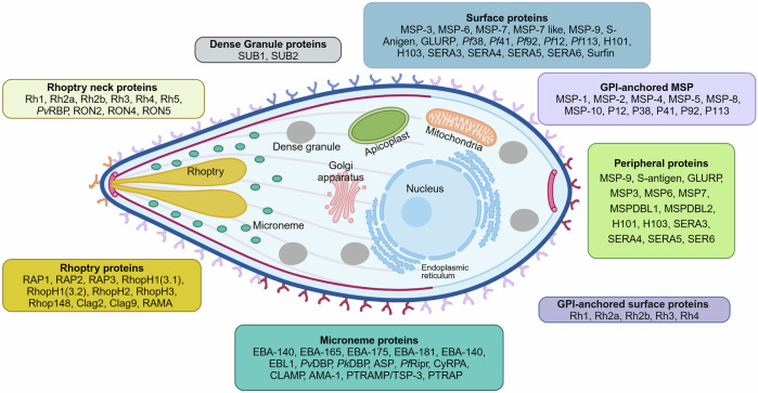
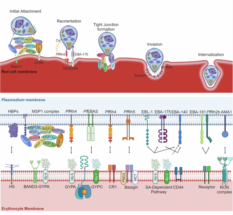
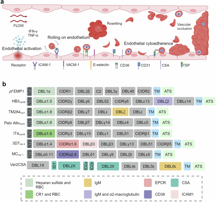
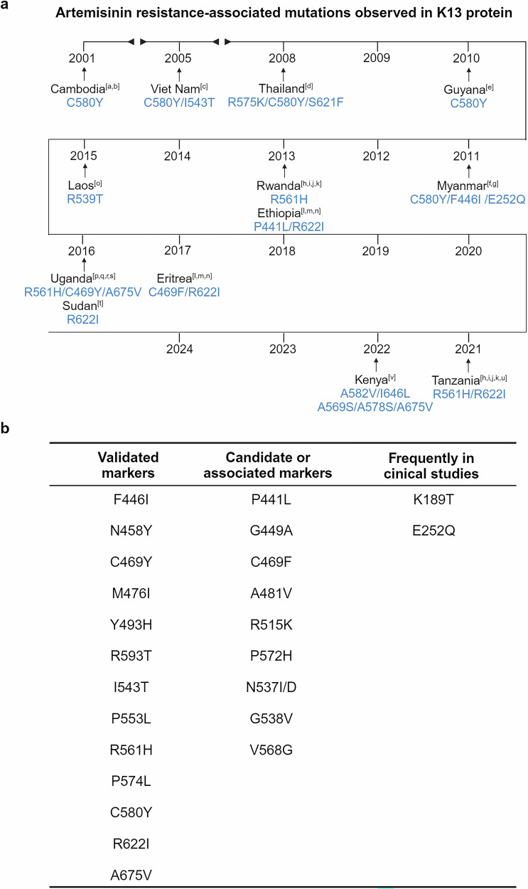
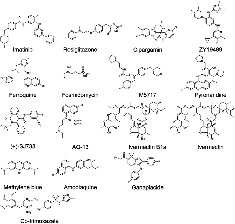
Malaria, caused by Plasmodium parasites and transmitted by Anopheles mosquitoes, greatly impacts public health and socioeconomic development, particularly in sub-Saharan African countries. Despite advances in malaria treatment and prevention, the number of clinical cases and deaths have increased in recent years. The complex life cycle and genetic diversity of Plasmodium parasites pose significant challenges in drug and vaccine development, particularly due to the emerging partial resistance of parasites to artemisinin. With the availability and application of state-of-the-art biotechnology in recent years, knowledge in terms of parasite biology, pathogenicity, host-parasite interactions and pathogenesis has advanced tremendously. This review highlights the most recent research progress and understanding in Plasmodium biology, with a primary focus on P. falciparum and associated pathogenesis. The therapeutic targets and progress in the clinical application of anti-malaria drugs have also been summarized. The FDA-approved regimens like Artemether-Lumefantrine, Atovaquone-Proguanil, and Primaquine are discussed, and their benefits and limitations are highlighted, especially in terms of drug resistance. Perspectives in the development of novel vaccines and new drugs, such as Sevuparin, Imatinib, and Cipargamin, and combination therapies with promise in overcoming resistance has been proposed. Overall, this review provides a detailed summary of the latest progress in malaria research and emphasizes the need for continuous monitoring and innovation in malaria treatment.


Conflict of interest statement

Competing interests: The authors declare no competing interests.

Figures

Fig. 1
Fig. 1
Milestones in malaria. The key milestones in the history of malaria research and control are depicted in this timeline. It highlights major discoveries, the development of treatments and vaccines, and significant global initiatives from the identification of Plasmodium parasites in 1880 to the approval of the RTS, S/AS01 (Mosquirix) malaria vaccine in 2018. This figure was created with BioRender.com
Fig. 2
Fig. 2
scRNA-seq analysis of P. falciparum. a A single P. falciparum-infected erythrocyte was manually isolated from a rosette and held by a 5-mm micropipette under a light microscope. b The scRNA sequences of P. falciparum in the asexual and sexual blood stages.
Fig. 3
Fig. 3
Pathophysiology of CM. a Subcortical petechial hemorrhages and microthrombi formation in the brain that occur during CM often result in ring hemorrhages and microvascular damage. b The interaction between iRBCs and endothelial cells in the cerebral vasculature. This figure is created with BioRender.com
Fig. 4
Fig. 4
Comparison of healthy and injured alveoli in malaria-induced acute lung injury. The healthy alveoli show intact epithelial and endothelial barriers, clear alveolar air spaces, and functional type I and II cells. In contrast, the injured alveoli display sloughing of the bronchial epithelium, protein-rich edema fluid, and necrotic type I cells. This figure was created with BioRender.com
Fig. 5
Fig. 5
The life cycle of P. falciparum and the known regulatory proteins associated with parasite development and differentiation. The Plasmodium parasite has a complex life cycle in both human and mosquito hosts. This figure was created with BioRender.com
Fig. 6
Fig. 6
Merozoite proteins involved in erythrocyte invasion of P. falciparum parasites. The various protein groups associated with different organelles in the Plasmodium parasite were shown. It categorizes key proteins into distinct groups, including rhoptry proteins, dense granule proteins, surface proteins, GPI-anchored surface proteins, peripheral proteins, and microneme proteins. Each protein group is color-coded for clarity and shown in association with the relevant organelle or cellular structure. This figure was created with BioRender.com
Fig. 7
Fig. 7
Mechanisms of Plasmodium invasion in erythrocytes. In the upper panel, the sequence of invasion begins with the attachment of the merozoite to the erythrocyte surface, followed by the discharge of the microneme and rhoptry contents, leading to apical reorientation, tight junction formation and erythrocyte membrane invagination, and the eventual entry of the merozoite into the erythrocyte. The lower panel highlights the key protein interactions during this process, revealing two distinct invasion pathways. HBP heparin-binding proteins, HS heparan sulfate, SA sialic acid, SA sialic acid, GYPA glycophorin A, GYPA glycophorin C, CR1 complement receptor 1, MSPs merozoite surface proteins, EBA erythrocyte binding antigen, EBL erythrocyte binding ligand, Rh reticulocyte binding protein homolog, AMA1 apical membrane antigen 1. This figure was created with BioRender.com
Fig. 8
Fig. 8
Endothelial cell activation and P. falciparum erythrocyte membrane protein 1 (PfEMP1)-mediated sequestration of iRBCs. a Mechanistic overview of the process by which activated endothelial cells express receptors that mediate the rolling and eventual sequestration of iRBCs. b PfEMP1 variants interact with distinct endothelial cell receptors. The PfEMP1 variants have been reviewed by Mats Wahlgren.
Fig. 9
Fig. 9
The immune responses during Plasmodium infection. The immune responses within the spleen during Plasmodium infection are shown. This figure was created with BioRender.com
Fig. 10
Fig. 10
The drugs and structures of anti-malaria drugs were approved by FDA. The drugs and structures shown include well-known treatments such as Artemisinin, Artemether, Dihydroartemisinin, Artesunate, and others like Atovaquone, Pyrimethamine, and Chloroquine, which are currently used in the treatment of malaria
Fig. 11
Fig. 11
PfKelch13 mutations. a The global emergence of ART resistance-associated mutations in the P. falciparum K13 protein from 2001 to 2024.The references are shown in [a], [b], [c], [d], [e], [f], [g], [h], [i], [j], [k], [l], [m], [n], [o], [p], [q], [r], [s], [t], [u], [v]. b Markers of artemisinin resistance in pfkelch13 and commonly observed mutations in clinical studies. The data was from WHO (https://www.who.int/news-room/questions-and-answers/item/artemisinin-resistance). This figure was created with BioRender.com
Fig. 12
Fig. 12
The structures of new anti-malaria drugs. The drugs presented include novel and experimental compounds that have shown potential against Plasmodium infections, such as Imatinib, Rosiglitazone, Cipargamin, ZY19489, and Ferroquine, among others. These structures represent different classes of anti-malaria agents currently under investigation or development, encompassing a range of mechanisms of action aimed at combating malaria

References

    1. Kwiatkowski, D. P. How malaria has affected the human genome and what human genetics can teach us about malaria. Am. J. Hum. Genet.77, 171–192 (2005). - PMC - PubMed
    1. Miller, L. H., Good, M. F. & Milon, G. Malaria pathogenesis. Science264, 1878–1883 (1994). - PubMed
    1. Cowman, A. F., Healer, J., Marapana, D. & Marsh, K. Malaria: biology and disease. Cell167, 610–624 (2016). - PubMed
    1. White, N. J. Severe malaria. Malar. J.21, 284 (2022). - PMC - PubMed
    1. Phillips, R. S. Current status of malaria and potential for control. Clin. Microbiol. Rev.14, 208–226 (2001). - PMC - PubMed

MeSH terms