

Accuracy of intraoral scans of the edentulous maxilla - an in vitro study
- PMID: 40524076
- PMCID: PMC12170734
- DOI: 10.1007/s00784-025-06419-w
Accuracy of intraoral scans of the edentulous maxilla - an in vitro study
Abstract
Objective: Investigation of the accuracy of various digitalization methods and the accuracy of digitalization of different regions of the edentulous maxilla.
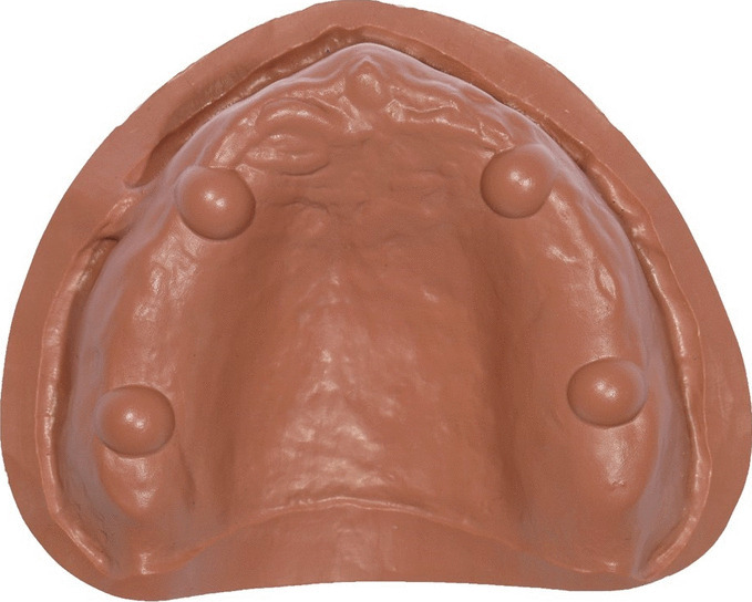
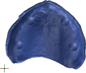
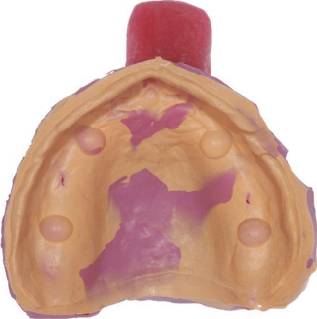
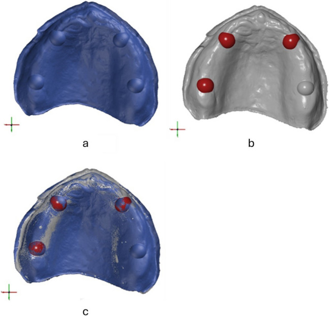
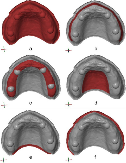
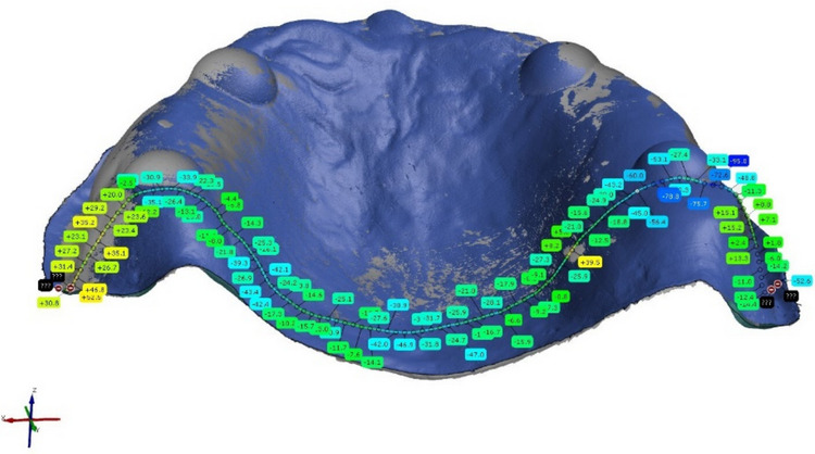
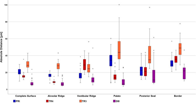
Material and method: A PEEK edentulous maxilla with four spherical reference geometries served as the testing model. A reference dataset (REF) was generated using a highly accurate 3D measuring instrument. The testing model was digitized as follows (n = 25/group). Direct digitalization (DD) with intraoral scanners (IOS): 1) Cerec AC Primescan (PRI), 2) Trios 4 Move (TR4), 3) Trios 3 Wireless (TR3), 4) Indirect digitalization of PVS impression with laboratory scanner 3Shape D810 (D8I). Three-dimensional deviations between REF and TEST were evaluated (GOM Inspect) in different areas of the model: 1) Complete Surface, 2) Alveolar Ridge, 3) Vestibular Ridge, 4) Palate, 5) Posterior Seal, 6) Border. Significant differences were analyzed with the Games-Howell test for trueness (p < 0.05) and multiple comparisons Levene's test for precision (for IOS: p < 0.008, for area: p < 0.003).
Results: Group D8I revealed the best trueness for Complete Surface (7.95 µm), Palate (9.11 µm), and Border (20.22 µm). Alveolar Ridge showed for PRI (16.45 µm) and TR4 (8.96 µm) the highest trueness. Groups TR4 and PRI resulted in significantly higher precision for Alveolar Ridge. Groups TR4 and D8I demonstrated the highest precision for Palate. Complete Surface and Alveolar Ridge showed for all digitalization methods significantly higher precision.
Conclusions: Indirect digitalization of impressions remains the most accurate approach for capturing edentulous jaws, whereas IOS deliver datasets with clinically acceptable accuracy. Peripheral regions characterized by limited accessibility and smooth surface morphology tend to demonstrate increased deviations in the resulting digital datasets.
Clinical relevance: Indirect digitalization of the impression still appears to be the most appropriate technique to access the clinical workflow for full dentures due to the superior digitalization trueness and inclusion of functional movements. Direct and indirect digitalization show nearly equal values for precision.
Keywords: Accuracy; Coordinate-based data analysis; Digital dentistry; Digital full-arch impression; Edentulous; Intraoral scanner; Precision; Trueness.
© 2025. The Author(s).
Conflict of interest statement
Declarations. Ethical approval: Not applicable. Informed consent: Not applicable. Competing interests: The authors declare no competing interests.
Figures
Similar articles
-
Feasibility, trueness and precision of intraoral scanners in digitizing maxillectomy defects with exposed zygomatic implants in situ: An in vitro 3D comparative study.J Dent. 2025 Feb;153:105557. doi: 10.1016/j.jdent.2025.105557. Epub 2025 Jan 10. J Dent. 2025. PMID: 39798233
-
Impact of implant scan body material and angulation on the trueness and precision of digital implant impressions using four intraoral scanners-an in vitro study.BMC Oral Health. 2025 Jul 31;25(1):1288. doi: 10.1186/s12903-025-06502-4. BMC Oral Health. 2025. PMID: 40745640 Free PMC article.
-
Trueness and precision of complete denture digital impression compared to conventional impression: an in vitro study.PeerJ. 2025 Feb 26;13:e19075. doi: 10.7717/peerj.19075. eCollection 2025. PeerJ. 2025. PMID: 40028199 Free PMC article.
-
Evaluation of the accuracy of intraoral scanners for complete-arch scanning: A systematic review and network meta-analysis.J Dent. 2023 Oct;137:104636. doi: 10.1016/j.jdent.2023.104636. Epub 2023 Jul 27. J Dent. 2023. PMID: 37516338
-
Accuracy of the digital implant impression with splinted and non-splinted intraoral scan bodies: A systematic review.J Indian Prosthodont Soc. 2025 Jan 1;25(1):3-12. doi: 10.4103/jips.jips_261_24. Epub 2025 Jan 3. J Indian Prosthodont Soc. 2025. PMID: 40654117 Free PMC article.
References
-
- Schweiger J et al (2018) Systematics and concepts for the digital production of complete dentures: risks and opportunities. Int J Comput Dent 21(1):41–56 - PubMed
-
- Wulfman C et al (2020) Digital removable complete denture: a narrative review. Fr J Dent Med 10:1–9
-
- Baba NZ et al (2021) CAD/CAM Complete denture systems and physical properties: a review of the literature. J Prosthodont 30(S2):113–124 - PubMed
-
- AlHelal A et al (2017) Comparison of retention between maxillary milled and conventional denture bases: A clinical study. J Prosthet Dent 117(2):233–238 - PubMed
MeSH terms
LinkOut - more resources
Full Text Sources
Research Materials
