Review
. 2025 Jun 16;30(1):72.
doi: 10.1186/s11658-025-00746-2.

The crucial role of metabolic reprogramming in driving macrophage conversion in kidney disease

Affiliations
Review

The crucial role of metabolic reprogramming in driving macrophage conversion in kidney disease

Na Gong et al. Cell Mol Biol Lett. .

Abstract

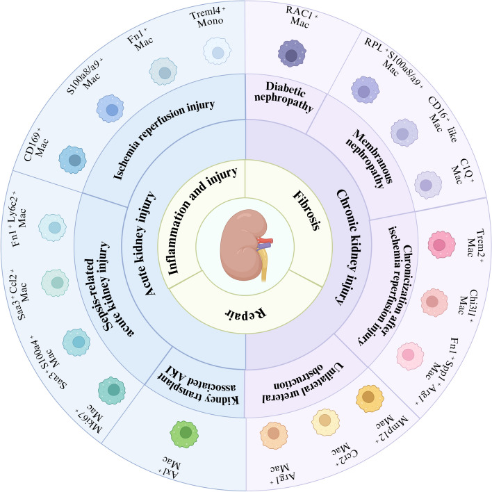
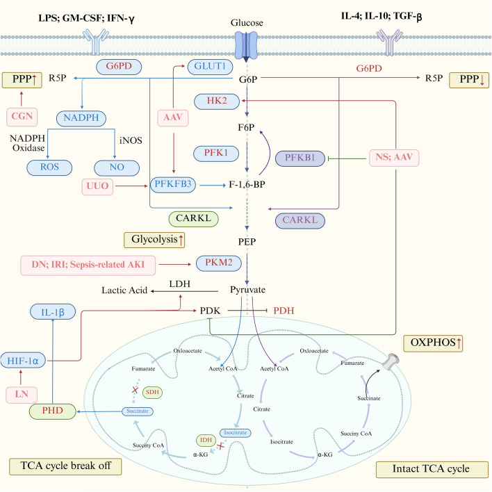
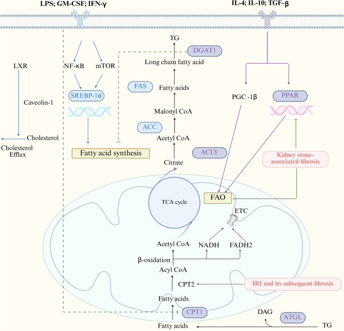
Interstitial fibrosis after acute kidney injury is an ongoing pathological process of chronic inflammatory injury and repair. Macrophages participate in renal inflammation, repair and fibrosis by continuously changing their phenotype and function. The tissue microenvironment of kidney injury induces changes in key metabolic enzymes, pathways and metabolites in macrophages, leading to phenotypic and functional conversions, but the detailed mechanisms are unclear. However, in the early phase of acute kidney injury, macrophages shift to a pro-inflammatory role relying on glycolysis and pentose phosphate pathways. The tissue microenvironment regulates the suppression of glycolysis-related genes and the up-regulation of oxidative phosphorylation and tricarboxylic acid cycle genes in macrophages, resulting in a gradual shift to an anti-inflammatory phenotype, which is involved in tissue repair and remodelling. In the late stage of injury, if macrophages continue to be overactive, they will be involved in renal fibrosis. The concomitant enhancement of nucleotide and amino acid metabolism, especially arginine and glutamine metabolism, is critical for the macrophage function and phenotypic transition during the above injury process. Macrophage metabolic reprogramming therefore provides new therapeutic targets for intervention in inflammatory injury and interstitial fibrosis in kidney disease.

Keywords: Inflammation; Injury and repair; Kidney fibrosis; Macrophages; Metabolic reprogramming.


Conflict of interest statement

Declarations. Ethics approval and consent to participate: Not applicable. This manuscript does not contain any studies with human participants or animals performed by any of the authors. Consent for publication: Not applicable. This manuscript does not include details, images, or videos relating to an individual person. Competing interests: All the authors declared no competing interests.

Figures

Fig. 1
Fig. 1
Molecular signatures of macrophage activation and transformation in kidney disease. Created in BioRender. na, g. (2025) https://BioRender.com/o8zi5tp. The innermost ring represents the core pathological processes, mainly including inflammation and injury, repair as well as fibrosis; the second ring denotes the different disease stages corresponding to the core pathological processes in the inner ring, namely the courses of acute kidney injury and chronic kidney disease; the third ring indicates the various aetiologies inducing acute kidney injury and chronic kidney disease models; the outermost ring signifies the analysis of major macrophage subtypes and marker genes in disease models of different aetiologies using single-cell RNA sequencing
Fig. 2
Fig. 2
Comparison of glucose metabolism reprogramming in macrophages under different functional states. Created in BioRender. na, g. (2025) https://BioRender.com/l11x496. 1. Left side of the figure: under the stimulation of LPS, GM—CSF, IFN -γ, etc., the pentose phosphate pathway and glycolysis are enhanced in pro-inflammatory macrophages, but the tricarboxylic acid cycle is interrupted. 2. Right side of the figure: under the stimulation of IL-4, IL-10, TGF -β, etc., the pentose phosphate pathway and glycolysis in macrophages decline, but there is a complete tricarboxylic acid cycle. 3. Among them, in NS and AAV, HK2 expression is upregulated while PFKB1 and PDK activities are inhibited. Notably, PKM2-mediated glycolysis is activated in DN, IRI and sepsis-induced AKI. Additionally, PPP is activated in CGN, PFKFB3 activity increases in UUO and AAV, GLUT1 expression is enhanced in AAV, and HIF-1α shows an upregulation trend in LN. 4. Blue boxes represent up-regulation in a pro-inflammatory state, purple boxes represent substances up-regulation in an anti-inflammatory state, green boxes represent substances down-regulation, red boxes represent kidney disease and red lines represent promotion and green lines represent inhibition. α-KG α-ketoglutarate, AAV Anti-neutrophil cytoplasmic antibody-associated vasculitis, CARKL Carbohydrate kinase-like, CGN Chronic glomerulonephritis, GLUT1 Glucose transporter 1, G6PD Glucose-6-phosphate dehydrogenease, HIF-1α Hypoxia inducible factor-1α, HK2 Hexokinase 2, IDH Isocitrate dehydrogenase, LDH Lactate dehydrogenase, LN Lupus nephritis, NS Glomerulonephritis, such as nephrotic syndrome, OXPHOS Oxidative phosphorylation, PEP Phosphoenolpyruvate, PFK1 Phosphofructokinase 1, PFKB1 Phosphofructokinase B1, PFKFB3 6-phosphofructo-2-kinase/fructose-2,6-bisphosphatase 3, PDH Pyruvate dehydrogenase, PDK Pyruvate dehydrogenase kinase, PHD Prolyl hydroxylase domain, PKM2 Pyruvate kinase M2, PPP Pentose phosphate pathway, ROS Reactive oxygen species, SDH Succinate dehydrogenase, TCA Tricarboxylic acid cycle, UUO Unilateral ureteral obstruction
Fig. 3
Fig. 3
Overview of lipid metabolic pathways in macrophages under different functional states. Created in BioRender. na, g. (2025) https://BioRender.com/t09f146. 1. Left side of the figure: under the stimulation of LPS, GM-CSF, IFN -γ, etc., fatty acid synthesis increases in pro-inflammatory macrophages; 2. Right side of the figure: under the stimulation of IL-4, IL-10, TGF-β, etc., fatty acid oxidation increases in anti-inflammatory macrophages. 3. Among them, PPAR and FAO are inhibited in kidney stone-associated fibrosis, and CPT2 is activated in IRI and its subsequent fibrosis.4. Blue boxes represent up-regulation in a pro-inflammatory state, purple boxes represent substances up-regulation in an anti-inflammatory state, green boxes represent substances down-regulation, red boxes represent kidney disease and red lines represent promotion and green lines represent inhibition. ACC Acetyl-CoA carboxylase, ACLY ATP citrate lyase, ATGL Adipose triglyceride lipase, CGI-58 Comparative gene identification-58, CPT1 Carnitine palmitoyltransferase 1, CPT2 Carnitine palmitoyl-transferase 2, DAG Diacylglycerol, DGAT-1 Diacyl glycerolacyltransferase 1, DN Diabetic nephropathy, FAO Fatty acid oxidation, FAS Fatty acid synthase, IRI Ischaemia–reperfusion injury, LXRs Liver X receptors, PGC-1β Peroxisome proliferator-activated receptor gamma coactivator 1β, PPAR-γ Peroxisome proliferator-activated receptor γ, SREBP-1α Sterol regulatory element-binding protein 1α, TG Triglyceride, UCP2 Uncoupling protein 2, UUO Unilateral ureteral obstruction

Similar articles

References

    1. Setten E, Castagna A, Nava-Sedeño JM, Weber J, Carriero R, Reppas A, et al. Understanding fibrosis pathogenesis via modeling macrophage-fibroblast interplay in immune-metabolic context. Nat Commun. 2022;13(1):6499. - PMC - PubMed
    1. Huang R, Fu P, Ma L. Kidney fibrosis: from mechanisms to therapeutic medicines. Signal Transduct Target Ther. 2023;8(1):129. - PMC - PubMed
    1. Wu CF, Chiang WC, Lai CF, Chang FC, Chen YT, Chou YH, et al. Transforming growth factor beta-1 stimulates profibrotic epithelial signaling to activate pericyte-myofibroblast transition in obstructive kidney fibrosis. Am J Pathol. 2013;182(1):118–31. - PMC - PubMed
    1. Lovisa S, Zeisberg M, Kalluri R. Partial epithelial-to-mesenchymal transition and other new mechanisms of kidney fibrosis. Trends Endocrinol Metab. 2016;27(10):681–95. - PubMed
    1. Lovisa S, Fletcher-Sananikone E, Sugimoto H, Hensel J, Lahiri S, Hertig A, et al. Endothelial-to-mesenchymal transition compromises vascular integrity to induce Myc-mediated metabolic reprogramming in kidney fibrosis. Sci Signal. 2020;13(635):eaaz2597. - PMC - PubMed