. 2025 Jun 16;31(1):240.
doi: 10.1186/s10020-025-01160-2.

4-Octyl itaconate alleviates endothelial cell inflammation and barrier dysfunction in LPS-induced sepsis via modulating TLR4/MAPK/NF-κB signaling : 4-Octyl itaconate alleviates endothelial dysfunction

Affiliations

4-Octyl itaconate alleviates endothelial cell inflammation and barrier dysfunction in LPS-induced sepsis via modulating TLR4/MAPK/NF-κB signaling : 4-Octyl itaconate alleviates endothelial dysfunction

Rong Li et al. Mol Med. .

Abstract

Aim: Sepsis-induced vascular injury is a major contributor to the high mortality rate of sepsis. However, effective treatments remain elusive due to limited knowledge regarding the underlying molecular mechanisms. Itaconic acid, an endogenous metabolite, involved in multiple inflammatory diseases, but its role in sepsis-induced vascular injury remains unclear. The current study investigates the effect of 4-octyl itaconate (4-OI), a cell-permeable derivative of itaconic acid, on sepsis-induced vascular injury and organ damage.

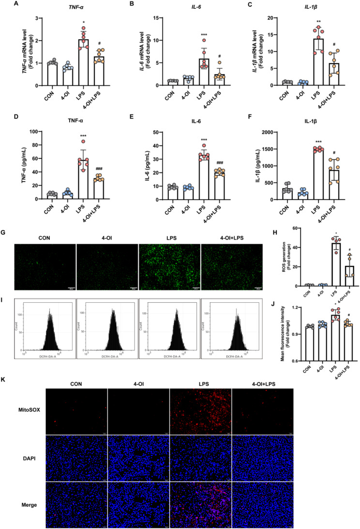
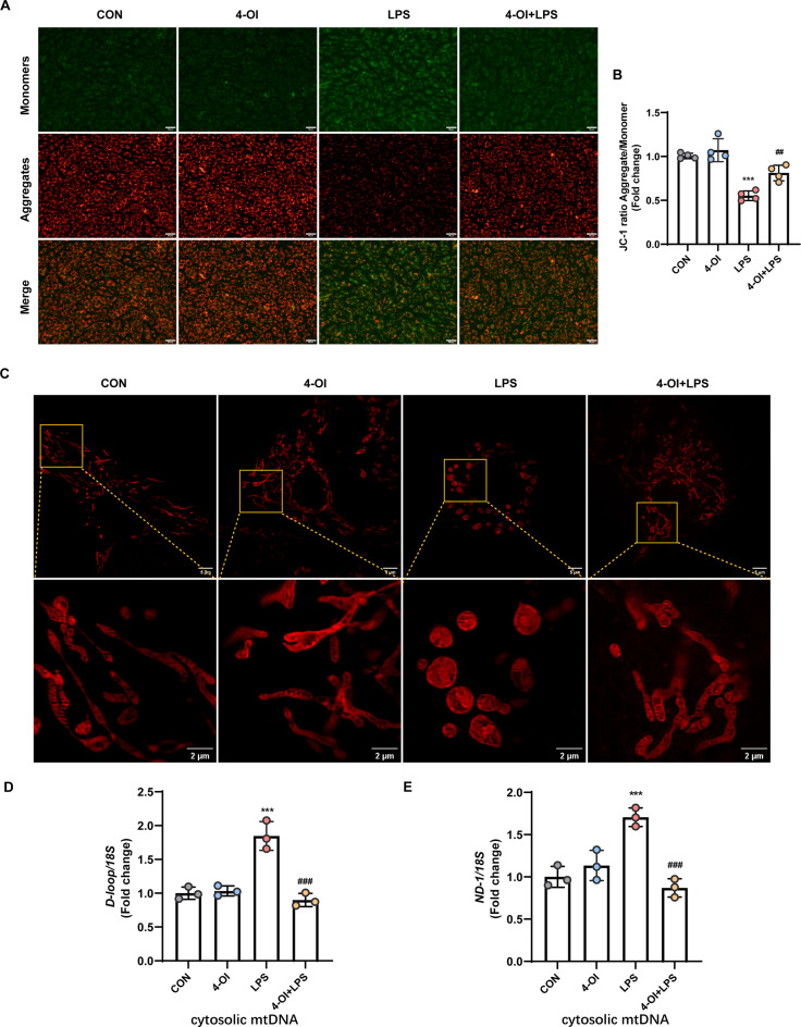
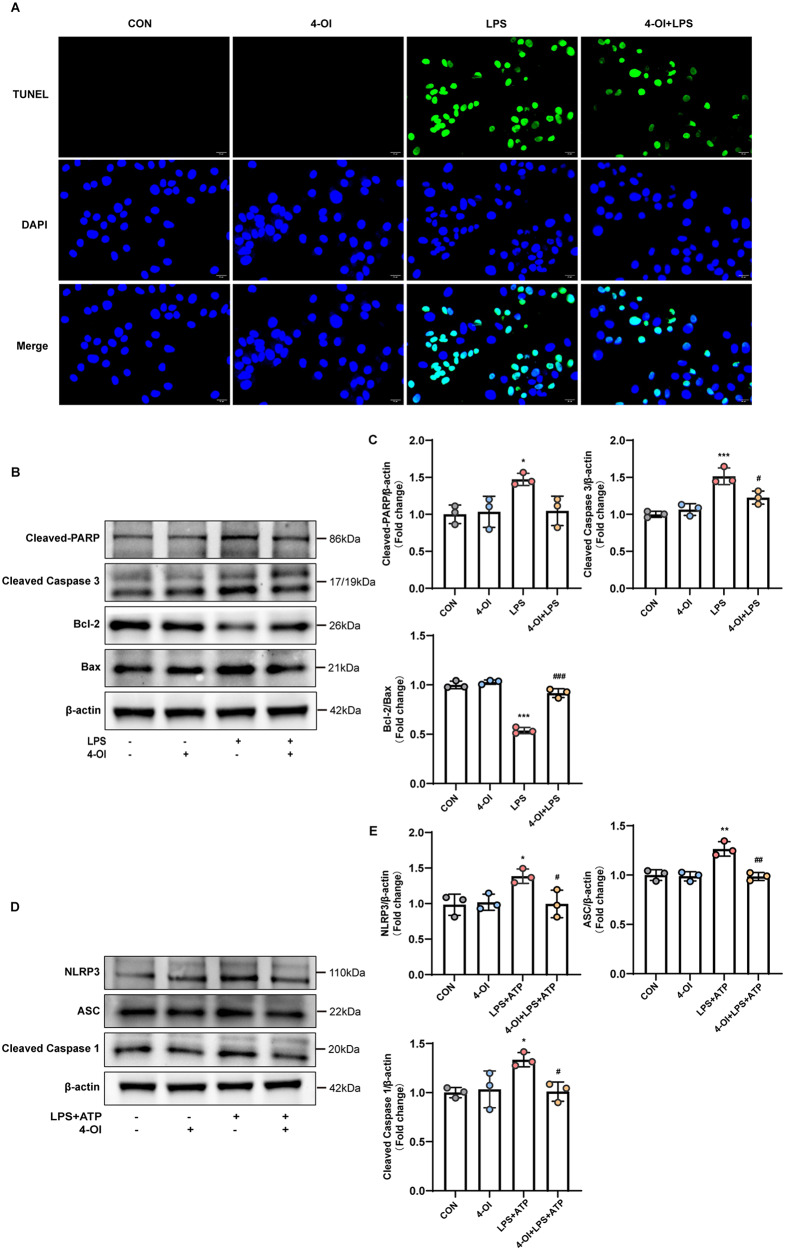
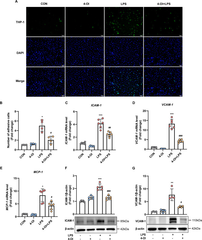
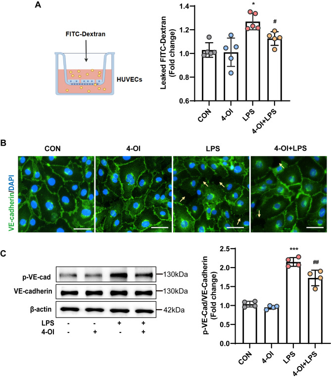
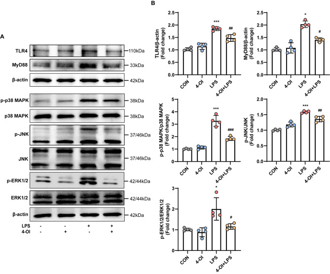
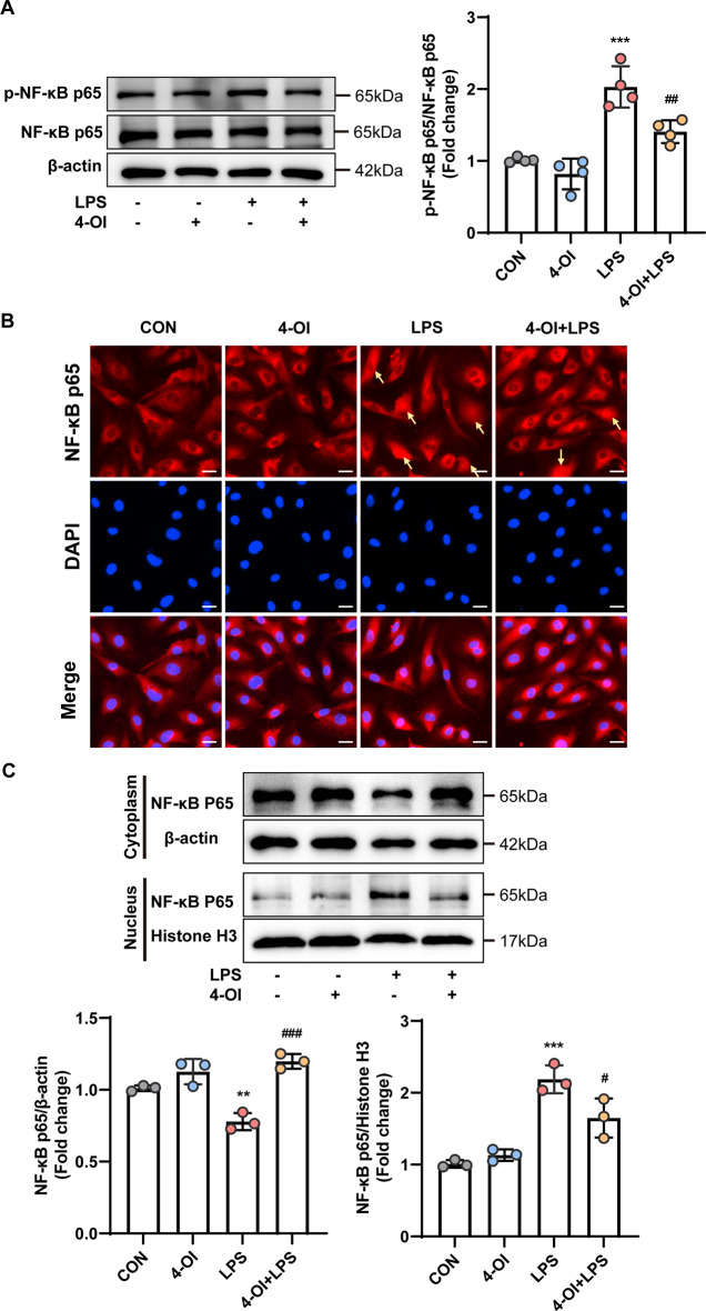
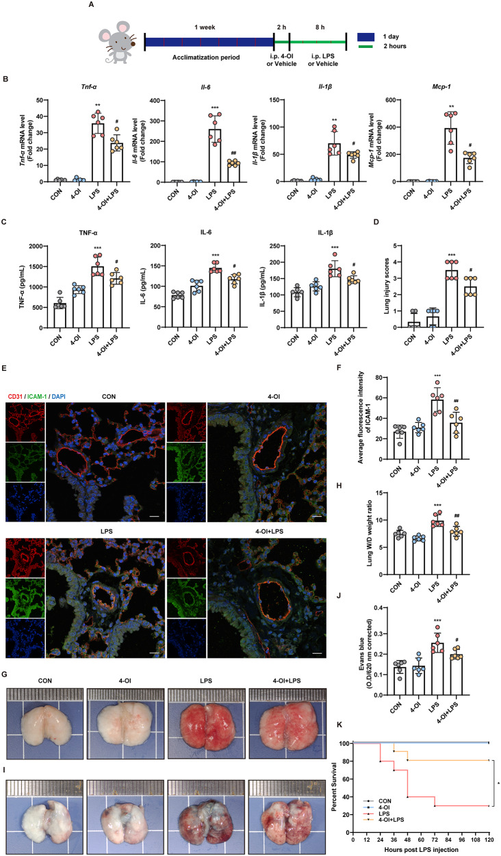
Methods and results: An in vitro cell model was established by treating human umbilical vein endothelial cells (HUVECs) with lipopolysaccharide (LPS). Quantitative reverse transcription polymerase chain reaction (qRT-PCR) and enzyme-linked immunosorbent assay (ELISA) revealed that 4-OI inhibited the LPS-induced increases in TNF-α, IL-6, and IL-1β levels. Cellular reactive oxygen species (ROS) levels, measured using the fluorescent probe DCFH-DA, mitochondrial ROS (mtROS) levels, measured by MitoSOX, and mitochondrial membrane potential (ΔΨ), detected by the fluorescent indicator JC-1, were all reduced following 4-OI treatment. Additionally, mtDNA release, detected by qRT-PCR, were decreased. Mitochondrial morphology, assessed by PK Mito Orange, was preserved by 4-OI treatment. Furthermore, 4-OI suppressed HUVECs apoptosis and pyroptosis, as detected by TUNEL staining and western blotting. 4-OI treatment also significantly inhibited LPS-induced cell adhesion, as shown in THP-1 attachment assay, by decreasing ICAM-1 and VCAM-1 expression. Cell permeability, determined by FITC-Dx-70 leakage, revealed that 4-OI effectively suppressed LPS-induced increases in cell permeability. Furthermore, 4-OI inhibited LPS-induced phosphorylation and internalization of VE-cadherin protein, preserving the adhesion junctions between endothelial cells. Network pharmacology and molecular docking analysis suggested the involvement of TLR4/MAPK/NF-κB signaling pathway as a key mechanism by which 4-OI ameliorated sepsis-induced vascular cell inflammation and injury, which was confirmed by western blotting. The in vitro results were subsequently verified in vivo in an LPS-induced sepsis mouse model. 4-OI pretreatment substantially decreased inflammatory cytokine levels in serum and lung tissues, inhibited pulmonary oedema and pulmonary vascular leakage, as evidenced by the wet-to-dry weight ratio and Evans blue staining of lung tissues, and alleviated tissue damage, as shown by histological analysis. Survival analysis indicated that 4-OI post-sepsis treatment improved the overall survival rate in LPS-induced ALI mice.

Conclusion: 4-OI protects against sepsis-induced vascular injury and tissue damage by suppressing endothelial inflammation, oxidative stress, and preserving endothelial barrier integrity.

Keywords: 4-Octyl Itaconate; Acute lung injury; Endothelial cell; Sepsis; Vascular injury.


Conflict of interest statement

Declarations. Ethical approval and consent to participate: All the animal procedures were approved by the Institutional Animal Care and Use Committee of Southwest Medical University and conformed to the Guide for the Care and Use of Laboratory Animals published by the US National Institutes of Health (NIH Publication No. 85-23, revised 1996). Ethical approval was granted by the Animal Experiment Ethics Committee of Southwest Medical University (No. 20221118-037). Consent for publication: Not applicable. Competing interests: The authors declare no competing interests.

Figures

Fig. 1
Fig. 1
4-OI alleviated LPS-induced inflammatory response and oxidative stress in HUVECs. Cells were pretreated with 4-OI (125 µM) for 3 h and then stimulated with 1 µg/mL LPS. (A-C) After LPS stimulation, the mRNA expression of the inflammatory cytokines TNF-α (A), IL-6 (B), and IL-1β (C) in HUVECs was measured by qRT-PCR (n = 6 samples per group). (D–F) After 8 h of LPS stimulation, ELISA kits were used to measure the levels of TNF-α (D), IL-6 (E), and IL-1β (F) in the culture supernatants of HUVECs (n = 6 samples per group). (G-H) DCFH-DA, a cell-permeable fluorescent probe, was added to the culture medium at a final concentration of 10 µM. DCFH reacts with ROS in the cells and is metabolized to generate fluorescent DCF. The fluorescence intensity of DCF was observed under a fluorescence microscope (n = 4 fields of view per group) (scale bar = 200 μm). (I-J) Flow cytometry was performed to evaluate the mean fluorescence intensity of DCF in each group, the fold increase in intracellular ROS levels relative to the CON group were visualized on a histogram (n = 6 samples per group). (K) Representative images of MitoSOX-stained mitochondrial ROS in HUVECs. After the indicated treatments, HUVECs were stained with 5 µM MitoSOX for 10 min, and images were captured with a fluorescence microscope (scale bar = 50 μm). MitoSOX (red), DAPI (blue). The p value was generated by one-way ANOVA followed by Turkey’s post hoc test. (*P < 0.05, **P <0.01, ***P < 0.001 compared with the CON group; #P < 0.05, ###P < 0.001 compared with the LPS group)
Fig. 2
Fig. 2
4-OI preserve mitochondrial function and structure. (A) HUVECs were treated with 4-OI, stimulated with LPS, and incubated with the JC-1 probe for 20 min, and images were captured with a fluorescence microscope (scale bar = 50 μm). (B) The fluorescence intensity of JC-1 monomers (490/530 nm) and aggregates (525/590 nm) was measured with a microplate reader, and the aggregate-to-monomer ratio was calculated (n = 4 samples per group). (C) Following treatment, HUVECs were washed and incubated with 125 nM PKMito Orange at 37 °C for 15 min in the dark. Super-resolution structured illumination microscopy (SIM) images were captured using a Multi-SIM X imaging system. (Upper: scale bar = 5 μm; Lower: magnified images, scale bar = 2 μm) (D-E) qRT-PCR analysis of cytosolic mtDNA (D-loop, ND-1) normalized to nuclear DNA (18 S) in HUVECs (n = 3 samples per group). The p value was generated by one-way ANOVA followed by Turkey’s post hoc test. (***P < 0.001 compared with the CON group; ##P < 0.01, ###P < 0.001 compared with the LPS group)
Fig. 3
Fig. 3
4-OI suppresses HUVECs apoptosis and pyroptosis. (A) TUNEL staining was applied to assess the effects of 4-OI on LPS-induced apoptosis of HUVECs. The cells are grown on the coverslip in 6 well plates, treated with 4-OI or LPS or both. The coverslips were fixed with paraformaldehyde followed by precooled ethanol and acetic acid. The coverslips were labeled with TUNEL (green) and counterstained with DAPI nuclear stain (blue). The pictures were taken with a fluorescence microscope (scale bar = 20 μm). (B-C) Western blotting was used to detect the protein expression of Cleaved-PARP, Cleaved-Caspase 3, Bcl-2, and Bax in whole cell lysates (n = 3 samples per group). (D-E) Western blotting was used to detect the protein expression of NLRP3, ASC, Cleaved-Caspase 1 in whole cell lysates (n = 3 samples per group). Representative images of 3 independent experiments are shown and the ration of the density of β-actin normalized to each protein was analyzed (n = 3 samples per group). For the above, all data shown are presented as fold changes. Error bar represents the standard deviation and p value was generated by one-way ANOVA followed by Turkey’s post hoc test. (*P < 0.05, **P < 0.01, ***P < 0.001 compared with the CON group; #P < 0.05, ##P < 0.01, ###P < 0.001 compared with the LPS+ATP group)
Fig. 4
Fig. 4
4-OI inhibited LPS-induced monocyte–endothelial cell adhesion. (A) Adhesion of THP-1 cells (green) labeled with the live cell tracer CMFDA to HUVECs; the cells were allowed to adhere for 30 min at 37 ℃ and imaged under a microscope (n = 4 fields of view per group) (scale bar = 100 μm). (B) Quantification of adhesion was normalized to that in the control group (without LPS stimulation). (C–E) qRT-RCR was used to measure the mRNA expression of ICAM-1 (C), VCAM-1 (D), and MCP-1 (E) in HUVECs (n = 6 samples per group). (F–G) Western blotting was used to detect the protein expression of ICAM-1 (F) and VCAM-1 (G) in whole cell lysates; Representative images of 3 independent experiments are shown and the expression of ICAM-1 and VCAM-1 relative to that of β-actin based on density analysis is shown above the blot (ICAM-1: n = 5 samples per group; VCAM-1: n = 4 samples per group). For the above, all data shown are presented as fold changes. Error bar represents the standard deviation and p value was generated by one-way ANOVA followed by Turkey’s post hoc test. (*P < 0.05, **P < 0.01, ***P < 0.001 compared with the CON group; #P < 0.05, ##P < 0.01, ###P < 0.001 compared with the LPS group)
Fig. 5
Fig. 5
4-OI mitigated the LPS-induced impairment of endothelial barrier integrity in vitro. (A) Endothelial permeability was assessed by measuring FITC-Dx-70 flux through HUVEC monolayers within 1 h in a transwell system (n = 5 samples per group). (B) Representative images of immunostaining for VE-cadherin (green) in HUVECs (n = 4 fields of view per group). Nuclei were stained with DAPI (blue) (scale bar = 5 μm). Yellow arrows: impaired inter-cellular adheren junctions between endothelial cells. (C) The levels of phospho-VE-cadherin in whole-cell lysates were detected by western blotting, and were normalized to those of VE-cadherin by density analysis (n = 4 samples per group). The p value was generated by one-way ANOVA followed by Turkey’s post hoc test. (*P < 0.05, ***P < 0.001 compared with the CON group; #P < 0.05, ##P < 0.01 compared with the LPS group)
Fig. 6
Fig. 6
4-OI inhibited LPS-induced TLR4/MAPK signaling pathway activation in HUVECs. (A) Western blotting was used to detect the protein expression of TLR4, MyD88, p-p38 MAPK, p-JNK, and p-ERK1/2 in whole cell lysates and representative images are shown. HUVECs in the 4-OI and 4-OI + LPS groups were pretreated with 4-OI exposure for 3 h, followed by LPS stimulation for various durations (TLR4 and MyD88, 24 h; p-ERK1/2, 15 min; p-p38 MAPK and p-JNK, 60 min) (B) Relative ratios of the band densities of TLR4/β-actin, MyD88/β-actin, p-p38 MAPK/p38 MAPK, p-ERK1/2/ERK1/2, and p-JNK/JNK (n = 4 samples per group). The p value was generated by one-way ANOVA followed by Turkey’s post hoc test. (*P < 0.05, ***P < 0.001 compared with the CON group; #P < 0.05, ##P < 0.01, ###P < 0.001 compared with the LPS group)
Fig. 7
Fig. 7
4-OI inhibited LPS-induced NF-κB signaling pathway activation in HUVECs. (A) The protein expression of p-NF-κB p65 in whole cell lysates was measured by western blotting and normalized to that of NF-κB p65 by density analysis (n = 4 samples per group). (B) Immunofluorescence analysis of the nuclear translocation of NF-κB p65 in HUVECs after 1 h of LPS stimulation. The arrow indicates the translocation of NF-κB p65 from the cytoplasm to the nucleus (n = 4 samples per group) (scale bar = 20 μm). (C) Western blotting was performed to detect the protein expression of NF-κB p65 in the cytoplasm and nucleus, with β-actin and histone H3 serving as the internal references in the cytoplasm and nucleus, respectively (n = 3 samples per group). The p value was generated by one-way ANOVA followed by Turkey’s post hoc test. (**P < 0.01, ***P < 0.001 compared with the CON group; #P < 0.05, ##P < 0.01, ###P < 0.001 compared with the LPS group)
Fig. 8
Fig. 8
4-OI alleviated lung injury and improved survival in sepsis-induced ALI mice. (A) Schematic diagram of the in vivo experimental design. (B) qRT-PCR was performed to measure the relative mRNA expression of Tnf-a, Il-6, Il-1β, and Mcp-1 in mouse lung tissues (n = 6 mice/group). (C) ELISA was performed to detect the protein levels of TNF-α, IL-1β, and IL-6 in mouse serum (n = 6 mice/group). (D) Semiquantitative analysis of lung injury in each group (n = 6 mice/group). (E-F) Immunofluorescence co-staining for CD31 (red) and ICAM-1 (green) in pulmonary vascular endothelial cells (n = 6 mice/group). Cell nuclei were stained with DAPI (blue) (scale bar = 200 μm). (G) Images of mouse lung tissues from each group (n = 6 mice/group). (H) The dissected lung tissues were weighed and dried at 60 ℃ for 72 h, and the wet weight -dry weight (W/D) ratio was calculated (n = 6 mice/group). (I) Images of mouse lung tissues after the injection of Evans blue. (J) Statistical analysis of the results of Evans blue staining using a microplate reader after formamide extraction (n = 6 mice/group). (K) Induction of ALI by a high dose of LPS (20 mg/kg, i.p.). The mortality of the mice was monitored every 12 h, and the percent survival rate was expressed as a Kaplan-Meier survival curve (n = 10 mice per group). The p value was generated by one-way ANOVA followed by Turkey’s post hoc test. (*P < 0.05, **P < 0.01, ***P < 0.001 compared with the CON group; #P < 0.05, ##P < 0.01 compared with LPS group in A-I; * P < 0.05 compared with the LPS group in K)

Similar articles

Cited by

References

    1. Akira S, Takeda K. Toll-like receptor signalling. Nat Rev Immunol. 2004;4(7):499–511. - PubMed
    1. Bambouskova M, Gorvel L, Lampropoulou V, Sergushichev A, Loginicheva E, Johnson K, Korenfeld D, Mathyer ME, Kim H, Huang LH, Duncan D, Bregman H, Keskin A, Santeford A, Apte RS, Sehgal R, Johnson B, Amarasinghe GK, Soares MP, Satoh T, Akira S, Hai T, de Strong G, Auclair C, Roddy K, Biller TP, Jovanovic SA, Klechevsky M, Stewart E, Randolph KM, Artyomov GJ. Electrophilic properties of Itaconate and derivatives regulate the IkappaBzeta-ATF3 inflammatory axis. Nature. 2018;556(7702):501–504. - PMC - PubMed
    1. Chen Y, Tang D, Zhu L, Yuan T, Jiao Y, Yan H, Yu W. hnRNPA2/B1 Ameliorates LPS-Induced Endothelial Injury through NF-kappaB Pathway and VE-Cadherin/beta-Catenin Signaling Modulation In Vitro. Mediators Inflamm 2020; 2020:6458791. - PMC - PubMed
    1. Cheng KT, Xiong S, Ye Z, Hong Z, Di A, Tsang KM, Gao X, An S, Mittal M, Vogel SM, Miao EA, Rehman J, Malik AB. Caspase-11-mediated endothelial pyroptosis underlies endotoxemia-induced lung injury. J Clin Invest. 2017;127(11):4124–4135. - PMC - PubMed
    1. Choi I, Son H, Baek JH. Tricarboxylic Acid (TCA) Cycle Intermediates: Regulators of Immune Responses. Life (Basel). 2021;11(1):69. - PMC - PubMed

LinkOut - more resources